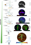
Diving head-first into brain intravital microscopy
- PMID: 38817606
- PMCID: PMC11137164
- DOI: 10.3389/fimmu.2024.1372996
Diving head-first into brain intravital microscopy
Abstract
Tissue microenvironments during physiology and pathology are highly complex, meaning dynamic cellular activities and their interactions cannot be accurately modelled ex vivo or in vitro. In particular, tissue-specific resident cells which may function and behave differently after isolation and the heterogenous vascular beds in various organs highlight the importance of observing such processes in real-time in vivo. This challenge gave rise to intravital microscopy (IVM), which was discovered over two centuries ago. From the very early techniques of low-optical resolution brightfield microscopy, limited to transparent tissues, IVM techniques have significantly evolved in recent years. Combined with improved animal surgical preparations, modern IVM technologies have achieved significantly higher speed of image acquisition and enhanced image resolution which allow for the visualisation of biological activities within a wider variety of tissue beds. These advancements have dramatically expanded our understanding in cell migration and function, especially in organs which are not easily accessible, such as the brain. In this review, we will discuss the application of rodent IVM in neurobiology in health and disease. In particular, we will outline the capability and limitations of emerging technologies, including photoacoustic, two- and three-photon imaging for brain IVM. In addition, we will discuss the use of these technologies in the context of neuroinflammation.
Keywords: brain; imaging; intravital microscopy; neuroinflammation; stroke.
Copyright © 2024 Suthya, Wong and Bourne.
Conflict of interest statement
The authors declare that the research was conducted in the absence of any commercial or financial relationships that could be construed as a potential conflict of interest.
Figures


References
-
- Waller A. XLIV. Microscopic examination of some of the principal tissues of the animal frame, as observed in the tongue of the living Frog, Toad, &c. London Edinburgh Dublin Philos Magazine J Science. (1846) 29:271–87. doi: 10.1080/14786444608645504 - DOI
-
- Cohnheim J. Ueber entzündung und eiterung. Archiv für pathologische Anatomie und Physiologie und für klinische Medicin. (1867) 40:1–79. doi: 10.1007/BF02968135 - DOI
Publication types
MeSH terms
LinkOut - more resources
Full Text Sources

