. 2024 May 16:15:1406438.
doi: 10.3389/fimmu.2024.1406438. eCollection 2024.

MRGPRX2 antagonist GE1111 attenuated DNFB-induced atopic dermatitis in mice by reducing inflammatory cytokines and restoring skin integrity

Affiliations

MRGPRX2 antagonist GE1111 attenuated DNFB-induced atopic dermatitis in mice by reducing inflammatory cytokines and restoring skin integrity

Trevor K Wong et al. Front Immunol. .

Abstract

Introduction: Atopic dermatitis (AD) is a chronic inflammatory skin disorder characterised by itching, erythema, and epidermal barrier dysfunction. The pathogenesis of AD is complex and multifactorial; however,mast cell (MC) activation has been reported to be one of the crucial mechanisms in the pathogenesis of AD. The MC receptor Mas related G protein-coupled receptor-X2 (MRGPRX2) has been identified as a prominent alternative receptor to the IgE receptor in causing MC activation and the subsequent release of inflammatory mediators. The current study aimed to evaluate the therapeutic effect of a novel small molecule MRGPRX2 antagonist GE1111 in AD using in vitro and in vivo approaches.

Methods: We developed an in vitro cell culture disease model by using LAD-2 MC, HaCaT keratinocytes and RAW 264.7 macrophage cell lines. We challenged keratinocytes and macrophage cells with CST-14 treated MC supernatant in the presence and absence of GE1111 and measured the expression of tight junction protein claudin 1, inflammatory cytokines and macrophage phagocytosis activity through immunohistochemistry, western blotting, RT-qPCR and fluorescence imaging techniques. In addition to this, we developed a DFNB-induced AD model in mice and evaluated the protective effect and underlying mechanism of GE1111.

Results and discussion: Our in vitro findings demonstrated a potential therapeutic effect of GE1111, which inhibits the expression of TSLP, IL-13, MCP-1, TNF-a, and IL-1ß in MC and keratinocytes. In addition to this, GE1111 was able to preserve the expression of claudin 1 in keratinocytes and the phagocytotic activity of macrophage cells. The in vivo results demonstrated that GE1111 treatment significantly reduced phenotypic changes associated with AD (skin thickening, scaling, erythema and epidermal thickness). Furthermore, immunohistochemical analysis demonstrated that GE1111 treatment preserved the expression of the tight junction protein Involucrin and reduced the expression of the inflammatory mediator periostin in the mouse model of AD. These findings were supported by gene and protein expression analysis, where GE1111 treatment reduced the expression of TSLP, IL-13, and IL-1ß, as well as downstream signalling pathways of MRGPRX2 in AD skin lesions. In conclusion, our findings provide compelling in vitro and in vivo evidence supporting the contribution of MRGPRX2-MC interaction with keratinocytes and macrophages in the pathogenesis of AD.

Keywords: MRGPRX2; allergy; atopic dermatitis; eczema; inflammation; small molecule antagonist.


Conflict of interest statement

The authors declare that the research was conducted in the absence of any commercial or financial relationships that could be construed as a potential conflict of interest. The author(s) declared that they were an editorial board member of Frontiers, at the time of submission. This had no impact on the peer review process and the final decision.

Figures

Figure 1
Figure 1
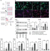
Effect of GE1111 on downstream CST-14- MRGPRX2 signalling pathway and inflammatory cytokine expression in LAD-2 MC. (A) LAD-2 MC were treated with GE1111 (50µM) or vehicle for 30 minutes, followed by CST-14 (5.81 µM) for 2 hours. [(A) (i)] Representative western blots of ERK 1/2 (P-ERK 1/2) and STIM1 protein expression. [(A) (ii & iii)] Bar graphs representing the relative band intensities for ERK 1/2 and STIM1. (B) Bar graphs representing the relative mRNA expression (normalised to housekeeping gene β actin) of (i) IL-13, (ii) IL-31, (iii) MCP-1 and (iv) TNF-α in LAD-2 MC measured by RT-qPCR. Data from 3–4 independent experiments are shown as means ± SEM. Statistical significance was determined by one-way ANOVA and Tukey’s multiple comparisons post-hoc test: *P < 0.05, ***P < 0.001, ****P < 0.0001.
Figure 2
Figure 2
GE1111 treatment recovered tight junction protein and inhibited inflammatory cytokines in HaCaT human keratinocyte cell- LAD-2 MC supernatant in vitro model. (A) Schematic representation of the design of HaCaT human keratinocyte cell- LAD-2 MC supernatant in vitro model. HaCaT human keratinocytes were challenged with different LAD-2 MC supernatants (vehicle/GE1111/CST-14 treated LAD-2 MC) for 2 hours, followed by immunofluorescence imaging, western blotting and RT-qPCR (created by BioRender.com). (B) Immunofluorescence imaging of HaCaT keratinocytes showing claudin 1 (green) and TSLP (red) expression. [(C) (i)] Representative western blots of claudin 1 and TSLP [(C) (ii, iii)] Bar graphs representing the relative band intensities for claudin 1 and TSLP. (D) Bar graphs representing the relative mRNA expression (normalised to housekeeping gene β actin) of (i) MCP-1, (ii) TNF-α, (iii) TSLP and (iv) IL-1ß in HaCaT keratinocytes quantified by RT-qPCR. Data from 3–5 independent experiments are shown as means ± SEM. Statistical significance was determined by one-way ANOVA and Tukey’s multiple comparisons post-hoc test: *P < 0.05, **P < 0.01, ***P < 0.001.
Figure 3
Figure 3
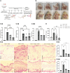
GE1111 treatment restores the phagocytotic response of RAW 264.7 macrophage-LAD-2 MC supernatant culture model. As shown in (A), RAW 264.7 macrophages were challenged with different LAD-2 MC supernatants (vehicle/GE1111/CST-14 treated LAD-2 MC) for 14–16 hours, followed by phagocytosis assay (created by BioRender.com). [(B) (i)] Fluorescence imaging showing phagocytosis of yellow beads by RAW 264.7 macrophages [(B) (ii)] Bar graph representing the ratio of beads to RAW 264.7 macrophage cells. Data from 3 independent experiments are shown as means ± SEM. Statistical significance was determined by one-way ANOVA and Tukey’s multiple comparisons post-hoc test: **P < 0.01, ****P < 0.0001.
Figure 4
Figure 4
GE1111 treatment ameliorates AD symptoms and inflammation in the DFNB-induced AD model. (A) Schematic representation of the design and timeline of the DFNB-induced AD model (created by BioRender.com). (B) Representative images of the mice's skin and ear at 1 week and at 4 weeks. (C) Bar graphs representing the (i) skin thickening, (ii) scaling, and (iii) erythema observed at the experimental endpoint of 4 weeks. ns, non significant. These symptoms of each mouse were scored on a scale of 0 to 3 [0 (none), 1 (mild), 2 (moderate), and 3 (severe)] (D) Quantification of serum MCP-1 in the experimental mice by ELISA kit. (E) Representative images of hematoxylin and eosin (H&E) staining of mouse skin and ear tissue. [(E) (ii, iii)] Bar graphs represent the mean thickness of the epidermis of the skin and ears of mice, respectively. The arrow indicates the epidermal region and inflammation, showing the hyperkeratosis of skin tissue. Data from 6–7 mice are shown as means ± SEM. Statistical significance was determined by one-way ANOVA and Tukey’s multiple comparisons post-hoc test: ns is non significant; **P < 0.01, ***P < 0.001, ****P < 0.0001.
Figure 5
Figure 5
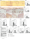
GE1111treatemnt inhibited in vivo MC degranulation, expression of inflammatory cytokines, downstream signalling and rescued the epidermal barrier protein expression in DFNB-induced AD mice. Male BALB/c mice aged 6–8 weeks (n=6–7) were treated with vehicle (VC), 50µl 0.3% DNFB (DC), 50µl 0.3% DNFB + 10 mg/kg GE1111 (T10) and 50µl 0.3% DNFB + 20mg/kg GE1111 (T20) once a week for 4 weeks. (A) (i) Representative images of toluidine blue stained ear sections of experimental mice. (ii) Bar graphs representing % degranulated MC in toluidine blue stained ear sections. (B) Representative immunohistochemistry images of skin tissue for expression of Involucrin and periostin. [(B) (ii, iii)] Bar graphs representing quantification of % area expresses Involucrin and periostin, respectively. (C) Bar graphs representing quantification of mRNA expression of (i) TSLP, (ii) IL-13 and (iii) IL-1β in the skin tissue of experimental mice by RT-qPCR. (D) Representative western blot images of STIM1, AKT, claudin, and TSLP protein expression. (D) Bar graphs representing the relative band intensities for (i) STIM1, (ii) p-AKT, (iii) claudin, and (iv) TSLP. Data from 3–7 independent experiments are shown as means ± SEM. Statistical significance was determined by one-way ANOVA and Tukey’s multiple comparisons post-hoc test: *P < 0.05, **P < 0.01, ***P < 0.001, ****P < 0.0001; ns, non significant.

Similar articles

Cited by

References

    1. Hadi HA, Tarmizi AI, Khalid KA, Gajdács M, Aslam A, Jamshed S. The epidemiology and global burden of atopic dermatitis: a narrative review. Life. (2021) 11:936. doi: 10.3390/life11090936 - DOI - PMC - PubMed
    1. Thomsen SF. Atopic dermatitis: natural history, diagnosis, and treatment. Int Scholarly Res Notices. (2014) 2014. doi: 10.1155/2014/354250 - DOI - PMC - PubMed
    1. Lopez Carrera YI, Al Hammadi A, Huang Y-H, Llamado LJ, Mahgoub E, Tallman AM. Epidemiology, diagnosis, and treatment of atopic dermatitis in the developing countries of Asia, Africa, Latin America, and the Middle East: a review. Dermatol Ther. (2019) 9:685–705. doi: 10.1007/s13555-019-00332-3 - DOI - PMC - PubMed
    1. Frazier W, Bhardwaj N. Atopic dermatitis: diagnosis and treatment. Am Fam Physician. (2020) 101:590–8. - PubMed
    1. Reynolds M, Gorelick J, Bruno M. Atopic dermatitis: a review of current diagnostic criteria and a proposed update to management. J Drugs Dermatol. (2020) 19:244–8. doi: 10.36849/JDD.2020 - DOI - PubMed

MeSH terms