. 2024 May 16:15:1400553.
doi: 10.3389/fimmu.2024.1400553. eCollection 2024.

Dynamic changes in immune cell populations by AXL kinase targeting diminish liver inflammation and fibrosis in experimental MASH

Affiliations

Dynamic changes in immune cell populations by AXL kinase targeting diminish liver inflammation and fibrosis in experimental MASH

Sturla Magnus Grøndal et al. Front Immunol. .

Abstract

Background and aims: Metabolic dysfunction-associated steatohepatitis (MASH) is a significant health concern with limited treatment options. AXL, a receptor tyrosine kinase activated by the GAS6 ligand, promotes MASH through activation of hepatic stellate cells and inflammatory macrophages. This study identified cell subsets affected by MASH progression and the effect of AXL inhibition.

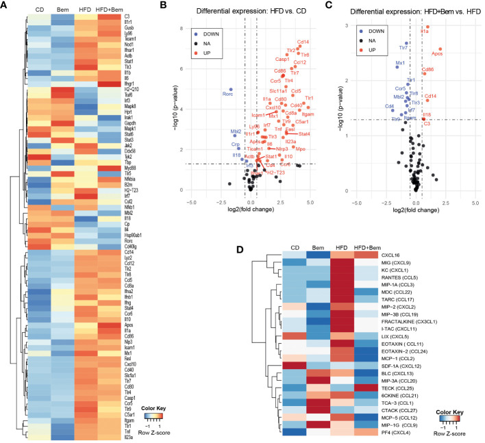
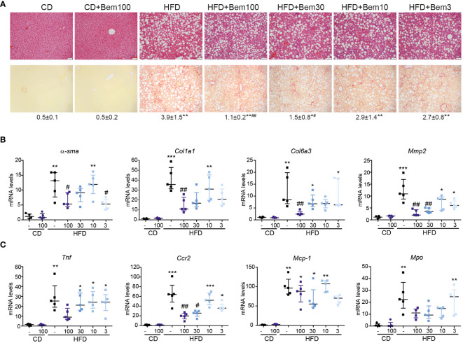
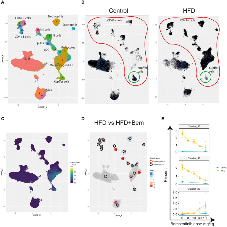
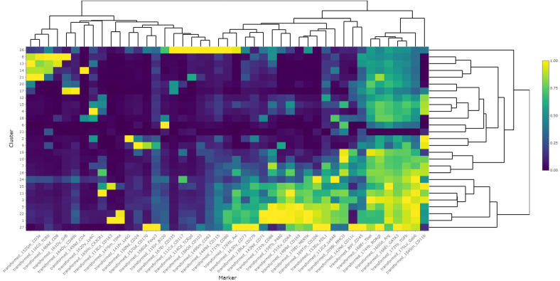
Methods: Mice were fed chow or different fat-enriched diets to induce MASH, and small molecule AXL kinase inhibition with bemcentinib was evaluated. Gene expression was measured by qPCR. Time-of-flight mass cytometry (CyTOF) used single cells from dissociated livers, acquired on the Fluidigm Helios, and cell populations were studied using machine learning.

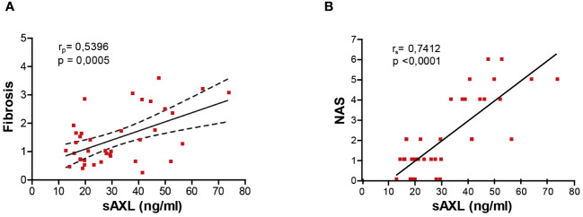
Results: In mice fed different fat-enriched diets, liver steatosis alone was insufficient to elevate plasma soluble AXL (sAXL) levels. However, in conjunction with inflammation, sAXL increases, serving as an early indicator of steatohepatitis progression. Bemcentinib, an AXL inhibitor, effectively reduced proinflammatory responses in MASH models, even before fibrosis appearance. Utilizing CyTOF analysis, we detected a decreased population of Kupffer cells during MASH while promoting infiltration of monocytes/macrophages and CD8+ T cells. Bemcentinib partially restored Kupffer cells, reduced pDCs and GzmB- NK cells, and increased GzmB+CD8+ T cells and LSECs. Additionally, AXL inhibition enhanced a subtype of GzmB+CD8+ tissue-resident memory T cells characterized by CX3CR1 expression. Furthermore, bemcentinib altered the transcriptomic landscape associated with MASH progression, particularly in TLR signaling and inflammatory response, exhibiting differential cytokine expression in the plasma, consistent with liver repair and decreased inflammation.

Conclusion: Our findings highlight sAXL as a biomarker for monitoring MASH progression and demonstrate that AXL targeting shifted liver macrophages and CD8+ T-cell subsets away from an inflammatory phenotype toward fibrotic resolution and organ healing, presenting a promising strategy for MASH treatment.

Keywords: GAS6; MERTK; TAM receptors; immune response; inflammation; liver fibrosis; mass cytometry.


Conflict of interest statement

These authors disclose the following: JL is a co-founder of BerGenBio. MB, LH, and GG were employed by BerGenBio. AJ is employed by BerGenBio. PG, MM, and AM received research funding from BerGenBio. MR has served as a consultant or on advisory boards: AstraZeneca, Bayer, BMS, Eli Lilly, Geneos, Ipsen, Merck, Roche, Universal DX; Speaking: AstraZeneca, Bayer, BMS, Eli Lilly, Gilead, ROCHE; Grant Research Support to the institution: Bayer, Ipsen; Educational Support to the institution: Bayer AstraZeneca, Eisai-MSD, ROCHE, Ipsen, Eli Lilly, Terumo, Next, Boston, Scientific, Ciscar Medical; Principal or sub-investigator of a drug under development: AbbVie, BMS, Adaptimmune, Nerviano, Bayer, Ipsen, AstraZeneca, Terumo, Incyte, ROCHE, Boston Scientific; Travel support: Terumo, AstraZeneca. The remaining authors declare that the research was conducted in the absence of any commercial or financial relationships that could be construed as a potential conflict of interest. The author(s) declared that they were an editorial board member of Frontiers, at the time of submission. This had no impact on the peer review process and the final decision.

Figures

Figure 1
Figure 1
Soluble AXL (sAXL) increases in the sera of steatotic mice before the onset of histological MASH. Representative images of liver sections after H&E and Sirius Red staining (A, E) non-alcoholic fatty liver disease (NAFLD) activity score (NAS) (B, F), ALT levels (C, G), and serum sAXL levels measured by ELISA (D, H) of mice fed with Western diet (WD) plus glucose/sucrose solution for 1 year (n = 5) and mice fed with a high-fat with fructose (HFHF) diet for 3 months (n = 8) compared with the respective control diet (CD) fed mice (n = 6, n = 4). Scale bar, 200 μm. H&E, hematoxylin and eosin. Mann–Whitney test; **p < 0.01 vs. CD.
Figure 2
Figure 2
Serum sAXL increases in concert with MASLD progression. Liver sections of mice fed with a high-fat, choline-deficient, and methionine-restricted diet (HFD) for 2 and 4 weeks were stained with H&E and Sirius Red (A). Triglycerides in liver extracts (B) and serum alanine aminotransferase (ALT) transaminases (C) were measured. mRNA expression level of Ccr2 and Col1a1 was measured in the liver samples (D, E). NAS was assessed (F), and serum sAXL was quantified by ELISA (G). Scale bar, 200 μm. H&E, hematoxylin and eosin; NAS, non-alcoholic fatty liver disease activity score. Kruskal–Wallis followed by Dunn multiple comparisons; *p ≤ 0.05, **p ≤ 0.01, and ***p ≤ 0.001 vs. control diet (CD), n = 5.
Figure 3
Figure 3
Bemcentinib administration prevents MASLD progression in 2-week HFD-fed mice. Liver sections of mice fed HFD for 2 weeks and treated with vehicle (n = 5) or bemcentinib 100 mg/kg (n = 5) for the last week, while on HFD, stained with H&E and Sirius Red (A). Triglycerides in liver extracts (B) and NAS (C) were determined. RNA expression of Col1a1 and Ccr2 in liver samples (D, E). Scale bar, 200 μm. H&E, hematoxylin and eosin; NAS, non-alcoholic fatty liver disease activity score. Mann–Whitney test; *p ≤ 0.05 vs. vehicle.
Figure 4
Figure 4
sAXL level is an early marker of liver fibrosis. Correlation of serum sAXL levels with liver fibrosis ImageJ quantification (A) and NAS (B). The coefficient of determination (r) and statistical significance (p-value) were determined using Pearson’s coefficient (A) since both variables are continuous and Spearman’s coefficient (B) since one variable is continuous and the other is ordinal.
Figure 5
Figure 5
Changes in immune response mediators induced by bemcentinib. Heatmap of row-normalized transcriptomic changes (row Z-score) related to the innate and adaptive immune response in liver samples of mice fed with HFD or chow diet (8 weeks) with the addition of bemcentinib or vehicle gavages for the last 2 weeks while continuing on HFD (A). Significantly upregulated genes are shown in red, while downregulated genes are in blue in the experimental MASH (B) and following bemcentinib treatment (C). Cytokines and chemokines in mice sera were determined by a glass slide-based protein array, and relative levels were plotted in a heatmap (row Z-score) (D). n = 2–5/group.
Figure 6
Figure 6
Dose–response effect of bemcentinib (Bem) in liver fibrosis in HFD-fed mice. Representative images of liver sections after H&E and Sirius Red staining from mice fed for 8 weeks with control diet (CD) and HFD diet that received vehicle or Bem for the last 2 weeks, while continuing on HFD, at different concentrations (100, 30, 10, and 3 mg/kg), n = 5 mice/group. Sirius Red quantifications are shown under representative pictures (mean ± SEM) (A). Fibrogenic (B) and inflammatory (C) gene expression in the liver (fold change vs. CD). Kruskal–Wallis test; *p < 0.05, **p ≤ 0.01, and ***p < 0.001 vs. CD; #p < 0.05 and ##p ≤ 0.01vs. HFD; n = 5/group.
Figure 7
Figure 7
Single-cell analysis from dissociated liver reveals changes in immune cell populations induced by bemcentinib in experimental MASH. Clusters and cell types identified in the livers using CyTOF (A). Comparison of UMAP density, immune cells (red), and Kupffer cells (green) in vehicle-treated mice fed control chow or HFD chow (B). UMAP colored by AXL signal intensity (C) with cluster labels colored by the effect of bemcentinib treatment in HFD-fed mice (blue: higher in bemcentinib-treated, red: lower in bemcentinib-treated). The red circle indicates adjusted p-value <0.05 (D). The percentages (mean ± SE) of the total selected clusters make up each sample across different dosages and diets (E).
Figure 8
Figure 8
Heatmap of scaled marker expression of each cluster. The mean asinh-transformed signal intensity was calculated for each cluster. The means of each channel in each cluster were then column-scaled from 0 to 1. A higher value indicates a higher mean expression in the given cluster compared with other clusters.
Figure 9
Figure 9
Proportionality between clusters suggests cell types that increase or decrease together. Heatmap colored by proportionality (rho) between clusters. Red is positive proportionality and blue is negative. A, B, C, and D label different groups of clusters that are proportional to each other. A and B are generally reduced in mice fed HFD, while C and D are increased.

Similar articles

Cited by

References

    1. Rinella ME. Nonalcoholic fatty liver disease: a systematic review. JAMA. (2015) 313:2263–73. doi: 10.1001/jama.2015.5370 - DOI - PubMed
    1. Younossi ZM, Koenig AB, Abdelatif D, Fazel Y, Henry L, Wymer M. Global epidemiology of nonalcoholic fatty liver disease-Meta-analytic assessment of prevalence, incidence, and outcomes. Hepatol Baltim. Md. (2016) 64:73–84. doi: 10.1002/hep.28431 - DOI - PubMed
    1. Schuppan D, Surabattula R, Wang XY. Determinants of fibrosis progression and regression in NASH. J Hepatol. (2018) 68:238–50. doi: 10.1016/j.jhep.2017.11.012 - DOI - PubMed
    1. Tutusaus A, de Gregorio E, Cucarull B, Cristóbal H, Aresté C, Graupera I, et al. . A functional role of GAS6/TAM in nonalcoholic steatohepatitis progression implicates AXL as therapeutic target. Cell. Mol. Gastroenterol. Hepatol. (2020) 9:349–68. doi: 10.1016/j.jcmgh.2019.10.010 - DOI - PMC - PubMed
    1. Cai B, Dongiovanni P, Corey KE, Wang X, Shmarakov IO, Zheng Z, et al. . Macrophage merTK promotes liver fibrosis in nonalcoholic steatohepatitis. Cell Metab. (2020) 31:406–421.e7. doi: 10.1016/j.cmet.2019.11.013 - DOI - PMC - PubMed

MeSH terms