

Single-Cell RNA Sequencing Analysis of Steroidogenesis and Spermatogenesis Impairment in the Testis of db/db Mice
- PMID: 38817616
- PMCID: PMC11139535
- DOI: 10.1155/2024/8797972
Single-Cell RNA Sequencing Analysis of Steroidogenesis and Spermatogenesis Impairment in the Testis of db/db Mice
Abstract
Objective: The mechanism of steroidogenesis and spermatogenesis impairment in men with type 2 diabetes remains unclear. We aimed to explore the local changes of steroidogenesis and spermatogenesis in the testis of db/db mice. Research Design and Methods. We performed single-cell RNA sequencing analysis in the testis of db/db and C57BL/6J mice. The differentially expressed genes were then confirmed by real-time PCR. The histopathological characteristics of testis in db/db mice and C57BL/6J control were also performed.
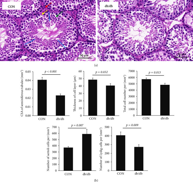
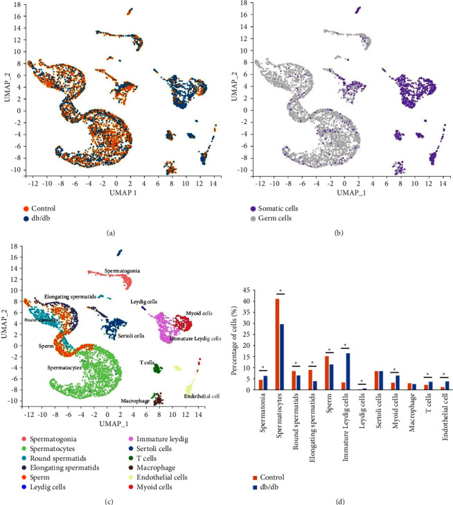
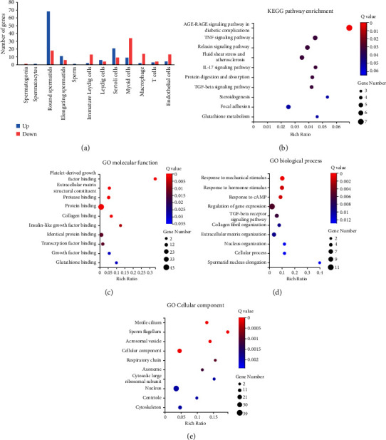
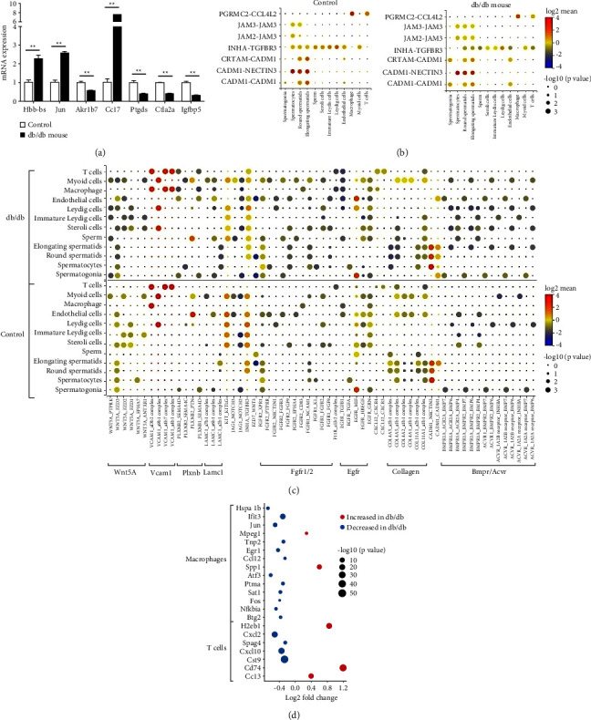
Results: The 20-week-old db/db mice had significantly higher blood glucose and body weight (both p < 0.001). The serum testosterone levels (4.4 ± 0.8 vs. 9.8 ± 0.7 ng/ml, p=0.001) and weight of the testis (0.16 ± 0.01 vs. 0.24 ± 0.01 g, p < 0.001) were significantly lower in db/db mice than that in C57BL/6J controls. db/db mice had a lower cross-sectional area of seminiferous tubules and thickness of the cell layer (both p < 0.05). The numbers of Sertoli cells and Leydig cells decreased in db/db mice (both p < 0.01). Single-cell RNA sequencing analysis showed that compared with the control group, the percentage of spermatogonia was significantly higher in the db/db mouse (p < 0.001), while the proportions of spermatocytes, round and elongating spermatids, and sperms were all lower in the db/db mouse (p all < 0.001). The most differentially expressed genes were found in round spermatids (n = 86), which were not found in spermatogonia, spermatocyte, and sperm. Igfbp5 was the most significantly decreased gene in Leydig cells of the db/db mouse, while the expression of Cd74, H2-Aa, and H2-Eb1 was elevated. Ccl7 and Ptgds were the most significantly increased and decreased genes in Sertoli cells of the db/db mouse.
Conclusions: The present study indicates spermiogenesis and steroidogenesis defects in db/db mice. The mechanism of steroidogenesis impairment in the testis of db/db mice deserves further investigation.
Copyright © 2024 Yun Hu et al.
Conflict of interest statement
The authors declare that there are no conflicts of interest.
Figures
Similar articles
-
Luteinizing hormone receptor-mediated effects on initiation of spermatogenesis in gonadotropin-deficient (hpg) mice are replicated by testosterone.Biol Reprod. 2004 Jan;70(1):32-8. doi: 10.1095/biolreprod.103.019398. Epub 2003 Sep 3. Biol Reprod. 2004. PMID: 12954730
-
Bcl-w forms complexes with Bax and Bak, and elevated ratios of Bax/Bcl-w and Bak/Bcl-w correspond to spermatogonial and spermatocyte apoptosis in the testis.Mol Endocrinol. 2000 May;14(5):682-99. doi: 10.1210/mend.14.5.0443. Mol Endocrinol. 2000. PMID: 10809232
-
Localization and expression of Orexin A and its receptor in mouse testis during different stages of postnatal development.Gen Comp Endocrinol. 2017 Jan 15;241:50-56. doi: 10.1016/j.ygcen.2016.05.006. Epub 2016 May 9. Gen Comp Endocrinol. 2017. PMID: 27174745
-
Ultrastructure of the aging human testis.J Electron Microsc Tech. 1991 Oct;19(2):241-60. doi: 10.1002/jemt.1060190209. J Electron Microsc Tech. 1991. PMID: 1748904 Review.
-
Spermatogenesis by Sisyphus: proliferating stem germ cells fail to repopulate the testis after 'irreversible' injury.Adv Exp Med Biol. 2001;500:421-8. doi: 10.1007/978-1-4615-0667-6_64. Adv Exp Med Biol. 2001. PMID: 11764975 Review.
References
-
- Barkabi-Zanjani S., Ghorbanzadeh V., Aslani M., Ghalibafsabbaghi A., Chodari L. Diabetes mellitus and the impairment of male reproductive function: possible signaling pathways. Diabetes and Metabolic Syndrome: Clinical Research Reviews . 2020;14(5):1307–1314. doi: 10.1016/j.dsx.2020.07.031. - DOI - PubMed
LinkOut - more resources
Full Text Sources
Miscellaneous
