. 2023 Jun 13;2(2):141-158.
doi: 10.1002/mlf2.12063. eCollection 2023 Jun.

A host E3 ubiquitin ligase regulates Salmonella virulence by targeting an SPI-2 effector involved in SIF biogenesis

Affiliations

A host E3 ubiquitin ligase regulates Salmonella virulence by targeting an SPI-2 effector involved in SIF biogenesis

Kun Meng et al. mLife. .

Abstract

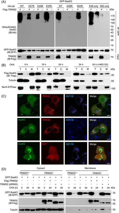
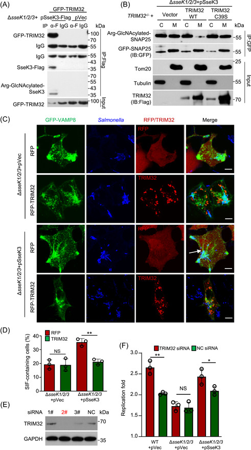
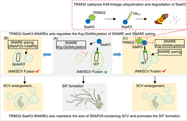
Salmonella Typhimurium creates an intracellular niche for its replication by utilizing a large cohort of effectors, including several that function to interfere with host ubiquitin signaling. Although the mechanism of action of many such effectors has been elucidated, how the interplay between the host ubiquitin network and bacterial virulence factors dictates the outcome of infection largely remains undefined. In this study, we found that the SPI-2 effector SseK3 inhibits SNARE pairing to promote the formation of a Salmonella-induced filament by Arg-GlcNAcylation of SNARE proteins, including SNAP25, VAMP8, and Syntaxin. Further study reveals that host cells counteract the activity of SseK3 by inducing the expression of the E3 ubiquitin ligase TRIM32, which catalyzes K48-linked ubiquitination on SseK3 and targets its membrane-associated portion for degradation. Hence, TRIM32 antagonizes SNAP25 Arg-GlcNAcylation induced by SseK3 to restrict Salmonella-induced filament biogenesis and Salmonella replication. Our study reveals a mechanism by which host cells inhibit bacterial replication by eliminating specific virulence factors.

Keywords: E3 ligase; SIF biogenesis; Salmonella; T3SS SPI‐2 effector.


Conflict of interest statement

The authors declare no conflict of interests.

Figures

Figure 1
Figure 1
SseK3 promotes Salmonella‐induced filament (SIF) formation by modifying SNARE proteins during Salmonella Typhimurium infection. (A and B) SseK3 promotes SIF formation during S. Typhimurium infection. HeLa cells stably expressing EGFP‐VAMP8 were infected with the indicated S. Typhimurium strains for 10 h and analyzed for SIFs. Arrow indicates the SIF structure. pVec indicates the pET28a empty vector. The pSseK3 DxD indicates the pET28a plasmid expressing glycosyltransferase motif mutant of SseK3. The ΔssaV (an SPI‐2‐deficient mutant) strain was used as a control in this model. (A) Representative images showing the distribution of VAMP8 (green) and S. Typhimurium (red). Scale bar, 10 μm. (B) The rates of VAMP8‐positive tubules for each sample are indicated. At least 50 cells were counted for samples from experiments conducted in triplicate. **p < 0.01. (C) Detection of enriched Arg‐GlcNAcylated proteins by silver staining. Lysates of 293T cells transfected to express GFP or GFP‐SseK3 were subjected to immunoprecipitation (IP) with Arg‐GlcNAc‐specific antibodies, and precipitates separated by sodium dodecyl sulfate‐polyacrylamide gel electrophoresis (SDS‐PAGE) were detected by silver staining. (D) Scatter plots of protein ratios as a function of their relative abundance. Proteins immunoprecipitated with an anti‐Arg‐GlcNAc antibody were subjected to liquid chromatography with tandem mass spectrometry (LC‐MS/MS) analysis. The ratio was calculated as spectral counts in SseK3‐transfected samples divided by those in GFP‐transfected samples. Large ratios indicate preferential detection and modification in 293T cells transfected to express SseK3. Red dots correspond to SNARE proteins, and green dots correspond to Rab GTPase proteins. (E) SseK3 but not SseK1 or SseK2 modifies SNARE proteins during S. Typhimurium infection. The 293T cells expressing the indicated Flag‐tagged SNARE proteins were infected with relevant S. Typhimurium strains. Lysates immunoprecipitated with the Flag‐specific antibody were detected by immunoblotting with the indicated antibodies. Similar results were obtained from at least three independent experiments. (F) SNARE proteins are modified by SseK3 during S. Typhimurium infection. The 293T cells expressing the indicated Flag‐tagged SNARE‐associated proteins were infected with S. Typhimurium strain ΔsseK1/2/3(pSseK3). The level of Arg‐GlcNAcylation was obtained by measuring the band intensity of Arg‐GlcNAcylated proteins to total protein using ImageJ software.
Figure 2
Figure 2
SseK3 interferes with the interactions between SNAP25 and VAMPs. (A) Schematic representation of the domain structure of known SNARE proteins. Each protein was represented by a rectangular bar and the localization of the t‐SNARE coiled‐coil domains and the v‐SNARE coiled‐coil domains are shown. (B) SseK3 modifies SNAP25 within the N‐terminal domain during Salmonella Typhimurium infection. Cells expressing the indicated domains of SNAP25 were infected with the indicated S. Typhimurium strains and the modification was detected by immunoblotting (IB). #IgG in the IP blot. (C) Mass spectrometric (MS) detection of modified peptides of SNAP25. The modification sites are indicated in red. Extracted ion chromatograms are shown with peak intensities indicating the relative amounts of either the modified or unmodified peptides in Figure S5. (D) Determination of modification sites by MS analysis. The tandem MS (MS/MS) spectrum of modified peptide 28STRRMLQLVEE30 is shown. The fragment ions b6 to b10 have a mass increase of 406 Da corresponding to the addition of two GlcNAc, while y1 to y6 fragments lack such a mass shift. SseK3 catalyzes GlyNAcylation on arginines and the 203‐Da increase corresponds to the attachment of one GlcNAc molecule, so the modification sites were mapped to Arg30 and Arg31 by MS/MS analyses. (E) Validation of Arg30 and Arg31 as the main modification sites of SNAP25 by SseK3. The 293T cells expressing the indicated Flag‐tagged SNAP25 mutants were infected with S. Typhimurium strain ΔsseK1/2/3(pSseK3); samples lysed were immunoprecipitated (IP) with anti‐Flag beads and detected with antibodies specific for the Arg‐GlcNAcylation. The levels of modification were quantified by measuring the ratio of band intensity for modified and total proteins with ImageJ. (F) Three‐dimensional (3D) structure visualization of Arg30 and Arg31 in SNAP25 (PBD number: 5LOW) and in the SNAP25–Syntaxin1 complex (PBD number: 3RK2). Note the role of the two residues in interactions between these two proteins. (G) The R30/R31 SNAP25 mutation inhibited the binding of SNAP25 with VAMP8. The 293T cells were transfected to express GFP‐VAMP8 and Flag‐SNAP25 or its R30/R31K mutant. At 18 h after transfection, co‐immunoprecipitation was performed with anti‐GFP antibodies, followed by standard immunoblotting analysis with the indicated antibodies. (H) Quantification of SNAP25‐binding proteins in cells expressing SseK3. Proteins that potentially bind SNAP25 were obtained by IP from cells transfected to express SseK3 and were analyzed by MS; cells transfected with the vector were used as controls. Scatter plots of protein ratios as a function of their relative abundance (denoted by MS/MS spectral counts). The ratio is calculated as spectral counts in SseK3‐transfected samples divided by those in controls. Lower ratios indicate decreased binding efficiency with SNAP25. Green dots correspond to Snapin and VAMPs, and the red dot corresponds to immunoprecipitated SNAP25. Results shown are representative of three independent experiments with similar results. (I) SseK3 interferes with the interactions between SNAP25 with VAMP8. Lysates of cells transfected to express the indicated protein combinations were subjected to IP with a Flag‐specific antibody. The products were detected for the presence of the binding partners by immunoblotting. Similar results were obtained in three independent experiments. WT, wild type.
Figure 3
Figure 3
SseK3 limits the size of SNAP25‐labeled Salmonella‐containing vacuoles (SCVs). (A) Effects of the SseK family proteins on the size of the SNAP25‐containing vacuole (SNAP25‐CV) during S. Typhimurium infection. HeLa cells transfected to express GFP‐SNAP25 were infected with the indicated S. Typhimurium strains. The diameter of SNAP25‐CV was measured at the indicated time points. (B and C) Effects of SseK3 on the size of SNAP25‐CV. HeLa cells transfected to express GFP‐SNAP25 were infected with the indicated S. Typhimurium strains for 6 h. The distributions of SNAP25 (green) and S. Typhimurium (red) are shown in (B). Statistical data of the diameter of SCVs positive for GFP‐SNAP25 are shown in (C). (D and E) Ectopic expression of SseK3 limits the size of SNAP25‐CV. HeLa cells transfected to express GFP‐SNAP25 and Flag‐SseK3 were infected with the indicated S. Typhimurium strains for 6 h. The distribution of GFP‐SNAP25 (green), S. Typhimurium (red), and Arg‐GlcNAcylated proteins (blue) is shown (D). Statistical data of the diameter of the SNAP25‐positive SCVs are shown in (E). At least 30 cells in (A), (C), and (E) were analyzed for each experiment. Scale bar, 10 μm. *p < 0.05; **p < 0.01. (F) Enlarged co‐localization of SNAP25 with Arg‐GlcNAcylation is shown. Arrows indicate the co‐localization of SNAP25 and Arg‐GlcNAcylation on the vacuoles. (G) Line scan was obtained from images in (D). Data show the localization of Arg‐GlcNActlated proteins (blue) relative to SNAP25 (green). Line scan shows the fluorescence intensity along a portion of the yellow line overlaying the image in (D). WT, wild type.
Figure 4
Figure 4
Expression of the host E3 ligase gene TRIM32 is induced in response to Salmonella Typhimurium infection. (A) Diagrams of the TRIM32 locus based on RNA‐sequencing (RNA‐seq) reads. Enriched RNA‐seq signals visualized by Integrated Genome Browser are representative of three independent experiments. (B) Quantitative real‐time PCR (qRT‐PCR) detection of TRIM32 expression during S. Typhimurium infection. HeLa cells infected with S. Typhimurium strain SL1344 for the indicated time points were probed for the mRNA levels of TRIM32. The statistical data are expressed as means ± SD from three independent experiments. *p < 0.05; **p < 0.01. (C) Induction of TRIM32 in response to S. Typhimurium infection was measured by detecting proteins. HeLa cells infected with S. Typhimurium strain SL1344 for the indicated time points were probed with TRIM32‐specific antibodies. Tubulin was detected as a loading control. Data shown are one representative of three independent experiments with similar results. (D and E) Effects of TRIM32 on Salmonella‐induced filament (SIF) biogenesis. HeLa cells transfected to express GFP‐VAMP8 and the indicated proteins for 12 h were infected with the indicated S. Typhimurium for 10 h. (D) The distribution of GFP‐VAMP8 (green), S. Typhimurium (blue), and RFP or RFP‐TRIM32 is shown. (E) Quantification of cells showing VAMP8‐positive tubules is indicated. At least 50 cells were counted for each experiment and the statistical data shown are from three independent experiments. Arrows indicate the SIF structure. Scale bar, 10 μm. *p < 0.05. h.p.i., hours postinfection; ns, not significant; UTR, the untranslated region.
Figure 5
Figure 5
TRIM32 interacts with and ubiquitinates SseK3. (A) Schematic representation of TRIM32 domain structure and the several TRIM32 mutants used in this study. TRIM32 truncation mutants that retain the ability to interact with SseK3 are shown in red. (B) Mapping the domain important for TRIM32 to interact with SseK3. Flag‐tagged full‐length or several deletion mutants of TRIM32 co‐expressed with GFP‐SseK3 in 293T cells and the interactions were determined by immunoprecipitation (IP) with beads coated with the Flag‐specific antibody. Binding was detected by immunoblotting (IB) with GFP‐specific antibodies. #IgG in the IP blot. (C) Overexpression of wild‐type (WT) but not the mutant TRIM32 promotes ubiquitination of SseK3. The 293T cells were transfected to express GFP‐SseK3 and TRIM32 or its mutants in the presence or absence of HA‐ubiquitin (Ub). At 18 h after transfection, coimmunoprecipitation was performed with anti‐GFP antibodies, followed by standard immunoblotting analysis with the indicated antibodies. (D) TRIM32 is required for the ubiquitination of SseK3. The indicated cell lines, TRIM32 +/+, TRIM32 −/−, and TRIM32 −/− complemented with TRIM32 were transfected to express GFP‐SseK3 for 16 h. Cells were treated with or without 25 μM MG132 for 12 h before probing for the ubiquitination levels of GFP‐SseK3 by immunoblotting. (E) TRIM32‐mediated ubiquitination of SseK3 occurs at the membrane components of SseK3. The 293T cells were transfected with the indicated plasmids. Total membrane and cytosol proteins were isolated and immunoblotted with the corresponding antibodies. (F) TRIM32 catalyzes SseK3 ubiquitination in vitro. Recombinant His‐TRIM32, GST‐SseK3, ubiquitin (Ub), E1 (huBE1), and E2 (UBCH5c) were added as indicated for ubiquitination assays. After the GST pull‐down, ubiquitin‐conjugated proteins were detected by immunoblot with a ubiquitin‐specific antibody. The input levels of TRIM32 proteins were detected by immunoblots. Data shown are representative of three independent experiments with similar results. C, cytoplasmic fraction; M, membrane fraction; T, total protein.
Figure 6
Figure 6
TRIM32 catalyzes K48‐linked ubiquitination to degrade membrane‐associated SseK3. (A) TRIM32 catalyzes K48‐linked polyubiquitination of SseK3. The 293T cells were transfected to express GFP‐SseK3 and the indicated proteins. Lysates of the samples were subjected to immunoprecipitation (IP) and immunoblot (IB) analysis with the indicated antibodies. (B) The stability of SseK3 was probed using the cycloheximide (CHX) pulse‐chase assay. The 293T cells transfected to express Flag‐SseK3 were treated with CHX for the indicated time points. Cells were treated with 25 μM MG132 for 12 h before cell lysis (the right panel). Total membrane and cytosol proteins were isolated and immunoblotted with the corresponding antibodies. (C) Co‐localization of TRIM32 and SseK3 on the Golgi apparatus in HeLa cells. HeLa cells transfected to express the indicated proteins were fixed with 4% paraformaldehyde and analyzed by confocal microscopy. The distribution of SseK3 or GFP (green), RFP‐TRIM32 or RFP (red), and GM130 (blue) is shown. Scale bar, 20 µm. (D) Effects of TRIM32 on the stability of SseK3. TRIM32 +/+, TRIM32 −/−, and TRIM32 −/− cells complemented with TRIM32 were transfected to express GFP‐SseK3. Samples were treated with 100 μg ml−1 CHX for 24 h and were separated into membrane and cytosolic fractions. The presence of the relevant proteins in these fractions was probed by immunoblotting with the appropriate antibodies. Data shown are representative of three independent experiments with similar results. C, cytoplasmic fraction; M, membrane fraction; T, total lysates; Ub, ubiquitin.
Figure 7
Figure 7
TRIM32 antagonizes SseK3‐catalyzed‐Arg‐GlcNAcylation on SNAP25 and restricts SseK3‐SNARE‐mediated Salmonella‐induced filament (SIF) biogenesis and Salmonella replication. (A) TRIM32 targets SseK3 during S. Typhimurium infection. The 293T cells transfected to express GFP‐TRIM32 were infected with the indicated bacterial strains for 16 h. The interactions between TRIM32 and SseK3 were detected by immunoprecipitation (IP). (B) Effects of TRIM32 on the SseK3‐catalyzed‐Arg‐GlcNAcylation on SNAP25 during S. Typhimurium infection. TRIM32 −/− cells transfected to express GFP‐SNAP25 and the indicated proteins were infected with strain ∆sseK1/2/3(pSseK3) for 10 h. Chloramphenicol was added to inhibit the bacteria protein synthesis for another 12 h. Lysed cells were separated into soluble and membrane fractions and the presence of SseK3 was probed by immunoblotting (IB). Data shown are representative of three independent experiments with similar results. (C and D) Effects of TRIM32 on SseK3‐SNARE‐mediated SIF biogenesis. HeLa cells transfected to express GFP‐VAMP8 and the indicated proteins for 12 h were infected with the indicated S. Typhimurium strains for 10 h. (C) The distribution of VAMP8 (green), S. Typhimurium (blue), and RFP or RFP‐TRIM32 was determined by confocal microscopic analysis. Arrows indicate the SIF structure. Scale bar, 10 μm. (D) Rates of cells showing VAMP8‐positive tubules are indicated. At least 50 cells were counted for each sample done in triplicate and the statistical data shown are from three independent experiments. **p < 0.01. (E and F) Effects of TRIM32 knockdown on Salmonella replication in macrophage cells. (E) Knockdown efficiency of TRIM32 siRNA was detected by immunoblotting. (F) RAW264.7 cells were transfected with 2# siRNA for TRIM32 for 48 h, and then subjected to infection with the indicated S. Typhimurium at a multiplicity of infection of 10. Replication fold was determined by comparing bacterial counts at 2 and 24 h postinfection. Results shown are mean values ± SD (error bar) from three independent experiments. *p < 0.05; **p < 0.01. α‐F, anti‐Flag antibody; NC, negative control; NS, not significant; WT, wild type.
Figure 8
Figure 8
Schematic diagram of the regulation of Salmonella Typhimurium intracellular lifecycle by SseK3 and TRIM32. Soon after Salmonella entry, nascent Salmonella‐containing vacuole (SCV) fuses with surrounding infection‐associated macropinosomes (IAMs) to form a larger bacterial vacuole, which is mediated by SNAREs proteins (e.g., SNAP25–VAMP8 pairing). At the late stage of infection, SPI‐2 type III secretion system (T3SS) effector SseK3 Arg‐GlcNAcylates SNARE proteins, blocks SNARE pairing, and prevents further fusion events. The decrease in the SCV size may favor Salmonella‐induced filament (SIF) biogenesis and Salmonella replication (A). Lack of SseK3 leads to a significant reduction in this capacity (B). On the other hand, the expression of TRIM32 is induced during Salmonella infection. TRIM32 catalyzes K48‐linked ubiquitination on SseK3 and targets it for degradation, which counteracts SseK3's functions and restricts SIF biogenesis (C).

Similar articles

Cited by

References

    1. Acheson D, Hohmann EL. Nontyphoidal salmonellosis. Clin Infect Dis. 2001;32:263–9. - PubMed
    1. Dos Santos AMP, Ferrari RG, Conte‐Junior CA. Type three secretion system in Salmonella Typhimurium: the key to infection. Genes Genom. 2020;42:495–506. - PubMed
    1. Lou L, Zhang P, Piao R, Wang Y. Salmonella pathogenicity island 1 (SPI‐1) and its complex regulatory network. Front Cell Infect Microbiol. 2019;9:270. - PMC - PubMed
    1. Jennings E, Thurston TLM, Holden DW. Salmonella SPI‐2 type III secretion system effectors: molecular mechanisms and physiological consequences. Cell Host Microbe. 2017;22:217–31. - PubMed
    1. Knuff K, Finlay BB. What the SIF is happening—the role of intracellular Salmonella‐induced filaments. Front Cell Infect Microbiol. 2017;7:335. - PMC - PubMed