. 2023 Jun 26;2(2):159-177.
doi: 10.1002/mlf2.12066. eCollection 2023 Jun.

The secretory Candida effector Sce1 licenses fungal virulence by masking the immunogenic β-1,3-glucan and promoting apoptosis of the host cells

Affiliations

The secretory Candida effector Sce1 licenses fungal virulence by masking the immunogenic β-1,3-glucan and promoting apoptosis of the host cells

Hongyu Wu et al. mLife. .

Abstract

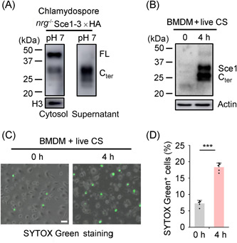
Candida albicans deploys a variety of mechanisms such as morphological switch and elicitor release to promote virulence. However, the intricate interactions between the fungus and the host remain poorly understood, and a comprehensive inventory of fungal virulence factors has yet to be established. In this study, we identified a C. albicans secretory effector protein Sce1, whose induction and secretion are associated with vagina-simulative conditions and chlamydospore formation. Sequence alignment showed that Sce1 belongs to a Pir family in C. albicans, which is conserved across several fungi and primarily characterized as a β-glucan binding protein in the Saccharomyces cerevisiae. Mechanically, Sce1 is primarily localized to the cell wall in a cleaved form as an alkali-labile β-1,3-glucan binding protein and plays a role in masking β-glucan in acidic environments and chlamydospores, a feature that might underline C. albicans' ability to evade host immunity. Further, a cleaved short form of Sce1 protein could be released into extracellular compartments and presented in bone marrow-derived macrophages infected with chlamydospores. This cleaved short form of Sce1 also demonstrated a unique ability to trigger the caspases-8/9-dependent apoptosis in various host cells. Correspondingly, genetic deletion of SCE1 led to dampened vaginal colonization of C. albicans and diminished fungal virulence during systemic infection. The discovery of Sce1 as a versatile virulence effector that executes at various compartments sheds light on the fungus-host interactions and C. albicans pathogenesis.

Keywords: Candida albicans; apoptosis; effector; immune evasion; β‐glucan.


Conflict of interest statement

The authors declare no conflict of interests.

Figures

Figure 1
Figure 1
Sce1 is upregulated under vagina‐simulative conditions and contributes to the virulence of Candida albicans. (A, B) Wild‐type (SC5314) C. albicans cells grown in various SC media in pH 4 or pH 7, or VSM (15 h) at 25°C were harvested, and the relative transcription of SCE1 was analyzed by qRT‐PCR. The value of WT yeast cells cultured in YPD log phase was set to 1 as a reference. SCD, synthetic complete dextrose; SCDLA, SCD with low‐ammonium sulfate; SCLDLA (SCM), SCD with low‐dextrose and low‐ammonium sulfate. The ingredients of each medium are described in the Materials and Methods section. (C) C. albicans strain with endogenous Sce1 in‐frame fused with 3×HA was cultured in YPD (6 h), SCM, and VSM (15 h) at 25°C and lysed for protein lysates. The Sce1A‐3×HA proteins were detected by western blot analysis with the anti‐HA antibody; the inputs were blotted with the anti‐H3 antibody. (D) WT (SN250), sce1a−/− (orf19555−/− ), sce1b−/− (orf19654−/− ), and sce1−/− −/− double null mutant strains carrying a vector with ARG4 (pCPC20) (LEU2 +, HIS1 +, ARG4 +) and sce1−/− −/− +ADH1pSCE1 revertant strains (LEU2 +, HIS1 +, ARG4 +) were administered to C57BL/6 mice (n = 6) via vaginal infection. Vaginal fungal burdens were shown as log10 (CFU/ml) in lavage fluid, mean ± SEM. (E) ICR mice were intravenously infected with the strains used in (D) (male mice weighing 17–19 g were inoculated with 5 × 105 C. albicans cells/mouse, n = 5). The survival percentages (left) and weight changes (right) were determined. The data are representative of three independent experiments (A, B) and shown as mean ± SD. One‐way ANOVA with Tukey's multiple‐comparison test (A, D) or the two‐tailed unpaired Student's t test (B) or the log‐rank test (E) was used for comparison between groups. *p < 0.05, **p < 0.01, ***p < 0.001; ns, not significant (p > 0.05). The experiments were repeated three times, with similar results. ANOVA, analysis of variance; SC, synthetic complete; qRT‐PCR, quantitative reverse‐transcription polymerase chain reaction; VSM, vagina simulative medium.
Figure 2
Figure 2
Sce1 is secreted as cleaved forms under neutral and alkaline conditions. (A) Domain annotation of CaSce1, CaPir32, CaPir1, and ScPir4 using Blast and Interproscan. (B) Schematic presentation of various Sce1‐HA constructs ectopically expressed in Candida albicans. Sce1‐HA(C), the HA‐tag fused to the C terminus of Sce1; Sce1ΔSP‐HA, Sce1‐HA(C) with its signal peptide deleted; and Sce1‐HA(M), the HA‐tag inserted right after Asp101. Sequence alignment reveals a conserved PIR motif in C. albicans CaSce1, CaPir1, CaPir32, and Saccharomyces cerevisiae ScPir4. (C) C. albicans Sce1‐HA(C) was cultured in YPD. Whole‐cell proteins (whole cell), cytosolic proteins (Cytosol; Cyto.), cell wall proteins (cell wall), and culture supernatants (Sup.) were obtained as described in the Materails and Methods section and assessed by western blot analysis. (D) C. albicans Sce1‐HA(C) and Sce1ΔSP‐HA were cultured in YPD. Whole‐cell proteins (whole cell), cytosolic proteins (Cytosol; Cyto.), and culture supernatants (Sup.) were obtained as described in the Methods section and assessed by western blot analysis. (E) Exogenous Sce1‐HA(M) and Sce1‐HA(C) from C. albicans cultured in YPD were detected by western blot analysis. (F) Sce1‐HA(M) overexpressed by C. albicans WT or the kex2 mutant strain cultured in YPD were detected by western blot analysis. (G) C. albicans expressing Sce1‐HA(M) or Sce1R124A were cultured in YPD, and the indicated proteins were detected by western blot analysis. (H) C. albicans expressing Sce1‐HA(M) or Sce1R124A were cultured in pH‐varied YPDs and the indicated proteins were detected by western blot analysis. (I) C. albicans expressing Sce1‐HA(M) was cultured in pH‐varied YPDs and the cells were collected and equally divided into two fractions. One fraction was lysed for whole‐cell proteins. The other fraction was subjected to 30 mM NaOH treatment. After treatment, the alkali solutions containing stripped Sce1 proteins from the cell wall and the pellet containing all the cytosolic proteins and the remaining cell wall proteins refractory to alkali treatment were subjected to western blot analysis. (J) C. albicans expressing Sce1‐HA(M) or Sce1Q74A were cultured in pH‐varied YPDs and the indicated proteins were detected by western blot analysis. Cter, C terminal; FL, full length; Nter, N terminal.
Figure 3
Figure 3
Sce1 on the cell wall plays a role in β‐glucan masking. (A) Representative immunofluorescent images of β‐glucan exposure of wild‐type (SN250) and sce1 −/− −/− mutant strains grown in SCM pH 7 or SCM pH 4. (B) The mean fluorescent intensity (MFI) of exposed β‐glucan in (A) was quantified by flow cytometry counting 100,000 events. (C) Representative immunofluorescent images of β‐glucan exposure of wild‐type and Sce1 overexpression strains grown in SCM pH 4. (D) The MFI of exposed β‐glucan in (C) was quantified by flow cytometry counting 50,000 events. (E–H) Immunofluorescent image and intensity of exposed β‐glucan of Candida albicans wild‐type cells grown in SCM (pH 7) or SCM (pH 4) media with or without 30 mM NaOH treatment. (I) NaOH‐treated SCM‐grown cells were incubated with the Escherichia coli purified recombinant wild‐type Sce1 or Sce1Q74A mutant, and immunofluorescent images of exposed β‐glucan were collected afterwards. (J) The MFI of exposed β‐glucan in (I) was quantified by flow cytometry counting 100,000 events. The scale bar represents 5 μm. The experiments were repeated two times, with similar results.
Figure 4
Figure 4
Decloaking alkali‐labile proteins enables Candida albicans chlamydospore to upregulate the host immunity. (A) Quantitative polymerase chain reaction (qPCR) analysis of SCE1 expression levels in C. albicans yeast (WT cells in YPD), pseudohyphae (PH, nrg1 −/− strain in YPD), and chlamydospore (CS, nrg1 −/− strain in VSM). (B) C. albicans yeast (WT Sce1‐3×HA in YPD) and chlamydospore (nrg1 −/− Sce1‐3×HA in VSM) were harvested. The cytosolic proteins (cytosol) and the cell wall proteins stripped off by alkali solution (cell wall) were subjected to western blot analysis. (C) Representative immunofluorescent images of anti‐HA. An Alexa Fluor® 488‐conjugated goat polyclonal to rabbit was used as the secondary antibody. The chlamydospores (nrg1 −/− Sce1‐3×HA) were cultured in VSM, treated with or without NaOH. Selected images are shown. (D) Representative immunofluorescent images of β‐glucan in chlamydospores (VSM) before or after alkali treatment. NaOH‐treated cells were incubated with Escherichia coli purified recombinant wild‐type Sce1. (E) BMDMs were infected with alkali‐treated or untreated UV‐killed chlamydospores for 6 h (MOI = 10). The expression levels of indicated cytokines and chemokines were quantified by qPCR. The data are representative of three independent experiments (A, E) and shown as mean ± SD. One‐way ANOVA with Tukey's multiple‐comparison test (A, E) were used for comparison between groups. *p < 0.05, **p < 0.01, ***p < 0.001. BMDM, bone marrow‐derived macrophage; VSM, vagina simulative medium. Scale bars: 5 μm.
Figure 5
Figure 5
Sce1 is required for chlamydospore formation. (A) Candida albicans nrg1 −/− and nrg1−/− sce1 −/− strains were cultured in corn meal media (supplemented with 1% Teween‐80) at pH 4, 25°C for 4 days and observed for chlamydospore formation (white arrow head). Scale bar represents 5 μm. (B) Chlamydospore formation of WT and sce1 −/− −/− strains overexpressed with Rme1 (WT+Rme1 and sce1 −/− −/−+Rme1) in corn meal media. (C) Expression of SCE1, CSP1, and CSP2 in WT, WT+Rme1, and sce1 −/− −/−+Rme1 strains. The cells were grown in YPD at 25°C for 6 h and subjected to qRT‐PCR analysis. R, Rme1. The data are shown as mean ± SD. (D) Representative immunofluorescent images of β‐glucan exposure of WT, WT+Rme1, and sce1 −/− −/− +Rme1 cells grown in YPD (pH 7 or pH 4). Scale bar represents 10 μm. (E) The MFI of exposed β‐glucan in (D) was quantified by flow cytometry. One‐way ANOVA with Tukey's multiple‐comparison test (C) was used for comparison between groups.  ***p < 0.001, ns, not significant. The experiments were repeated two times, with similar results.
Figure 6
Figure 6
Cleaved Sce1 induces caspase‐dependent apoptosis in cervical epithelial cells. (A) Coomassie blue staining of purified Sce1 proteins secreted by Candida albicans. Proteins were purified from the culture supernatants of the sce1−/−  mutant strain (Vec) and Sce1 overexpression strains based on the sce1−/− −/−  background (Sce1; Sce1R124A). The concentrations of Sce1 proteins were quantified over BSA. Two micrograms of secreted Sce1 proteins was supplied for transfection into 7.5 × 105 HeLa cells in 1 ml of cell culture (with final concentration of 2 μg/ml). (B, C) C. albicans‐secreted Sce1 proteins (Vec; Sce1; Sce1R124A) were transfected into HeLa cells by a protein delivery reagent PULSin. In 24 h, SYTOX Green was added to the HeLa cells and incubated for 10 min before harvest. The representative images were collected and the percentages of SYTOX Green+ cells (SYTOX Green+ vs. total cells per field) were calculated (n = 5). Scale bars: 10 μm. (D, E) HeLa cells were pretreated with z‐VAD‐FMK (z‐VAD, 20 μM), z‐DEVD‐FMK (DEVD, 30 μM), zIETD‐fmk (ZIETD, 30 μM), z‐YVAD‐FMK (YVAD, 50 μM), disulfiram (DSF, 50 μM), necrostatin 2 racemate (Nec‐1S, 30 μM), and ferrostatin‐1 (Fer‐1, 5 μM) for 30 min, respectively. The Sce1 proteins were delivered into HeLa cells and the SYTOX Green+ cells were calculated. Scale bars: 10 μm. (F) Sce1‐treated HeLa cells were harvested and stained with Annexin V and PI. The percentages of apoptotic cells were measured by FACS analysis. (G) WT or mutant Sce1‐treated HeLa cells were harvested, and the cleavage of caspases was analyzed by western blot analysis. The data are representative of three independent experiments (C, E) and shown as mean ± SD. One‐way ANOVA with Tukey's multiple‐comparison test (C, E) were used for comparison between groups. ***p < 0.001. ANOVA, analysis of variance; BSA, bovine serum albumin.
Figure 7
Figure 7
Chlamydospores can induce macrophage cell death. (A) Chlamydospores (nrg1−/− Sce1‐3×HA) cultured in VSM (pH 7) were harvested, and the cytosolic proteins and culture supernatants were collected for western blot analysis. (B) BMDMs were infected with live chlamydospores (nrg1−/− Sce1‐3×HA) for 4 h (MOI = 1) and gently washed with PBS and lysed for probing with the anti‐HA antibody. (C, D) After incubation with live chlamydospores for 4 h, BMDMs were stained with SYTOX Green for 10 min. The representative images were collected and percentages of SYTOX Green+ cells were calculated (n = 5). Scale bars: 20 μm. The data are representative of three independent experiments (D) and shown as mean ± SD. One‐way ANOVA with Tukey's multiple‐comparison test (D) was used for comparison between groups. ***p < 0.001. PBS, phosphate‐buffered saline.
Figure 8
Figure 8
Candida albicans Sce1 facilitates macrophage cell death and host infection. (A–D) Macrophage cell death assay. BMDMs were infected with live C. albicans yeast cells for 4 h (MOI = 1). The SYTOX Green was added to the BMDMs and incubated for 10 min before harvest. The representative images were collected and percentages of SYTOX Green+ cells were calculated. Scale bars: 20 μm. The C. albicans strains WT (SN250) carrying a vector with ARG4 or ectopically overexpressed Sce1 (ADH1p‐SCE1) were used in (A); WT+Rme1 (SN250 + ADH1p‐RME1) and sce1 −/− −/− +Rme1 (sce1 −/− −/− +ADH1p‐RME1) strains were used in (C); C. albicans cells were grown in YPD at 25°C for 6 h and harvested for infection. (E–H) Systemic infection assay. ICR male mice (n = 5) weighing 18–21 g were intravenously infected with C. albicans strains. The survival percentages and weight changes were determined. Recoverable fungal CFUs in infected mouse tissues (kidney, dpi 2) were quantified, and results were presented as CFU per gram of the tissue (mean ± SEM). C. albicans WT (SN250 + ADH1p‐V) or Sce1 overexpression (SN250 + ADH1p‐SCE1) strains were used for infection in (E) and (F) (5 × 105 CA cells/mouse). Rme1 overexpressing in WT (SN250 + ADH1p‐RME1) or sce1 −/− −/− mutant (sce1 −/− −/− +ADH1p‐RME1) were used for infection in (G, H) (2 × 106 CA cells/mouse). (I) WT+Rme1 and sce1 −/− −/− +Rme1 strains were administered to C57BL/6 mice (n = 5) via vaginal infection. Vaginal fungal burdens were shown as log10 (CFU/ml) in lavage fluid (mean ± SEM). The data are representative of three independent experiments (B, D) and shown as mean ± SD. One‐way ANOVA with Tukey's multiple‐comparison test (B, D) or the two‐tailed unpaired Student's t test (F, H, and I) or log‐rank test (E, G) was used for comparison between groups. *p < 0.05, **p < 0.01, ***p < 0.001. The experiments were repeated three times, with similar results.
Figure 9
Figure 9
Sce1 represents a novel fungal secretory effector protein promoting Candida albicans virulence. Upon encountering the hostile conditions (such as low pH and poor nutrition) similar to that in the mammalian vaginal environment, C. albicans rapidly switches on Sce1 expression. Sce1, and its paralogs might primarily bind to the fungal cell wall β‐glucan, thus preventing its recognition by the host innate immune receptor dectin‐1 and ensuing cytokine and chemokine production. Upon phagocytosis by macrophages (phagosome pH at the range of 4.5–7), alkali‐labile Sce1 can be released from the cell wall and in turn trigger a caspase‐dependent apoptosis. Conceivably, both functions of Sce1 could contribute to C. albicans immune evasion.

Similar articles

References

    1. Mayer FL, Wilson D, Hube B. Candida albicans pathogenicity mechanisms. Virulence. 2013;4:119–28. - PMC - PubMed
    1. Ardizzoni A, Wheeler RT, Pericolini E. It takes two to tango: how a dysregulation of the innate immunity, coupled with Candida virulence, triggers VVC onset. Front Microbiol. 2021;12:692491. - PMC - PubMed
    1. Pekmezovic M, Hovhannisyan H, Gresnigt MS, Iracane E, Oliveira‐Pacheco J, Siscar‐Lewin S, et al. Candida pathogens induce protective mitochondria‐associated type I interferon signalling and a damage‐driven response in vaginal epithelial cells. Nat Microbiol. 2021;6:643–57. - PubMed
    1. Moyes DL, Wilson D, Richardson JP, Mogavero S, Tang SX, Wernecke J, et al. Candidalysin is a fungal peptide toxin critical for mucosal infection. Nature. 2016;532:64–8. - PMC - PubMed
    1. Moyes DL, Runglall M, Murciano C, Shen C, Nayar D, Thavaraj S, et al. A biphasic innate immune MAPK response discriminates between the yeast and hyphal forms of Candida albicans in epithelial cells. Cell Host Microbe. 2010;8:225–35. - PMC - PubMed