

18F-fluorodeoxyglucose Positron Emission Tomography/Computed Tomography and 68Ga-prostate-specific Membrane Antigen Positron Emission Tomography/Computed Tomography Imaging in the Evaluation of Rare Entity Adult Embryonal Rhabdomyosarcoma of Prostate
- PMID: 38817716
- PMCID: PMC11135377
- DOI: 10.4103/ijnm.ijnm_110_23
18F-fluorodeoxyglucose Positron Emission Tomography/Computed Tomography and 68Ga-prostate-specific Membrane Antigen Positron Emission Tomography/Computed Tomography Imaging in the Evaluation of Rare Entity Adult Embryonal Rhabdomyosarcoma of Prostate
Abstract
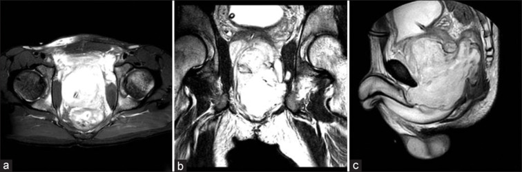
A 21-year-old male with embryonal rhabdomyosarcoma of the prostate was referred for 18F-fluorodeoxyglucose positron emission tomography/computed tomography (18F-FDG PET/CT) and 68Ga-prostate-specific membrane antigen (PSMA) PET/CT for initial disease staging. The PET scans revealed hypermetabolic and PSMA expressing lobulated mass involving both lobes of the prostate and weakly metabolic and PSMA expressing few bilateral pararectal and external iliac nodes, multiple bilateral lung nodules scattered over the lung parenchyma and multiple bone marrow lesions in both axial and appendicular skeleton. Magnetic resonance imaging prostate showed gross prostatomegaly with large lobulated T2 hyperintense heterogeneously enhancing mass lesion showing restricted diffusion, involving both lobes of the prostate with extraprostatic spread along anterior, posterior, and left lateral margins with evidence of lymph nodal and osseous metastases. The demonstration of increased uptake of 18F-FDG and 68Ga-PSMA in the primary as well as bilateral pararectal and external iliac nodes, multiple bilateral lung nodules, and multiple bone marrow lesions in both axial and appendicular skeleton indicates a potential role of 18F-FDG PET/CT and 68Ga-PSMA PET/CT in disease staging in this rare aggressive tumor of the prostate.
Keywords: 18F-fluorodeoxyglucose positron emission tomography/computed tomography; 68Ga-prostate-specific membrane antigen positron emission tomography/computed tomography; embryonal rhabdomyosarcoma prostate; rhabdomyosarcoma.
Copyright: © 2024 Indian Journal of Nuclear Medicine.
Conflict of interest statement
There are no conflicts of interest.
Figures


Similar articles
-
More advantages in detecting bone and soft tissue metastases from prostate cancer using 18F-PSMA PET/CT.Hell J Nucl Med. 2019 Jan-Apr;22(1):6-9. doi: 10.1967/s002449910952. Epub 2019 Mar 7. Hell J Nucl Med. 2019. PMID: 30843003
-
A Prospective Comparison of 18F-prostate-specific Membrane Antigen-1007 Positron Emission Tomography Computed Tomography, Whole-body 1.5 T Magnetic Resonance Imaging with Diffusion-weighted Imaging, and Single-photon Emission Computed Tomography/Computed Tomography with Traditional Imaging in Primary Distant Metastasis Staging of Prostate Cancer (PROSTAGE).Eur Urol Oncol. 2021 Aug;4(4):635-644. doi: 10.1016/j.euo.2020.06.012. Epub 2020 Jul 13. Eur Urol Oncol. 2021. PMID: 32675047 Clinical Trial.
-
68Ga-Labeled Prostate-specific Membrane Antigen Ligand Positron Emission Tomography/Computed Tomography for Prostate Cancer: A Systematic Review and Meta-analysis.Eur Urol Focus. 2018 Sep;4(5):686-693. doi: 10.1016/j.euf.2016.11.002. Epub 2016 Nov 15. Eur Urol Focus. 2018. PMID: 28753806
-
Comparison of 68Ga-labeled Prostate-specific Membrane Antigen Ligand Positron Emission Tomography/Magnetic Resonance Imaging and Positron Emission Tomography/Computed Tomography for Primary Staging of Prostate Cancer: A Systematic Review and Meta-analysis.Eur Urol Open Sci. 2021 Sep 28;33:61-71. doi: 10.1016/j.euros.2021.09.006. eCollection 2021 Nov. Eur Urol Open Sci. 2021. PMID: 34632423 Free PMC article. Review.
-
68Ga-PSMA-11 Positron Emission Tomography/Computed Tomography for Primary Lymph Node Staging Before Radical Prostatectomy: Central Review of Imaging and Comparison with Histopathology of Extended Lymphadenectomy.Eur Urol Focus. 2021 Mar;7(2):288-293. doi: 10.1016/j.euf.2021.01.004. Epub 2021 Jan 25. Eur Urol Focus. 2021. PMID: 33509671
References
-
- Russo P, Brady MS, Conlon K, Hajdu SI, Fair WR, Herr HW, et al. Adult urological sarcoma. J Urol. 1992;147:1032–6. - PubMed
LinkOut - more resources
Full Text Sources
Molecular Biology Databases
Miscellaneous