

Primary Leiomyosarcoma of Suprahepatic Inferior Vena Cava with Metastases
- PMID: 38817723
- PMCID: PMC11135366
- DOI: 10.4103/ijnm.ijnm_130_23
Primary Leiomyosarcoma of Suprahepatic Inferior Vena Cava with Metastases
Abstract
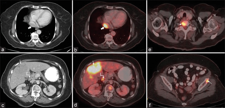
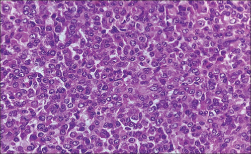
A 67-year-old female presented with shortness of breath, weight loss, abdomen, and back pain for 2 months. Ultrasound of the abdomen revealed multiple focal liver lesions. 18F-Fluorodeoxyglucose whole-body positron emission tomography/computed tomography revealed a hypermetabolic lesion in the suprahepatic inferior vena cava extending into the right atrium. Multiple hypermetabolic lesions were seen in liver, bones, and abdominal lymph nodes, suggestive of metastases. Histopathology and immunohistochemistry of the lesions revealed it to be metastatic leiomyosarcoma.
Keywords: Inferior vena cava; leiomyosarcoma; metastases; positron emission tomography/computed tomography; right atrium.
Copyright: © 2024 Indian Journal of Nuclear Medicine.
Conflict of interest statement
There are no conflicts of interest.
Figures

Similar articles
-
Incidentally Diagnosed Extraluminal Leiomyosarcoma of Infrarenal Inferior Vena Cava: A Case Report and Literature Review from a Radiologist's Perspective.Acta Med Litu. 2022;29(2):258-270. doi: 10.15388/Amed.2022.29.2.12. Epub 2022 Jun 29. Acta Med Litu. 2022. PMID: 37733410 Free PMC article.
-
Rare case of primary inferior vena cava leiomyosarcoma on F-18 fluorodeoxyglucose positron emission tomography-computed tomography scan: Differentiation from nontumor thrombus in a background of procoagulant state.Indian J Nucl Med. 2014 Oct;29(4):246-8. doi: 10.4103/0972-3919.142629. Indian J Nucl Med. 2014. PMID: 25400364 Free PMC article.
-
Primary Cardiac Leiomyosarcoma Extending From Right Atrium to Inferior Vena Cava Without Metastasis: A Case Report.Cureus. 2024 Nov 16;16(11):e73826. doi: 10.7759/cureus.73826. eCollection 2024 Nov. Cureus. 2024. PMID: 39687822 Free PMC article.
-
Primary leiomyosarcoma of the inferior vena cava with Budd-Chiari syndrome.Acta Pathol Jpn. 1989 Jan;39(1):73-7. doi: 10.1111/j.1440-1827.1989.tb02405.x. Acta Pathol Jpn. 1989. PMID: 2652979 Review.
-
Leiomyosarcoma of the inferior vena cava--a very rare case report.Rev Port Cardiol. 2002 Dec;21(12):1469-78. Rev Port Cardiol. 2002. PMID: 12621920 Review. English, Portuguese.
References
-
- Wachtel H, Gupta M, Bartlett EK, Jackson BM, Kelz RR, Karakousis GC, et al. Outcomes after resection of leiomyosarcomas of the inferior vena cava: A pooled data analysis of 377 cases. Surg Oncol. 2015;24:21–7. - PubMed
-
- Amin M, Edge SB, Greene F, editors, editors. 8th. Chicago: Springer International Publishing, American Joint Commission on Cancer; 2017. AJCC Cancer Staging Manual; pp. 517–21.
-
- Kulaylat MN, Karakousis CP, Doerr RJ, Karamanoukian HL, O’Brien J, Peer R. Leiomyosarcoma of the inferior vena cava: A clinicopathologic review and report of three cases. J Surg Oncol. 1997;65:205–17. - PubMed
LinkOut - more resources
Full Text Sources