Review
. 2024 Apr 1;6(3):245-267.
doi: 10.1016/j.jhsg.2024.01.015. eCollection 2024 May.

Scapholunate Ligament Injuries

Affiliations
Review

Scapholunate Ligament Injuries

Joanne Y Zhou et al. J Hand Surg Glob Online. .

Abstract

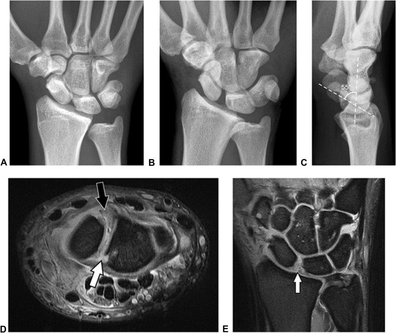
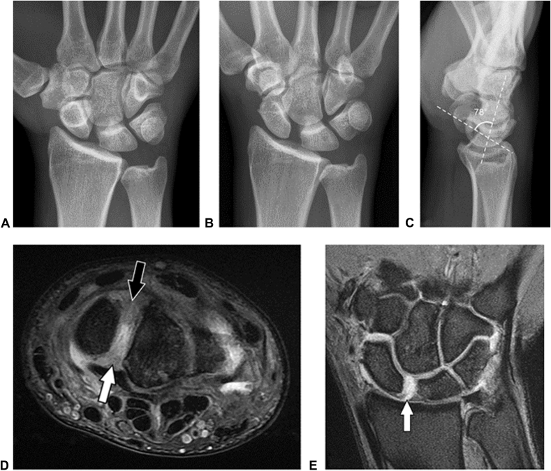
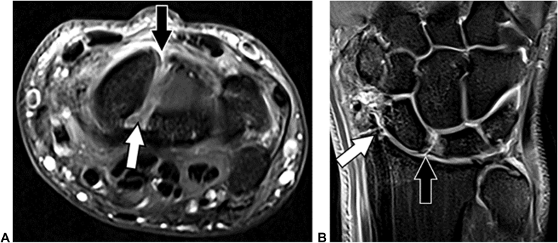
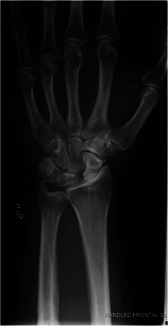
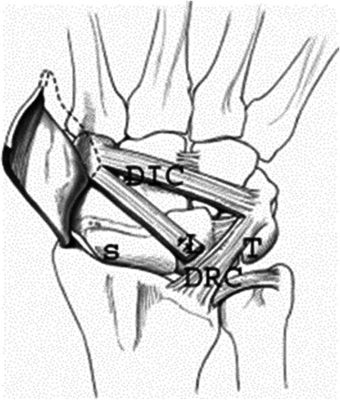
Injuries to the scapholunate interosseous ligament (SLIL) complex can result in a predictable cascade of incongruous motion in the carpus that leads to radiocarpal degeneration. Both acute traumatic impact and repetitive motion can render the SLIL insufficient. A thorough understanding of SLIL anatomy is required for appropriate diagnosis and treatment. Here, we review scapholunate ligament anatomy, prevention strategies, methods of diagnosis, nonoperative and operative treatments, and outcomes. A myriad of treatment options exist for each stage of the SLIL injury, and management should be an open discussion between the patient and physician.

Keywords: Anatomy; Diagnosis; Prevention; Scapholunate interosseous ligament complex injuries; Treatment.


Conflict of interest statement

No benefits in any form have been received or will be received related directly to this article.

Figures

Figure 1
Figure 1
Proximal row of the carpus demonstrating the scapholunate interosseous ligament between the scaphoid (S) and lunate (L) and the lunotriquetral interosseous ligament between the lunate (L) and triquetrum (T).
Figure 2
Figure 2
A The bones of the wrist: trapezium (1), trapezoid (2), capitate (3), hamate (4), scaphoid (5), lunate (6), triquetrum (7), pisiform (8), radius (9), ulna (10), and the bases of the metacarpals (11). B The dorsal wrist ligaments: dorsalcapitohamate (12), dorsal capitotrapezoid (13), dorsal intercarpal ligament (14), radiotriquetral (15), dorsal lunotriquetral (16), dorsal scapholunate (17), and dorsolateral scaphotrapeziotrapezoid (18). C The palmar superficial wrist ligaments: transverse carpal ligament (19), radioscaphoid (20), scaphocapitate (21), radioscaphocapitate (22), long radiolunate (23), ulnocapitate (24), and pisohamate (25). D The palmar deep wrist ligaments: palmar capitotrapezoid (26), scaphocapitate (27), triquetral-hamate-capitate (28), palmar scapholunate (29), palmar lunotriquetral (30), short radiolunate (31), ulnolunate (32), ulnotriquetral (33), and palmar capitohamate (34). Adapted with permission from Konopka et al.
Figure. 3
Figure. 3
Volar secondary stabilizers. A Diagram of the volar aspect of the wrist shows the three volar secondary stabilizers of the scapholunate joint. B Coronal PDW fat-suppressed MR image of the volar aspect of the wrist shows the STT ligament (arrowhead), radioscaphocapitate ligament (black arrow), and long radiolunate ligament (white arrow). (Adapted with permission from Palisch et al101).
Figure 4
Figure 4
Normal dorsal secondary stabilizers. A Diagram of the dorsal aspect of the wrist shows the two dorsal secondary stabilizers of the scapholunate joint. B Coronal PDW fat-suppressed MR image of the dorsal aspect of the wrist shows the DRT ligament (black arrow) and DIC ligament (white arrow). (Adapted with permission from Palisch et al101).
Figure 5
Figure 5
The scapholunate ligament is C-shaped and has dorsal (D) proximal (P), and volar (V) components. (Adapted with permission from Anderrson et al102).
Figure 6
Figure 6
Biomechanics of SLIL injury. With failure of the scapholunate interosseous ligament or partial tear of the dorsal intercarpal ligament, dorsal radiocarpal ligament, or a critical volar ligament, such as the scaphotrapeziotrapezoid ligament or the long radiolunate ligament, there is loss of the normal alignment and consequent movement between the scaphoid and lunate. With axial load, the scaphoid preferentially flexes volarly, while the lunate, being pulled by the triquetrum with an intact lunotriquetral ligament, dorsiflexes (arrows) (Used with permission from Flores et al, 2021103).
Figure 7
Figure 7
Scapholunate interval in a 20-year-old male collegiate football player. One should use caution when using upper limits in scapholunate interval measurement, and it is important to compare sides. A Static posteroanterior radiograph of the asymptomatic left wrist shows a scapholunate interval of 4.5 mm. B Static posteroanterior radiograph of the symptomatic right wrist shows a scapholunate interval of 6 mm. (Adapted with permission from Palisch et al101).
Figure 8
Figure 8
Posteroanterior view of the left wrist demonstrating SL widening (“Terry Thomas sign”) and a signet ring sign, suggesting SL injury with rotatory subluxation.
Figure 9
Figure 9
Pencil grip view demonstrating the following: A proper patient position and B the radiographic image demonstrating scapholunate interval widening on the right. (From Lee SK, Desai H, Silver B, Dhaliwal G, Paksima N. Comparison of radiographic stress views for scapholunate dynamic instability in a cadaver model. J Hand Surg 2011;36A:1149–1157; with permission).
Figure 10
Figure 10
Normal SLIL. A Axial proton-density–weighted (PDW) fat-suppressed MR image shows the dorsal band (black arrow) and volar band (white arrow) of the SLL. B Coronal PDW fat-suppressed MR image shows the triangular interosseous band (arrow). C Diagram shows the dorsal, volar, and interosseous bands of the SLL. (Adapted with permission from Palisch et al101).
Figure 11
Figure 11
Stage 1 SLIL injury in a 22-year-old professional American football player. The scapholunate interval and angle were within normal limits on static posteroanterior, dynamic clenched fist, and pallateral radiographs. A Axial PDW fat-suppressed MR image shows an intact SLIL dorsal band (arrowhead) and torn SLIL volar band (white arrow), which were confirmed at surgery. A normal long radiolunate ligament (black arrow) courses directly over the torn SLIL volar band and can be mistaken for an intact SLIL volar band. B Coronal PDW fat-suppressed MR image shows partial tearing (arrow) extending to the SLIL interosseus band. (Adapted with permission from Palisch et al101).
Figure 12
Figure 12
Stage 2 SLIL injury in a 26-year-old professional basketball player with a recent fall on an outstretched hand. The scapholunate interval and angle were within normal limits on static posteroanterior, dynamic clenched fist, and lateral radiographs. A Axial PDW fat-suppressed MR image shows a torn SLIL dorsal band (arrow) from the scaphoid. B Coronal PDW fat-suppressed MR image shows a torn SLIL interosseus band (white arrow) and an incidental tear of the central triangular fibrocartilage (black arrow). Reparability of the SLIL dorsal band can be suggested with imaging if the tear appears noncomplex and nonelongated but is ultimately determined at surgery. (Adapted with permission from Palisch et al101).
Figure 13
Figure 13
Stage 3 SLIL injury with normal static alignment in a 25-year-old professional American football player who experienced a fall 3 months earlier. A Static posteroanterior radiograph shows a scapholunate interval of 2.7 mm. B Dynamic clenched fist radiograph shows an increase in the scapholunate interval to 4 mm. C Lateral radiograph shows a normal scapholunate angle of 55°. D Axial PDW fat-suppressed MR image shows a torn SLIL dorsal band (black arrow) from the scaphoid attachment and a torn SLIL volar band (white arrow), which were confirmed at surgery. E Coronal PDW fat-suppressed MR image shows a torn SLIL interosseous band (arrow). Reparability of the SLIL dorsal band can be suggested with imaging if the tear appears noncomplex and nonelongated but is ultimately determined at surgery. (Adapted with permission from Palisch et al101).
Figure 14
Figure 14
Stage 3 reducible malalignment in a 23-year-old professional American football player with a remote injury. A Static posteroanterior radiograph shows a scapholunate interval of 4.5 mm. B Dynamic clenched fist radiograph shows an increase in scapholunate interval to 5 mm. C Lateral radiograph shows that the scapholunate angle is increased to 78° with dorsal tilting of the lunate, compatible with DISI. D Axial PDW fat-suppressed MR image shows a torn SLL dorsal band (black arrow) and torn SLL volar band (white arrow) with scar tissue. E Coronal PDW fat-suppressed MR image shows a tear (arrow) of the SLIL interosseous band and scapholunate interval widening. Stage 3 reducible malalignment and stage 4 irreducible malalignment injuries have similar imaging appearances and are differentiated on the basis of reducibility during fluoroscopic traction or surgery. If the malalignment is reducible during fluoroscopic traction or surgery, the patient can undergo SLIL reconstruction. If the malalignment is not reducible, the patient can undergo a salvage procedure. (Adapted with permission from Palisch et al101).
Figure 15
Figure 15
Stage 5 SLIL injury in a 30-year-old professional American football player. A Axial PDW fat-suppressed MR image shows a torn SLIL dorsal band (black arrow) and torn SLIL volar band (white arrow). B Coronal PDW fat-suppressed MR image shows cartilage loss, most prominent at the articulation of the radial styloid and scaphoid, with osteophyte formation (white arrow) and a torn SLIL interosseous band (black arrow). (Adapted with permission from Palisch et al101).
Figure 16
Figure 16
Scapholunate (SL) advanced collapse after an SL injury, with subsequent arthritic changes at the radiocarpal and mid-carpal joints (SL advanced collapse - SLAC III). Also note the volarly flexed scaphoid appearing triangular distally with the so-called “ring sign.” (Adapted with permission from Andersson et al).
Figure 17
Figure 17
SLIL viewed through the 3–4 portal with A normal appearance and B abnormal appearance with loss of concavity and laxity observed.
Figure 18
Figure 18
Scapholunate interval viewed through the midcarpal radial portal demonstrating A abnormal widening suggestive of SL injury determined to be B Geissler grade III on probing. C Normal SL interval for comparison with tight apposition of the scaphoid and lunate. (Adapted with permission from Gire et al, 2019104).
Figure 19
Figure 19
Arthroscopic view of the A–C normal dorsal capsuloscapholunate septum (stars) in 3 patients and D–F a torn dorsal capsuloscapholunate septum in 3 patients viewed from the 6R portal. (From Binder AC, Kerfant N, Wahegaonkar AL, et al. Dorsal Wrist Capsular Tears in Association with Scapholunate Instability: Results of an Arthroscopic Dorsal Capsuloplasty. J Wrist Surg 2013;2:160-167; with permission.)
Figure 20
Figure 20
Arthroscopic view of the scapholunate interval through the midcarpal radial portal demonstrating A Geissler grade III SL injury, B volar SLIL and capsule before thermal shrinkage, C golden yellow/tan color change, and D improvement in interval stability after shrinkage and K-wire fixation. (Adapted with permission from Burn et al, 201950).
Figure 21
Figure 21
A cannulated 3.2-mm drill is used to create a bone tunnel from the dorsal SL insertion to the scaphoid tuberosity along the central axis in preparation for ligament reconstruction. (From Paci GM, Yao J. Surgical Techniques for the Treatment of Carpal Ligament Injury in the Athlete. Clin Sports Med 2015;34:11-35; with permission).
Figure 22
Figure 22
The tendon graft is tensioned and attached to the lunate with a suture anchor and then passed through the radiotriquetral ligament and secured to itself. (From Paci GM, Yao J. Surgical Techniques for the Treatment of Carpal Ligament Injury in the Athlete. Clin Sports Med 2015;34:11-35; with permission).
Figure 23
Figure 23
Dorsal ligament-sparing capsulotomy with the dorsal intercarpal and dorsal radiocarpal ligaments outlined. (From Paci GM, Yao J. Surgical Techniques for the Treatment of Carpal Ligament Injury in the Athlete. Clin Sports Med 2015;34:11-35; with permission).
Figure 24
Figure 24
Repair of the scapholunate ligament back to the lunate using suture anchors. (From Paci GM, Yao J. Surgical Techniques for the Treatment of Carpal Ligament Injury in the Athlete. Clin Sports Med 2015;34:11-35; with permission).
Figure 25
Figure 25
The modified dorsal intercarpal (DIC) capsulodesis with a strip of the DIC maintained on the scaphoid (S) and transferred from the triquetrum (T) to the dorsal lunate (L). DRC, dorsoradiocarpal. (From Manuel J & Moran SL. The diagnosis of treatment of scapholunate instability. Hand Clinic 2010;26:129-144; used with permission of Mayo Foundation for Medical Education and Research, all rights reserved).
Figure 26
Figure 26
The radial approach through the anatomic snuffbox for the scapholunate axis method exposes the starting point midlateral on the right of the scaphoid as indicated by the probe. (From Paci GM, Yao J. Surgical Techniques for the Treatment of Carpal Ligament Injury in the Athlete. Clin Sports Med 2015;34:11-35; with permission).
Figure 27
Figure 27
The guidewire is placed along the central axis of the scaphoid and lunate as visualized on the posteroanterior A and lateral B radiographs, respectively, ending at the proximoulnar corner of the lunate A. (From Paci GM, Yao J. Surgical Techniques for the Treatment of Carpal Ligament Injury in the Athlete. Clin Sports Med 2015;34:11-35; with permission).
Figure 28
Figure 28
A cannulated step drill (2.9 leading, 3.8 trailing) is used to create the graft tunnel over the central guidewire. (From Paci GM, Yao J. Surgical Techniques for the Treatment of Carpal Ligament Injury in the Athlete. Clin Sports Med 2015;34:11-35; with permission).
Figure 29
Figure 29
The tendon graft is threaded through the bullet anchor and placed in the proximoulnar corner of the lunate. (From Paci GM, Yao J. Surgical Techniques for the Treatment of Carpal Ligament Injury in the Athlete. Clin Sports Med 2015;34:11-35; with permission).
Figure 30
Figure 30
The tendon graft is tensioned and secured with a 4-mm interference screw into the scaphoid. (From Paci GM, Yao J. Surgical Techniques for the Treatment of Carpal Ligament Injury in the Athlete. Clin Sports Med 2015;34:11-35; with permission).
Figure 31
Figure 31
The remaining graft is passed dorsal to the scaphoid and then secured to the lunate and the dorsal radiocarpal ligament. (From Paci GM, Yao J. Surgical Techniques for the Treatment of Carpal Ligament Injury in the Athlete. Clin Sports Med 2015;34:11-35; with permission).
Figure 32
Figure 32
MBR in a 42-year-old salesman with stage 3 chronic SLL injury. Preoperative posteroanterior radiography showed a widened scapholunate interval of 6 mm and DISI deformity. A Posteroanterior radiograph 10 weeks after surgery shows a scapholunate interval of 2.5 mm (arrow). B Posteroanterior radiograph 7 months after surgery shows a scapholunate interval of 4 mm (arrow). Postoperative widening of the scapholunate interval can occur over time after reconstruction and does not definitely indicate graft failure. The patient was asymptomatic over the scapholunate ligament with no instability at clinical examination. (Adapted with permission from Palisch et al101).
Figure 33
Figure 33
A Complication of the RASL procedure. Posteroanterior radiograph after the RASL procedure shows a fractured compression screw (arrow) across the scapholunate interval. B Complication of the SLAM procedure. Posteroanterior radiograph after the SLAM procedure shows lucency and collapse in the proximal scaphoid (arrow) from osteonecrosis. Similar changes from osteonecrosis are in the radial aspect of the lunate. (Adapted with permission from Palisch et al101).

Similar articles

Cited by

References

    1. Slade J.F., Milewski M.D. Management of carpal instability in athletes. Hand Clin. 2009;25(3):395–408. - PubMed
    1. Tosti R., Shin E. Wrist arthroscopy for athletic injuries. Hand Clin. 2017 Feb;33(1):107–117. - PubMed
    1. Watson H.K., Ballet F.L. The SLAC wrist: scapholunate advanced collapse pattern of degenerative arthritis. J Hand Surg. 1984;9(3):358–365. - PubMed
    1. Beck A. The integrity of the scapholunate ligament in competitive divers. Br J Sports Med. 2019;53(13):843. 1–843.
    1. Benjamin H.J., Engel S.C., Chudzik D. Wrist pain in gymnasts: a review of common overuse wrist pathology in the gymnastics athlete. Curr Sports Med Rep. 2017;16(5):322–329. - PubMed

LinkOut - more resources