. 2024 May 14;4(5):1911-1927.
doi: 10.1021/jacsau.4c00138. eCollection 2024 May 27.

CDK2 and CDK4: Cell Cycle Functions Evolve Distinct, Catalysis-Competent Conformations, Offering Drug Targets

Affiliations

CDK2 and CDK4: Cell Cycle Functions Evolve Distinct, Catalysis-Competent Conformations, Offering Drug Targets

Wengang Zhang et al. JACS Au. .

Abstract

Cyclin-dependent kinases (CDKs), particularly CDK4 and CDK2, are crucial for cell cycle progression from the Gap 1 (G1) to the Synthesis (S) phase by phosphorylating targets such as the Retinoblastoma Protein (Rb). CDK4, paired with cyclin-D, operates in the long G1 phase, while CDK2 with cyclin-E, manages the brief G1-to-S transition, enabling DNA replication. Aberrant CDK signaling leads to uncontrolled cell proliferation, which is a hallmark of cancer. Exactly how they accomplish their catalytic phosphorylation actions with distinct efficiencies poses the fundamental, albeit overlooked question. Here we combined available experimental data and modeling of the active complexes to establish their conformational functional landscapes to explain how the two cyclin/CDK complexes differentially populate their catalytically competent states for cell cycle progression. Our premise is that CDK catalytic efficiencies could be more important for cell cycle progression than the cyclin-CDK biochemical binding specificity and that efficiency is likely the prime determinant of cell cycle progression. We observe that CDK4 is more dynamic than CDK2 in the ATP binding site, the regulatory spine, and the interaction with its cyclin partner. The N-terminus of cyclin-D acts as an allosteric regulator of the activation loop and the ATP-binding site in CDK4. Integrated with a suite of experimental data, we suggest that the CDK4 complex is less capable of remaining in the active catalytically competent conformation, and may have a lower catalytic efficiency than CDK2, befitting their cell cycle time scales, and point to critical residues and motifs that drive their differences. Our mechanistic landscape may apply broadly to kinases, and we propose two drug design strategies: (i) allosteric Inhibition by conformational stabilization for targeting allosteric CDK4 regulation by cyclin-D, and (ii) dynamic entropy-optimized targeting which leverages the dynamic, entropic aspects of CDK4 to optimize drug binding efficacy.


Conflict of interest statement

The authors declare no competing financial interest.

Figures
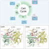
Figure 1
Figure 1
Cell cycle progression and cyclin-dependent kinase (CDK) complex structures. Upper panel illustrates the phases of the cell cycle, including G1, G1/S transition, and S, G2, and M phases, annotated with their corresponding cyclin and CDK complexes. The bottom panels display in silico modeled structures of the active cyclin-D/CDK4 and cyclin-E/CDK2 complexes, which are crucial for driving the cell cycle through the G1 and G1/S transition phases, respectively. ATP binding sites and structural features relevant to their kinase activity and regulatory mechanisms are labeled. Their sequence alignments are shown in Figure S1, which reveal high sequence similarity between CDK4 and CDK2, while low sequence similarity between cyclin-D and cyclin-E, yet their structural conformations in complex with their respective kinases are notably similar.
Figure 2
Figure 2
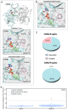
Differential ATP binding site dynamics between active CDK4 and CDK2 complexes suggest distinct catalytically competent states. (Upper panel) Structure of the active CDK4 (A) and CDK2 (B) complex in the active site. It shows some key structural determinants influencing ATP-binding dynamics: (i) Adenosine-CDK interaction: Adenosine ring of ATP forms hydrogen bonds with the hinge region of CDKs, which is important for ATP orientation and stability. (ii) Hydrophobic Val and Leu residues (V20 and L147 for CDK4; V18 and L134 for CDK2) on the top and bottom of adenosine ring in ATP; their proximity is linked to the ATP binding site’s open or closed conformation. (iii) G-loop enclosure: (G-loop) caps the ATP binding site, providing another layer of stabilization on ATP. (iv) Phosphate stabilization: Lysine on β3 (K35 for CDK4; K33 on CDK2) interacts with α/β-phosphate and Glu on the αC-helix, further securing ATP in its binding site. (Middle panel) Hydrogen bond stability: CDK2 exhibits a higher hydrogen bond occupancy with ATP, indicative of enhanced ATP stabilization compared to CDK4. (Middle panel) G-loop dynamics: the G-loop of CDK4 is more dynamic, suggesting a propensity to assume an open conformation. (Lower panel) Potential of mean force landscapes for two reaction coordinates—the distance between nitrogen on Lys (Lys-N) and α-phosphate (Pα) of ATP, and the distance between the valine and leucine residues. Both distances are consistently greater in CDK4 indicating that CDK4 has a broader range of accessible conformations when it binds to ATP, suggesting greater conformational flexibility. In contrast, ATP-bound CDK2 shows a more constrained energy landscape. Specifically, the larger Val–Leu distance suggests that the ATP binding site tends to adopt a more open and flexible conformation, while the shorter Lys-N–Pα distance suggests an enhanced ATP stabilization and potentially efficient phosphoryl transfer. The contour maps illustrate the energy profiles along these coordinates, showing that CDK4 has a shallow energetic barrier than does CDK2 for their ATP-bound states.
Figure 3
Figure 3
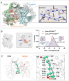
R-spine stability reveals CDK2’s enhanced propensity for an active and assembled conformation. Stability of the R-spine is quantified by monitoring the distance between key residues, RS1, histidine (in the HRD-motif) and RS2, phenylalanine (in the DFG-motif), within the R-spine. (A) R-spine dynamics: for CDK4, the R-spine occasionally adopts unstable conformations deviating from the canonical, fully assembled state. To quantify the stability of the R-spine, we monitored the distance between the center of mass of the imidazole ring in histidine and the phenyl ring in phenylalanine residues within the spine. (B) CDK2 consistently, in contrast, maintains a more stable and fully assembled R-spine conformation throughout the simulations. (C) Likelihood for both CDK4 and CDK2 to adopts either a fully assembled or an unstable R-spine conformation. (D) Distances between Phe and His for all available PDB structures of CDK2 from the Protein Data Bank in Angstrom (Å). The criteria for R-spine stability are based on the histidine-phenylalanine distance with the critical cutoff distances derived from an analysis of all publicly available PDB structures of CDK2 and CDK4, as well as numerous well-documented human kinases, including both inactive and active conformational states (Figure S11). Each data point represents a PDB structure. Through simulation data and PDB structure analysis, we identified a cutoff distance for R-spine stability: configurations are unstable above 7 Å and stable below it.
Figure 4
Figure 4
Differential interaction profiles between CDKs and their respective cyclins highlight CDK2’s robust interface with cyclin-E. Contacting residue maps on the top row offer a dynamic representation of residue-wise interaction propensities between the CDKs and their partnering cyclins. It captures both transient and persistent contacts formed between the proteins. (A) CDK4 and cyclin-D interaction dynamics: CDK4 primarily forms persistent contacts with cyclin-D at residues L49 and G48 of the loopβ3-αC. Notably, the A-loop of CDK4 engages in extensive interactions with the N-terminus of cyclin-D, a distinct structural feature that underscores the specificity of their interaction. (B) CDK2 and cyclin-E interaction dynamics: CDK2 exhibits a more extensive interaction profile with cyclin-E, especially within the loopβ3-αC region. (C) CDK cyclin binding free energies ΔGb for both CDK4 and CDK2 complexes. The box indicates 25–75% range, the whisker depicts the mean ±1.5 SD, and the labels show the mean binding free energies. Cyclin-E/CDK2 complex has a lower binding free energy than that of cyclin-D/CDK4. We further identify loopβ3-αC and the A-loop in CDK2 as contributing significantly to the lower binding affinity of CDK2 complex, compared to that of the CDK4 complex using alanine scanning in Figure S12.
Figure 5
Figure 5
Differential roles of cyclin partners in A-loop stabilization highlight cyclin-D’s importance in CDK4 activity regulation. We evaluate the chemical shift perturbations (CSPs) for active CDK complexes when their cyclin partners bind. (A) CDK4’s chemical shift landscape: CSPs for active CDK4 are evaluated with key peaks annotated to map to specific structural features. The A-loop region of CDK4 showcases more residues, exhibiting pronounced chemical shifts. The magnitudes of these shifts are indicative of the role cyclin-D plays in stabilizing CDK4’s activation loop. (B) In contrast to CDK2’s chemical shift landscape, active CDK2 displays fewer and relatively diminished CSPs in its A-loop region. This suggests a comparatively subdued role for cyclin-E in stabilizing CDK2’s A-loop, highlighting the unique regulatory dynamics between the two CDK-cyclin pairs. The red lines indicate the average CSP across all residues.
Figure 6
Figure 6
Cyclin-D’s N-terminus acts as an allosteric regulator, governing the activation loop and ATP binding site conformation in CDK4. (A) N-terminus interactions with A-loop: the N-terminus of cyclin-D and the A-loop of CDK4 form a β-strand and hydrogen bonds with the A-loop, suggesting a structural interdependence critical for maintaining CDK4’s active conformation. (B) Influence on the ATP binding site conformation: By evaluating the distance distribution between residues V20 and L147, we contrast the conformational behavior of the active cyclin-D/CDK4 complex with that of its counterpart, cyclin-DΔN-ter/CDK4 (cyclin-D lacking the N-terminus). Notably, the absence of cyclin-D’s N-terminus results in a pronounced shift in the ATP binding site’s dynamics. This is evident from the diminished probability of the complex assuming a closed ATP binding site conformation, as indicated by the vanishing peak near the 13.5 Å mark in the red curve. (C) Allosteric pathways between the N-terminus of cyclin-D (R14 and R15) and V20 of CDK4. The source residues are either R14 or R15 in cyclin-D, and the sink residue is V20 in CDK4. Red beads represent the source and sink residues, while green beads denote the allosteric signal nodes. The blue lines represent the optimal, shortest allosteric pathway; lines of other colors indicate suboptimal allosteric pathways. The residue labels on the left-hand side identify the residues involved in the optimal allosteric pathway. This pathway shows that the source residue (R14) transmits the allosteric signal first through A-loop residues (L171, A170, M169, and R163) and then αC-helix residues (R55, and E56), followed by K35 on the β3-strand, and finally reaches V20, a component of the C-spine. The residue labels with orange text indicate cyclin-D residues, while black text indicates CDK4 residues.
Figure 7
Figure 7
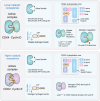
Differential conformational dynamics influence the stability of active cyclin-D/CDK4 and cyclin-E/CDK2 complexes and may affect their catalytically competent state. Key features of cyclin-D/CDK4/6: ATP binding site dynamics: CDK4’s active state has a more expansive ATP binding site characterized by a highly flexible G-loop and varying distance between hydrophobic residues that sandwich ATP. Suboptimal ATP binding: the adenosine ring of ATP exhibits less stable hydrogen bond interactions with CDK4’s hinge region, compared to the high occupancy of CDK2. R-spine instability: the hydrophobic core’s R-spine in CDK4 shows reduced stability compared to its counterpart in CDK2. Unique interactions with cyclin-D: while CDK4’s interactions with cyclin-D are limited (e.g., minimal contacts via loopβ3-αC), the contacts with cyclin-D’s N-terminus are vital. This is emphasized by its role in A-loop stabilization through β-strand formation and its allosteric modulation of the ATP binding site’s conformation.

Similar articles

Cited by

References

    1. Morgan D. O.The Cell Cycle: Principles of Control; New Science Press, 2007.
    1. Rubin S. M. Deciphering the retinoblastoma protein phosphorylation code. Trends Biochem. Sci. 2013, 38 (1), 12–19. 10.1016/j.tibs.2012.10.007. - DOI - PMC - PubMed
    1. Malumbres M.; Barbacid M. Cell cycle, CDKs and cancer: a changing paradigm. Nat. Rev. Cancer 2009, 9 (3), 153–166. 10.1038/nrc2602. - DOI - PubMed
    1. Rubin S. M.; Sage J.; Skotheim J. M. Integrating Old and New Paradigms of G1/S Control. Mol. Cell 2020, 80 (2), 183–192. 10.1016/j.molcel.2020.08.020. - DOI - PMC - PubMed
    1. Cornwell J. A.; Crncec A.; Afifi M. M.; Tang K.; Amin R.; Cappell S. D. Loss of CDK4/6 activity in S/G2 phase leads to cell cycle reversal. Nature 2023, 619 (7969), 363–370. 10.1038/s41586-023-06274-3. - DOI - PMC - PubMed

LinkOut - more resources