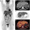
Multimodal imaging findings of primary liver clear cell carcinoma: a case presentation
- PMID: 38818401
- PMCID: PMC11137254
- DOI: 10.3389/fmed.2024.1408967
Multimodal imaging findings of primary liver clear cell carcinoma: a case presentation
Abstract
Primary clear cell carcinoma of liver (PCCCL) is a special and relatively rare subtype of hepatocellular carcinoma (HCC), which is more common in people over 50 years of age, with a preference for men and a history of hepatitis B or C and/or cirrhosis. Herein, we present a case of a 60-year-old woman who came to our hospital for medical help with right upper abdominal pain. The imaging examination showed a low-density mass in the right lobe of his liver. In contrast enhanced computed tomography (CT) or T1-weighted imaging, significant enhancement can appear around the tumor during the arterial phase, and over time, the degree of enhancement of the tumor gradually decreases. The lession showed obviously increased fluorine-18 fluorodeoxyglucose (18F-FDG) uptake on positron emission tomography/CT. These imaging findings contribute to the diagnosis of PCCCL and differentiate it from other types of liver tumors.
Keywords: MRI; PET/CT; clear cell carcinoma; imaging findings; liver.
Copyright © 2024 Hu, Li, Zhao, Cai and Wang.
Conflict of interest statement
The authors declare that the research was conducted in the absence of any commercial or financial relationships that could be construed as a potential conflict of interest.
Figures


Similar articles
-
More advantages in detecting bone and soft tissue metastases from prostate cancer using 18F-PSMA PET/CT.Hell J Nucl Med. 2019 Jan-Apr;22(1):6-9. doi: 10.1967/s002449910952. Epub 2019 Mar 7. Hell J Nucl Med. 2019. PMID: 30843003
-
Primary clear cell carcinoma in the liver: CT and MRI findings.World J Gastroenterol. 2011 Feb 21;17(7):946-52. doi: 10.3748/wjg.v17.i7.946. World J Gastroenterol. 2011. PMID: 21412505 Free PMC article.
-
Efficacy of contrast-enhanced FDG PET/CT in patients awaiting liver transplantation with rising alpha-fetoprotein after bridge therapy of hepatocellular carcinoma.Eur Radiol. 2018 Dec;28(12):5356-5367. doi: 10.1007/s00330-018-5425-z. Epub 2018 Jun 12. Eur Radiol. 2018. PMID: 29948070
-
Hepatic epithelioid angiomyolipoma mimicking hepatocellular carcinoma on MR and 18F-FDG PET/CT imaging: A case report and literature review.Hell J Nucl Med. 2022 May-Aug;25(2):205-209. doi: 10.1967/s002449912480. Epub 2022 Aug 3. Hell J Nucl Med. 2022. PMID: 35913867 Review.
-
CT findings of primary clear cell carcinoma of liver: with analysis of 19 cases and review of the literature.Abdom Imaging. 2014 Aug;39(4):736-43. doi: 10.1007/s00261-014-0104-2. Abdom Imaging. 2014. PMID: 24549879 Review.
Cited by
-
Artificial intelligence techniques in liver cancer.Front Oncol. 2024 Sep 3;14:1415859. doi: 10.3389/fonc.2024.1415859. eCollection 2024. Front Oncol. 2024. PMID: 39290245 Free PMC article. Review.
References
Publication types
LinkOut - more resources
Full Text Sources

