Review
. 2025;13(2):2361197.
doi: 10.1080/21688370.2024.2361197. Epub 2024 May 31.

The stratum corneum barrier: impaired function in relation to associated lipids and proteins

Affiliations
Review

The stratum corneum barrier: impaired function in relation to associated lipids and proteins

Jie Chen et al. Tissue Barriers. 2025.

Abstract

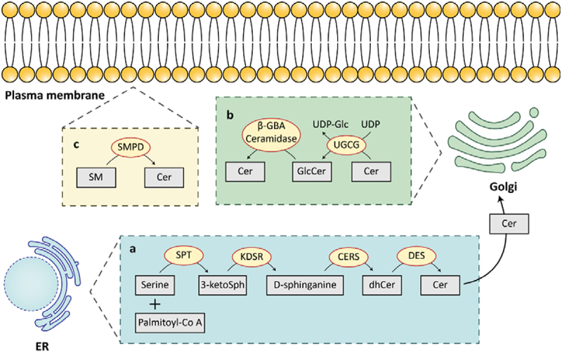
The skin is the largest organ of the human body and is widely considered to be the first-line defense of the body, providing essential protection against mechanical, physical, and chemical damage. Keratinocytes are the primary cells of the outer layer of the epidermis, which acts as a mechanical and permeability barrier. The epidermis is a permanently renewed tissue where undifferentiated keratinocytes located at the basal layer proliferate and migrate to the overlying layers. Here we report that some components of keratinocytes affect the formation and differentiation of the stratum corneum, which is the most specialized layer of the epidermis.

Keywords: Stratum corneum; ceramide; differentiation; filaggrin; keratin; keratinocyte.


Conflict of interest statement

No potential conflict of interest was reported by the author(s).

Figures

Figure 1.
Figure 1.
Relationship between multiple epidermal barriers. the epidermal barrier is centrally interconnected with the stratum corneum barrier. The epidermal commensal microbiota colonizes the outer stratum corneum and influences host metabolism between pathologies, and the microbiota is associated with the development of many autoimmune diseases. The lipids that make up the sebum barrier are secreted by the sebaceous glands and protect the stratum corneum from external aggressions, sebum provides the substrate required for lipid metabolism by the skin microbiota, and the sebum barrier further inhibits the growth of harmful bacteria. The stratum corneum barrier isolates immune stimulants, and damage to the stratum corneum causes an immune response.
Figure 2.
Figure 2.
FLG production process, factors regulating FLG expression, and FLG-induced lipid production. i. Profilaggrin is secreted into the cytoplasm by keratohyalin granules to break down into filaggrin. ii. It binds to keratin intermediate filaments to form a dense keratin matrix, which facilitates the mechanical strength and integrity of the stratum corneum and the skin barrier function. iii. Filaggrin monomers are further decomposed to hygroscopic amino acids and their derivatives in the stratum corneum by proteases for skin hydration. Lysophosphatidic acid (LPA) binding to LPAR1/5 activates the RHO-ROCK-SRF pathway via G12/13, leading to the activation of the transcription factor SRF, which induces FLG gene expression and protein production. Filaggrin acts to inhibit α-toxin pathogenicity by mediating the secretion of SMPD to reduce the number of α-toxin binding sites on the surface of keratinocytes.
Figure 3.
Figure 3.
Main pathways of ceramide synthesis. in the ER, serine and palmitoyl-CoA are condensed to 3-ketosphinganine (3-ketoSph) by serine palmitoyltransferase (SPT); 3-ketodihydrosphingosine reductase (KDSR) reduces it to D-sphingosine; D-sphingosine is produced as dihydroceramide (dhCer) by ceramide synthase (CERS); Dihydroceramide is desaturated by dihydroceramide desaturase (DES) to generate ceramide (Cer). In the Golgi, Cer is produced as glucose ceramides (GlcCer) via UDP-glucose ceramide glucosyltransferase (UGCG), and GlcCer are converted to Cer by β-glucocerebrosidase (β-GBA) and ceramidase. In the plasma membrane, sphingomyelin (SM) are decomposed by sphingomyelinase (SMPD) to ceramides.
Figure 4.
Figure 4.
Immunomodulatory factors in keratinocyte differentiation. Th2 cytokines IL-4 and IL-13 affect structural components and barrier function at early stages of keratinocyte differentiation; exogenous histamine prevents the initiation of late keratinocyte differentiation program through activation of histamine receptor-1; AKT1 is expressed at late stages of terminal differentiation and AKT1 may affect barrier function in the keratinocyte layer by regulating nuclear degradation.
Figure 5.
Figure 5.
Skin barrier changes during skin sensitization. Skin sensitization manifests as dry, flaky, painful and reddened skin. Dryness and increased permeability are caused by thinning of the sebum and stratum corneum; subsequent activation of inflammation causes redness and pain; and sensitive skin is often accompanied by a decrease in the diversity of the microbiome.

Similar articles

Cited by

References

    1. Kabashima K, Honda T, Ginhoux F, Egawa G.. The immunological anatomy of the skin. Nat Rev Immunol. 2019;19(1):1–12. doi: 10.1038/s41577-018-0084-5. - DOI - PubMed
    1. Tricarico PM, Mentino D, De Marco A, Del Vecchio C, Garra S, Cazzato G, Foti C, Crovella S, Calamita G. Aquaporins are one of the critical factors in the disruption of the skin barrier in inflammatory skin diseases. Int J Mol Sci. 2022;23(7):4020. doi: 10.3390/ijms23074020. - DOI - PMC - PubMed
    1. Chambers ES, Vukmanovic‐Stejic M. Skin barrier immunity and ageing. Immunology. 2020;160(2):116–125. doi: 10.1111/imm.13152. - DOI - PMC - PubMed
    1. Nestle FO, Di Meglio P, Qin J-Z, Nickoloff BJ. Skin immune sentinels in health and disease. Nat Rev Immunol. 2009;9(10):679–691. doi: 10.1038/nri2622. - DOI - PMC - PubMed
    1. Matsui T, Amagai M. Dissecting the formation, structure and barrier function of the stratum corneum. Int Immunol. 2015;27(6):269–280. doi: 10.1093/intimm/dxv013. - DOI - PubMed

LinkOut - more resources