. 2024 May 31:13:RP96065.
doi: 10.7554/eLife.96065.

Neutralizing gut-derived lipopolysaccharide as a novel therapeutic strategy for severe leptospirosis

Affiliations

Neutralizing gut-derived lipopolysaccharide as a novel therapeutic strategy for severe leptospirosis

Xufeng Xie et al. Elife. .

Abstract

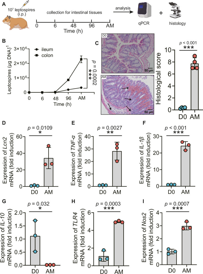
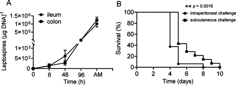
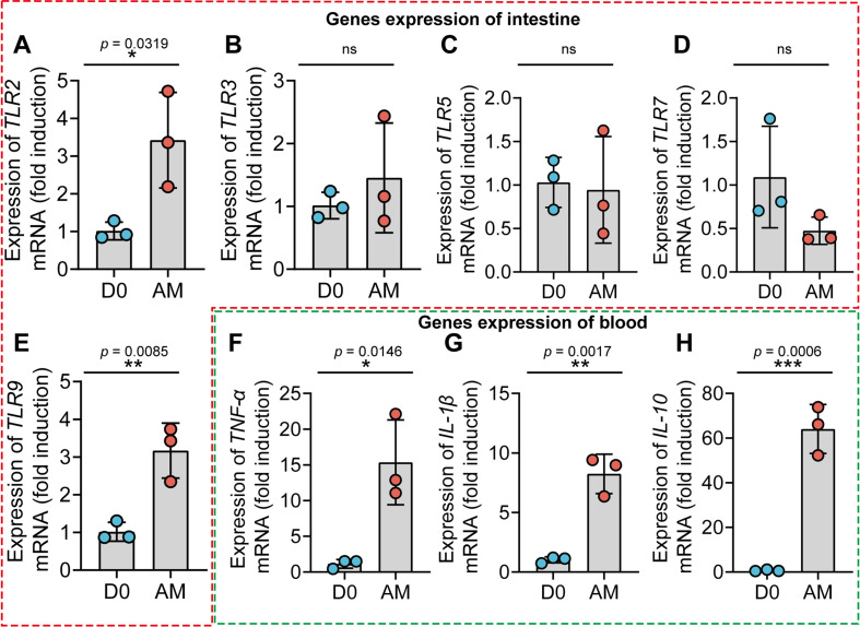
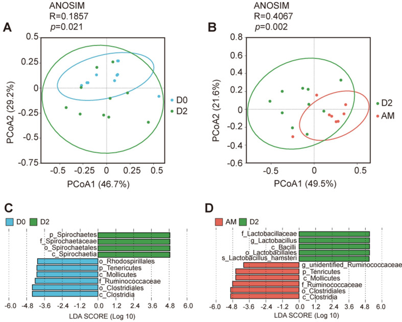
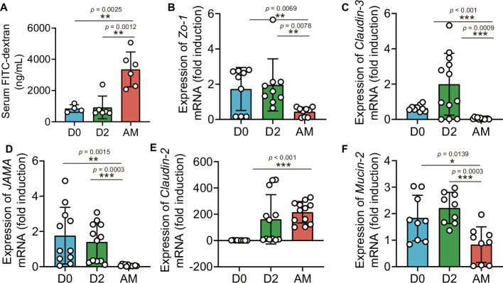
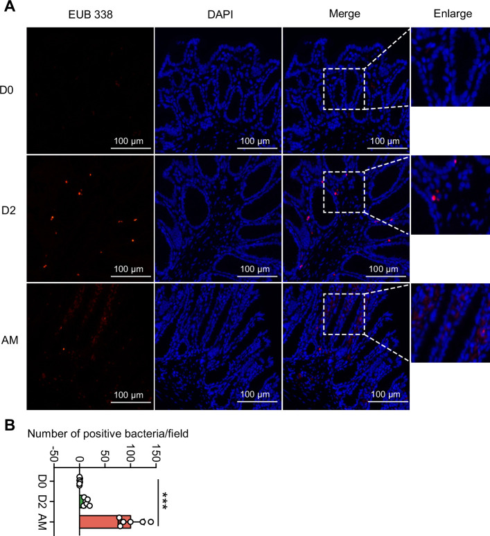
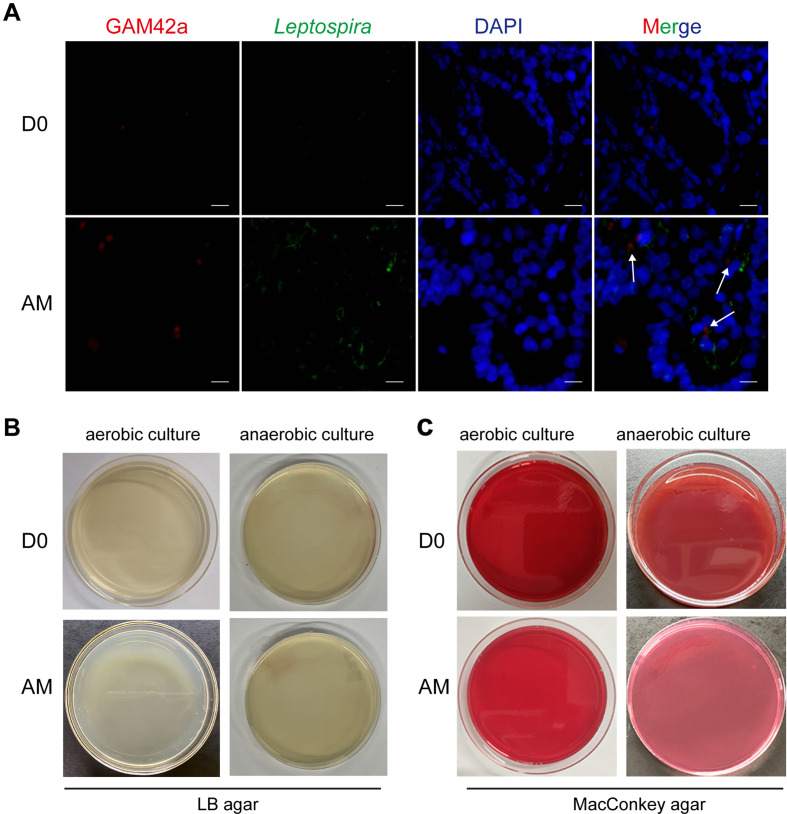
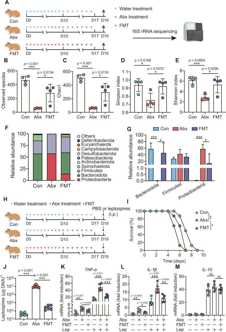
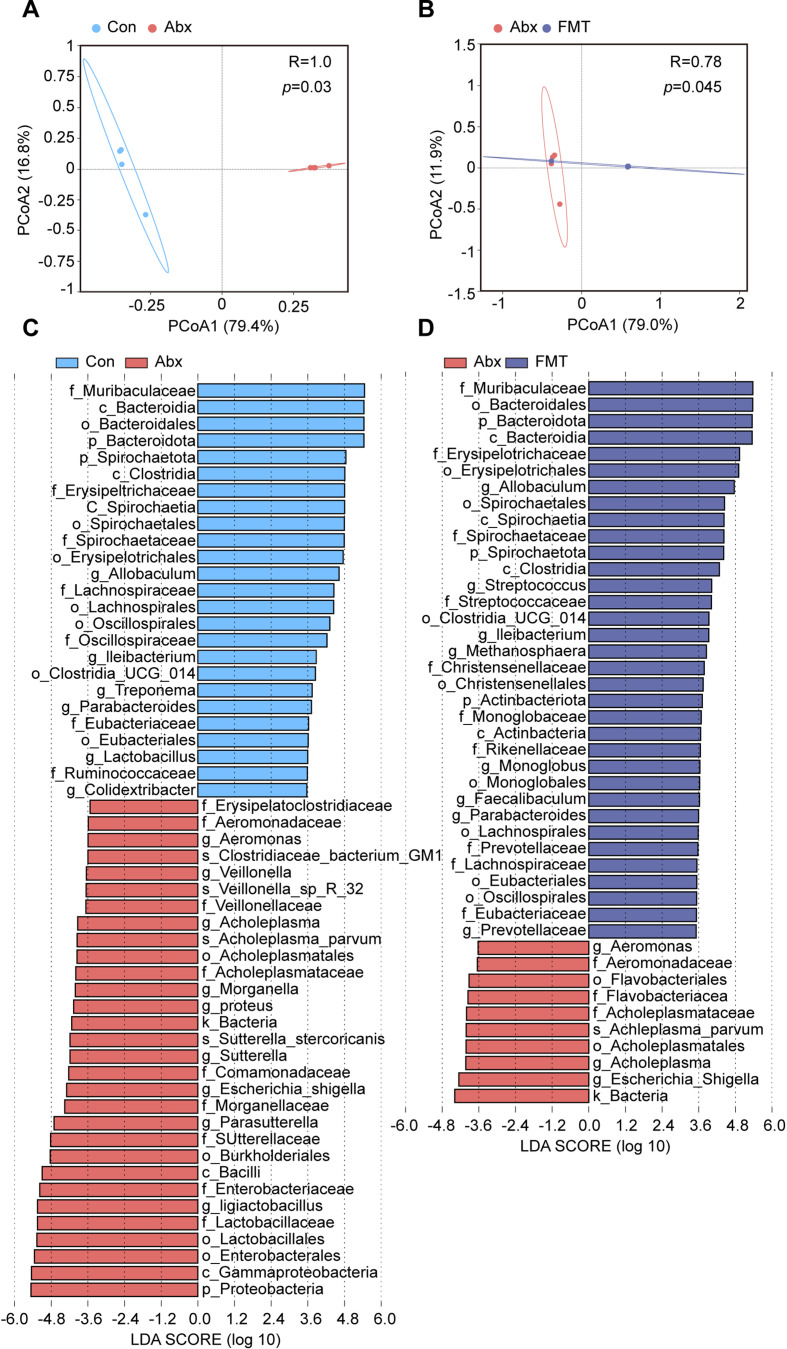
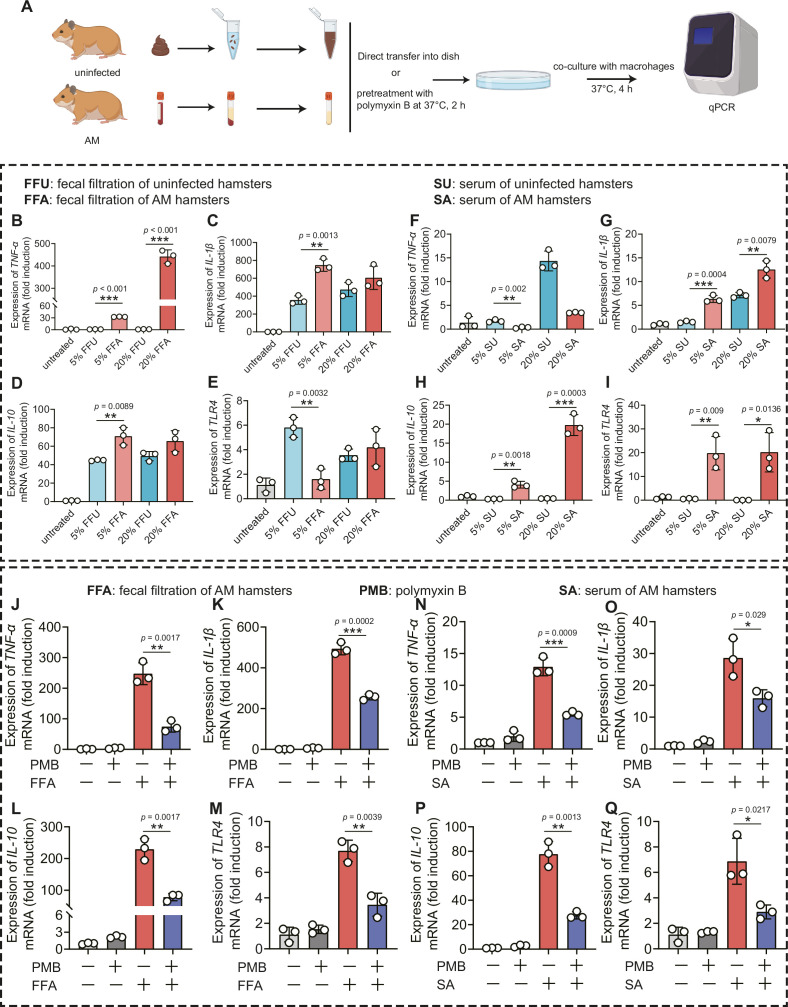
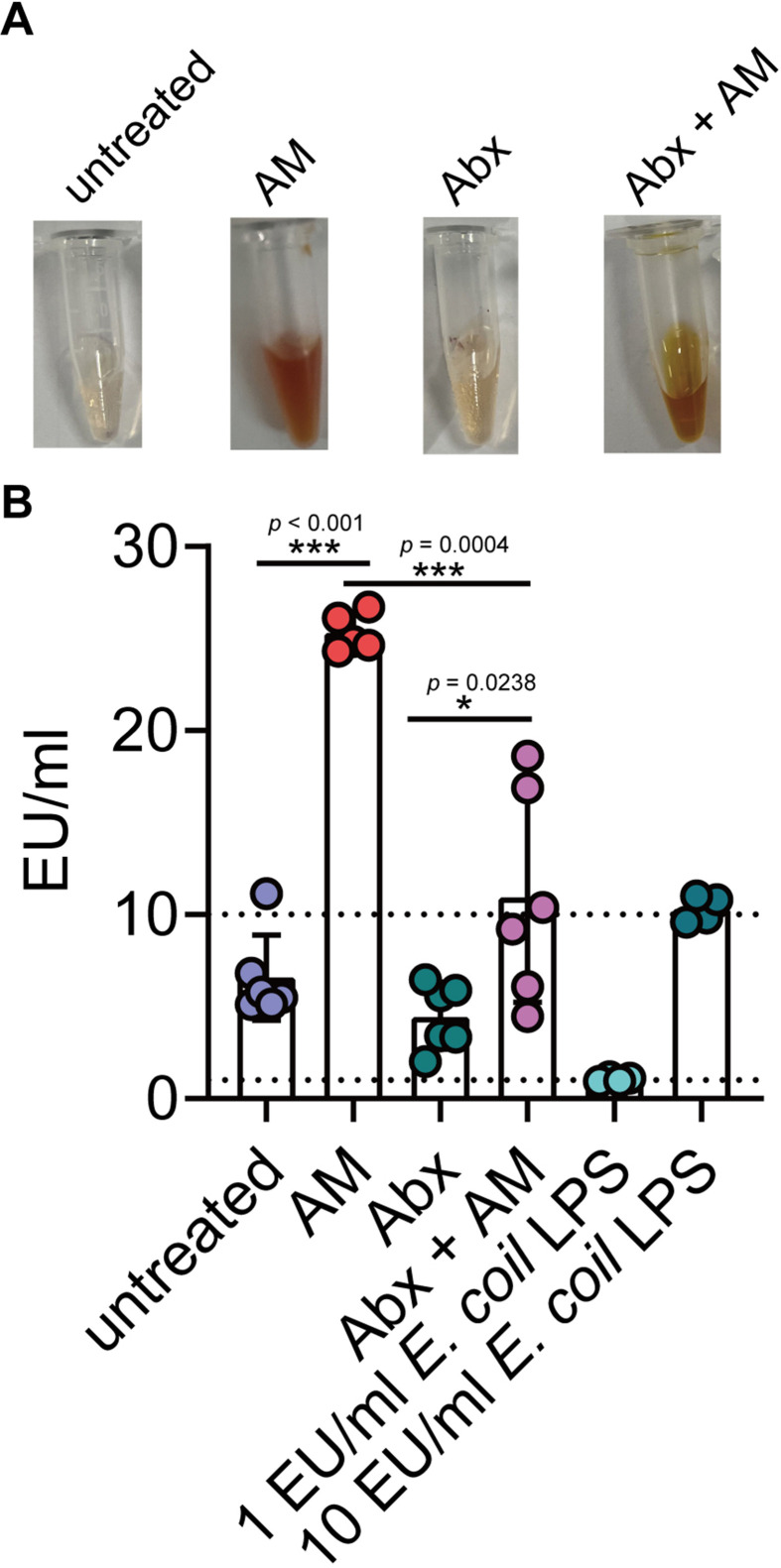
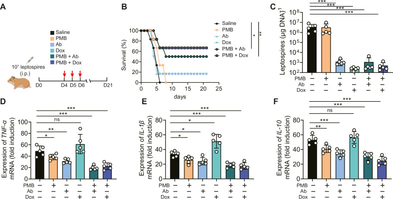
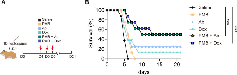
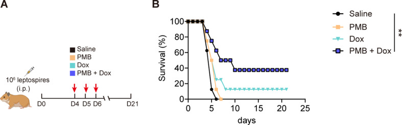
Leptospirosis is an emerging infectious disease caused by pathogenic Leptospira spp. Humans and some mammals can develop severe forms of leptospirosis accompanied by a dysregulated inflammatory response, which often results in death. The gut microbiota has been increasingly recognized as a vital element in systemic health. However, the precise role of the gut microbiota in severe leptospirosis is still unknown. Here, we aimed to explore the function and potential mechanisms of the gut microbiota in a hamster model of severe leptospirosis. Our study showed that leptospires were able to multiply in the intestine, cause pathological injury, and induce intestinal and systemic inflammatory responses. 16S rRNA gene sequencing analysis revealed that Leptospira infection changed the composition of the gut microbiota of hamsters with an expansion of Proteobacteria. In addition, gut barrier permeability was increased after infection, as reflected by a decrease in the expression of tight junctions. Translocated Proteobacteria were found in the intestinal epithelium of moribund hamsters, as determined by fluorescence in situ hybridization, with elevated lipopolysaccharide (LPS) levels in the serum. Moreover, gut microbiota depletion reduced the survival time, increased the leptospiral load, and promoted the expression of proinflammatory cytokines after Leptospira infection. Intriguingly, fecal filtration and serum from moribund hamsters both increased the transcription of TNF-α, IL-1β, IL-10, and TLR4 in macrophages compared with those from uninfected hamsters. These stimulating activities were inhibited by LPS neutralization using polymyxin B. Based on our findings, we identified an LPS neutralization therapy that significantly improved the survival rates in severe leptospirosis when used in combination with antibiotic therapy or polyclonal antibody therapy. In conclusion, our study not only uncovers the role of the gut microbiota in severe leptospirosis but also provides a therapeutic strategy for severe leptospirosis.

Keywords: Leptospira; hamster; infectious disease; microbiology; microbiota.


Conflict of interest statement

XX, XC, SZ, JL, WZ, YC No competing interests declared

Figures

Figure 1.
Figure 1.. L. interrogans proliferated in the intestine, destroyed the intestinal structure, and increased intestinal inflammation.
(A) Flow diagram of the experiment. Six-week-old female hamsters were injected intraperitoneally with 107 leptospires. Hamsters were euthanized at 0 hr, 6 hr, 48 hr, 96 hr, and AM post infection (p.i.). The ileums and colons were collected aseptically for further analysis. (B) Leptospiral burdens in the ileums and colons of Leptospira-infected hamsters were determined by qPCR (n = 6). (C) Left panel: histopathology of the colons of D0 hamsters (scale bar, 50 μm; n = 4) and AM hamsters (scale bar, 50 μm; n = 4). Representative photographs are presented. Right panel: histopathology scores of the colons. D0, uninfected hamster; AM, articulo mortis. (D–I) The gene expression of Lcn2 (D), TNF-α (E), IL-1β (F), IL-10 (G), TLR4 (H), and Nos2 (I) was analyzed by RT-qPCR. The mRNA levels of genes in the colons of the D0 group (n = 3) and the AM group (n = 3) were normalized to the expression of the housekeeping gene GAPDH. Each infection experiment was repeated three times. Data are shown as the mean ± SEM and analyzed by using the Wilcoxon rank-sum test. *p<0.05, **p<0.01, ***p<0.001.
Figure 1—figure supplement 1.
Figure 1—figure supplement 1.. The effect of challenge routes on leptospirosis.
Six-week-old female hamsters were injected intraperitoneally or subcutaneously with 107 leptospires. Then, hamsters were treated as described in ‘Materials and methods’. (A) Leptospiral load in the ileums and colons of hamsters (n = 4) infected subcutaneously was determined by qPCR. (B) Survival curves of hamsters infected intraperitoneally (n = 16) or subcutaneously (n = 14) with 107 leptospires. Each experiment was repeated three times. Data are shown as the mean ± SEM and analyzed by using the Wilcoxon rank-sum test. Survival differences was analyzed by using the Kaplan–Meier log-rank test. **p<0.01.
Figure 1—figure supplement 2.
Figure 1—figure supplement 2.. The gene expression of TLRs in the intestine and inflammatory cytokines in the blood.
Six-week-old female hamsters were injected intraperitoneally with 107 leptospires. Hamsters were euthanatized at 0 hr and AM post infection (p.i.). Colons and blood were collected aseptically for analysis of gene expression. D0, uninfected hamster; AM, articulo mortis. (A–E) The gene expression of TLR2 (A), TLR3 (B), TLR5 (C), TLR7 (D), and TLR9 (E) in the intestine was analyzed by RT-qPCR. (F–H) The gene expression of TNF-α (F), IL-1β (G), and IL-10 (H) in the blood was analyzed by RT-qPCR. The mRNA levels of genes of the D0 group (n = 3) and the AM group (n = 3) were normalized to the expression of the housekeeping gene GAPDH. The mRNA levels of cytokines in uninfected controls were set as 1.0. Each infection experiment was repeated three times. Data are shown as the mean ± SEM and analyzed by using the Wilcoxon rank-sum test. *p<0.05, **p<0.01, ***p<0.001. ns, not significant.
Figure 2.
Figure 2.. L. interrogans infection changed the composition of the gut microbiota.
(A) Flow diagram of the experiment. Six-week-old female hamsters were injected intraperitoneally with 107 leptospires. Fecal pellets were collected aseptically for 16S rRNA gene sequencing at 0 d, 2 d, and AM post infection (p.i.). D0, uninfected hamster; AM, articulo mortis. (B, C) Observed species (B) and ace (C) in the feces of hamsters. Observed species and ace indicate species richness. (D, E) Simpson index (D) and Shannon index (E) in the feces of hamsters. Simpson and Shannon indexes indicate species diversity. (F) Principal coordinate analysis (PCoA) of fecal samples based on 16S rRNA gene sequencing using weighted UniFrac. (G) PCoA of fecal samples based on 16S rRNA gene sequencing using weighted UniFrac. (H) Relative abundance of the top 10 phyla in the feces of hamster. (I) Linear discriminant analysis (LDA) of effect size (LEfSe) between D0 and AM hamsters (LDA score > 3). Cambridge blue bars indicate taxa enrichment in D0 hamsters, and pink bars indicate taxa enrichment in AM hamsters. (J) The ratio of Firmicutes to Bacteroidetes in the D0 and AM groups. (K–M) The relative abundance of Proteobacteria (K), Lactobacillus (L), and Allobaculum (M) in the D0 and AM groups (D0, n = 10; D2, n = 10; AM, n = 8). Data are shown as the mean ± SEM and analyzed by using the Wilcoxon rank-sum test. *p<0.05, **p<0.01.
Figure 2—figure supplement 1.
Figure 2—figure supplement 1.. The effect of Leptospira infection on the composition of the gut microbiota.
(A) Principal coordinate analysis (PCoA) of fecal samples based on 16S rRNA gene sequencing using weighted UniFrac (D0, n = 10; D2, n = 10). (B) PCoA of fecal samples based on 16S rRNA gene sequencing using weighted UniFrac (D2, n = 10; AM, n = 8). (C) Linear discriminant analysis (LDA) effect size (LEfSe) between D0 and D2 hamsters (LDA score > 4). Cambridge blue bars indicate taxa enrichment in D0 hamsters, and green bars indicate taxa enrichment in D2 hamsters. (D) LEfSe between D2 and AM hamsters (LDA score > 4). Green bars indicate taxa enrichment in D2 hamsters, and pink bars indicate taxa enrichment in AM hamsters. D0, uninfected hamster; AM, articulo mortis.
Figure 3.
Figure 3.. L. interrogans infection disrupted intestinal tight junctions.
Six-week-old female hamsters were injected intraperitoneally with 107 leptospires. Blood was collected to determine intestinal permeability at 0 d, 2 d, and AM post infection (p.i.). Colons were collected aseptically to measure gene expression at 0, 2, and AM p.i. (A) The intestinal permeability of Leptospira-infected hamsters was analyzed with a fluorescence spectrophotometer at the indicated time (D0, n = 4; D2, n = 6; AM, n = 6). (B–F) The relative gene expression of ZO-1 (B), Claudin-3 (C), JAMA (D), Claudin-2 (E), and Mucin-2 (F) in Leptospira-infected hamsters (n = 6–10 per group) was determined by qPCR. D0, uninfected hamster. AM, articulo mortis. Each infection experiment was repeated three times. Data are shown as the mean ± SEM and analyzed by using the Wilcoxon rank-sum test. *p<0.05, **p<0.01, ***p<0.001.
Figure 4.
Figure 4.. Translocated microbiota in the intestinal epithelium following L. interrogans infection.
Six-week-old female hamsters were injected intraperitoneally with 107 leptospires. Hamsters were euthanized at 0 d, 2 d, and AM post infection (p.i.). Colons were collected aseptically. The intestinal contents were excluded from the intestine with phosphate-buffered saline (PBS) and a segment of the colon was fixed in 4% paraformaldehyde solution overnight and analyzed by fluorescence in situ hybridization (FISH) with an EUB 338 probe (red). (A) The results are representative photographs of three groups. Scale bar, 100 μm. (B) Number of positive bacteria per field in the three groups. n = 6 per group. D0, uninfected hamster; AM, articulo mortis. Each infection experiment was repeated three times. Data are shown as the mean ± SEM and analyzed by using the Wilcoxon rank-sum test. ***p<0.001.
Figure 4—figure supplement 1.
Figure 4—figure supplement 1.. Translocation of the Proteobacteria during L. interrogans infection.
Six-week-old female hamsters were intraperitoneally infected with 107 leptospires. Colons of the D0 group and the AM group were collected and detected by fluorescence in situ hybridization (FISH) and immunofluorescence (IF). Blood from the D0 group and the AM group was plated on LB agar or MacConkey agar plates aerobically or anaerobically at 37°C for 36 hr. D0, uninfected hamster; AM, articulo mortis. (A) Colons of D0 (n = 4) and AM (n = 4) were examined by FISH with GAM42a probe (red) and by IF with anti-Leptospira serum (green). Scale bar, 20 μm. (B, C) The blood of the D0 (n = 4) hamsters and AM hamsters (n = 4) was aerobically or anaerobically cultured on LB plates (B) or MacConkey plates (C). Each experiment was repeated three times.
Figure 5.
Figure 5.. Microbiota depletion exacerbated leptospirosis.
(A) Flow diagram of the experiment. Hamsters were administered with Abx or water intragastrically once daily for 10 consecutive days. Then, fecal pellets were collected aseptically for 16S rRNA gene sequencing. (B, C) Observed species (B) and Chao1 (C) in the feces of hamsters. Observed species and Chao1 indicate species richness. (D, E) Simpson index (D) and Shannon index (E) in the feces of hamsters. Simpson and Shannon indexes indicate species diversity. (F) Relative abundance of the top 10 phyla in the feces of hamsters. (G) The relative abundance of Bacteroidetes, Firmicutes, and Proteobacteria in the Con, Abx, and fecal microbiota transplantation (FMT) groups. n = 4 per group. (H) Hamsters were treated with Abx as described above. After a 2-day washout period, hamsters were intraperitoneally infected with 106 L. interrogans. Then, the survival rate of the hamsters was recorded. n = 6 per group. (J) Leptospiral load in the blood of the Con, Abx, and FMT groups. n = 6 per group. (K, M) Gene expression of TNF-α (K), IL-1β (L), and IL-10 (M) in the blood of the three groups. n = 5 per group. Each infection experiment was repeated three times. Data are shown as the mean ± SEM and analyzed by using the Wilcoxon rank-sum test. Survival differences between the study groups were compared by using the Kaplan–Meier log-rank test. *p<0.05, **p<0.01, ***P<0.001. ns, not significant.
Figure 5—figure supplement 1.
Figure 5—figure supplement 1.. The effect of Abx treatment on the composition of the gut microbiota.
(A) Principal coordinate analysis (PCoA) of fecal samples based on 16S rRNA gene sequencing using weighted UniFrac (Con, n = 4; Abx, n = 4). (B) PCoA of fecal samples based on 16S rRNA gene sequencing using weighted UniFrac (Abx, n = 4; FMT, n = 4). (C) Linear discriminant analysis (LDA) effect size (LEfSe) between Con and Abx hamsters (LDA score > 4). Cambridge blue bars indicate taxa enrichment in Con hamsters, and pink bars indicate taxa enrichment in Abx hamsters. (D) LEfSe between FMT and Abx hamsters (LDA score > 4). Blue bars indicate taxa enrichment in FMT hamsters, and pink bars indicate taxa enrichment in Abx hamsters.
Figure 6.
Figure 6.. Leptopsira infection induced intestinal and systemic inflammation that was partially inhibited by lipopolysaccharide (LPS) neutralization.
(A) Flow diagram of the experiment. Fecal filtration or serum of uninfected hamsters and AM hamsters was directly added into dishes or pretreated with polymyxin B (PMB) at 37°C for 2 hr, and then cocultured with macrophages for 4 hr. RNA was extracted for gene expression analysis. (B–E) The gene expression of TNF-α (B), IL-1β (C), IL-10 (D), and TLR4 (E) of fecal filtration of uninfected hamsters (n = 3) and AM hamsters (n = 3) was analyzed by RT-qPCR. (F–I) The gene expression of TNF-α (F), IL-1β (G), IL-10 (H), and TLR4 (I) of serum of uninfected hamsters (n = 3) and AM hamsters (n = 3) was analyzed by RT-qPCR. (J–M) The effect of LPS neutralization on the gene expression of TNF-α (J), IL-1β (K), IL-10 (L), and TLR4 (M) in the fecal filtration of AM hamsters (n = 3) was analyzed by RT-qPCR. (N–Q) The effect of LPS neutralization on the gene expression of TNF-α (N), IL-1β (O), IL-10 (P), and TLR4 (Q) in the serum of AM hamsters (n = 3) was analyzed by RT-qPCR. The mRNA levels in untreated controls were set as 1.0. Each infection experiment was repeated three times. Data are shown as the mean ± SEM and analyzed by using the Wilcoxon rank-sum test. *p<0.05, **p<0.01, ***p<0.001.
Figure 6—figure supplement 1.
Figure 6—figure supplement 1.. Leptospira infection increased lipopolysaccharide (LPS) level in the blood.
Six-week-old female hamsters were intraperitoneally infected with 107 leptospires. The serum of untreated hamsters (n = 6), Abx-treated hamsters (n = 6), AM hamsters (n = 5), and Abx-treated AM hamsters (n = 6) was collected for LPS detection. E. coil LPS was used as a positive control to examine the quality of the kits. (A) Representative pictures of collected serum from untreated group, Abx-treated group, AM group, and Abx-treated AM group. (B) LPS level in the untreated group, Abx-treated group, AM group, Abx-treated AM group, 1 EU/ml E. coil LPS group, and 10 EU/ml E. coil LPS group. Each experiment was repeated three times. Data are shown as the mean ± SEM and analyzed by using the Wilcoxon rank-sum test. *p<0.05, ***p<0.001.
Figure 6—figure supplement 2.
Figure 6—figure supplement 2.. Determination minimum inhibitory concentrations of polymyxin B (PMB) and Dox on Leptospira 56601.
Leptospires at exponential phase were deposited at a final concentration of 2 × 10⁶ leptospires/ml in each well of 96-well plates with serial twofold dilutions of PMB and Dox ranging from 32 to 0.0156 μg/l in Ellinghausen–McCullough–Johnson–Harris (EMJH). The final volume in each well was 200 μl. The plates were incubated for 3 d at 30°C, then 20 μl of Alamar Blue was added to each well and the samples were incubated at 30°C for 2 d. Each strain–drug combination was tested in duplicate, and positive (bacteria and no antibiotic added) and negative (no bacteria added) controls were included in each plate. (A) Representative pictures of Alamar Blue test. (B) The percentage of resazurin reduction at different dosages of PMB and Dox incubated with leptospires. Each experiment was repeated three times. Data are shown as the mean ± SEM.
Figure 7.
Figure 7.. Lipopolysaccharide (LPS) neutralization combined with antibody therapy or antibiotic therapy improved the survival rate of female hamsters with severe leptospirosis.
(A) Flow diagram of the experiment. Six-week-old female hamsters were injected intraperitoneally with 107 leptospires. Group 1: saline control; group 2: polymyxin B (PMB) (1 mg/kg, i.p.); group 3: antibody against Leptospira (Ab) (16 mg/kg, subcutaneous injection); group 4: doxycycline (Dox) (5 mg/kg, i.p.); group 5: PMB (1 mg/kg, i.p.) and Ab (16 mg/kg, subcutaneous injection); group 6: PMB (1 mg/kg, i.p.) and Dox (5 mg/kg, i.p.). Hamsters were treated twice a day for three consecutive days. Hamsters were monitored daily for 21 d. (B) Survival rate of female hamsters of severe leptospirosis after different treatments. n = 6 per group. (C) Leptospiral load in the blood of different groups. n = 5 per group. (D–F) Gene expression of TNF-α (D), IL-1β (E), and IL-10 (F) in the blood of different groups. The mRNA levels of cytokines in uninfected controls were set as 1.0. n = 5 per group. Each infection experiment was repeated three times. Survival differences between the study groups were compared by using the Kaplan–Meier log-rank test. *p<0.05, **p<0.01, ***p<0.001. ns, not significant.
Figure 7—figure supplement 1.
Figure 7—figure supplement 1.. Lipopolysaccharide (LPS) neutralization combined with antibody therapy or antibiotic therapy improved the survival rate in male hamsters of severe leptospirosis.
(A) Flow diagram of the experiment. Six-week-old male hamsters were injected intraperitoneally with 107 leptospires. Group 1: saline control; group 2: polymyxin B (PMB) (1 mg/kg, i.p.); group 3: antibody against Leptospira (Ab) (16 mg/kg, subcutaneous injection); group 4: doxycycline (Dox) (5 mg/kg, i.p.); group 5: PMB (1 mg/kg, i.p.) and Ab (16 mg/kg, subcutaneous injection); group 6: PMB (1 mg/kg, i.p.) and Dox (5 mg/kg, i.p.). Hamsters were treated on three consecutive days, twice a day. (B) Survival rate of male hamsters of severe leptospirosis after different treatments Hamsters was monitored daily for 21 d. n = 8 per group. Each infection experiment was repeated three times. Survival differences between the study groups were compared by using the Kaplan–Meier log-rank test. *p<0.05, ***p<0.001.
Figure 7—figure supplement 2.
Figure 7—figure supplement 2.. Lipopolysaccharide (LPS) neutralization combined with antibiotic therapy improved the survival rate in female hamsters infected by 56606.
(A) Flow diagram of the experiment. Six-week-old female hamsters were injected intraperitoneally with 106 leptospires (56606). Group 1: saline control; group 2: polymyxin B (PMB) (1 mg/kg, i.p.); group 3: doxycycline (Dox) (5 mg/kg, i.p.); group 4: PMB (1 mg/kg, i.p.) and Ab (16 mg/kg, subcutaneous injection); Hamsters were treated on three consecutive days, twice a day. Hamsters were monitored daily for 21 d. (B) Survival rate of female hamsters of severe leptospirosis after different treatments. n = 8 per group. Each infection experiment was repeated three times. Survival differences between the study groups were compared by using the Kaplan–Meier log-rank test. **p<0.01.

Update of

  • doi: 10.1101/2024.01.17.576119
  • doi: 10.7554/eLife.96065.1
  • doi: 10.7554/eLife.96065.2

Similar articles

Cited by

References

    1. Alavi S, Mitchell JD, Cho JY, Liu R, Macbeth JC, Hsiao A. Interpersonal gut microbiome variation drives susceptibility and resistance to cholera infection. Cell. 2020;181:1533–1546. doi: 10.1016/j.cell.2020.05.036. - DOI - PMC - PubMed
    1. Aldape MJ, Bryant AE, Katahira EJ, Hajjar AM, Finegold SM, Ma Y, Stevens DL. Innate immune recognition of, and response to, Clostridium sordellii. Anaerobe. 2010;16:125–130. doi: 10.1016/j.anaerobe.2009.06.004. - DOI - PubMed
    1. Alventosa Mateu C. Gastrointestinal bleeding and acute hepatic failure by leptospirosis: an entity that should not be forgotten. J Rev Gastroenterol Peru. 2017;37:96–99. - PubMed
    1. Becattini S, Sorbara MT, Kim SG, Littmann EL, Dong Q, Walsh G, Wright R, Amoretti L, Fontana E, Hohl TM, Pamer EG. Rapid transcriptional and metabolic adaptation of intestinal microbes to host immune activation. Cell Host & Microbe. 2021;29:378–393. doi: 10.1016/j.chom.2021.01.003. - DOI - PMC - PubMed
    1. Bernard-Raichon L, Venzon M, Klein J, Axelrad JE, Zhang C, Sullivan AP, Hussey GA, Casanovas-Massana A, Noval MG, Valero-Jimenez AM, Gago J, Putzel G, Pironti A, Wilder E, Yale IMPACT Research Team. Thorpe LE, Littman DR, Dittmann M, Stapleford KA, Shopsin B, Torres VJ, Ko AI, Iwasaki A, Cadwell K, Schluter J. Gut microbiome dysbiosis in antibiotic-treated COVID-19 patients is associated with microbial translocation and bacteremia. Nature Communications. 2022;13:5926. doi: 10.1038/s41467-022-33395-6. - DOI - PMC - PubMed