. 2024 Oct;20(10):2314-2322.
doi: 10.1080/15548627.2024.2360345. Epub 2024 Jun 4.

Atg44/Mdi1/mitofissin facilitates Dnm1-mediated mitochondrial fission

Affiliations

Atg44/Mdi1/mitofissin facilitates Dnm1-mediated mitochondrial fission

Kentaro Furukawa et al. Autophagy. 2024 Oct.

Abstract

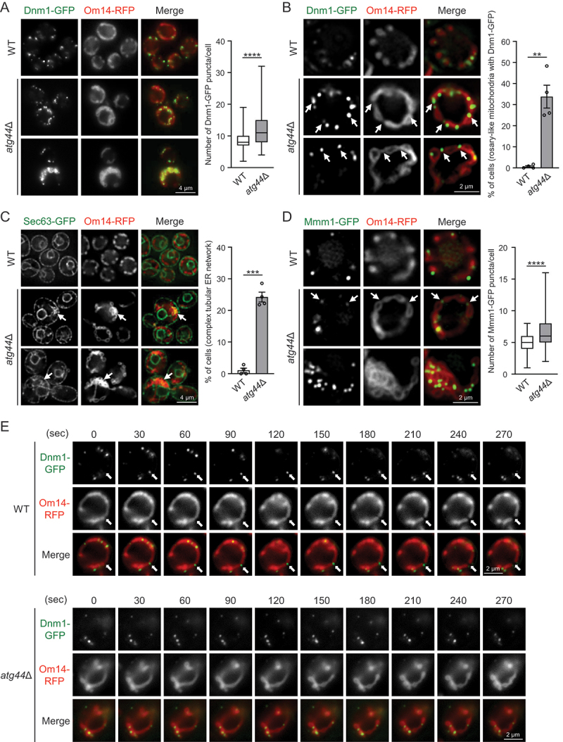
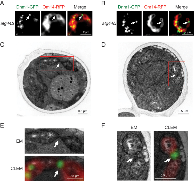
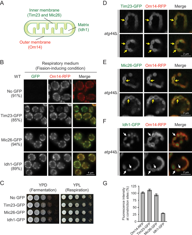
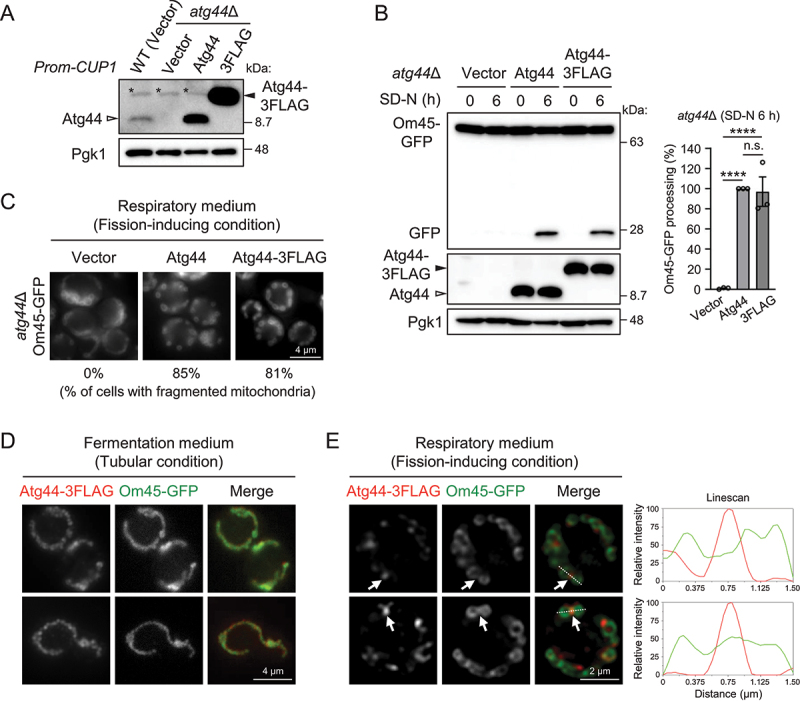
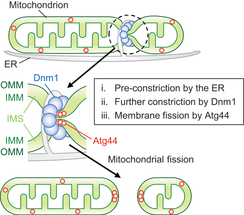
Mitochondria undergo fission and fusion, and their coordinated balance is crucial for maintaining mitochondrial homeostasis. In yeast, the dynamin-related protein Dnm1 is a mitochondrial fission factor acting from outside the mitochondria. We recently reported the mitochondrial intermembrane space protein Atg44/mitofissin/Mdi1/Mco8 as a novel fission factor, but the relationship between Atg44 and Dnm1 remains elusive. Here, we show that Atg44 is required to complete Dnm1-mediated mitochondrial fission under homeostatic conditions. Atg44-deficient cells often exhibit enlarged mitochondria with accumulated Dnm1 and rosary-like mitochondria with Dnm1 foci at constriction sites. These mitochondrial constriction sites retain the continuity of both the outer and inner membranes within an extremely confined space, indicating that Dnm1 is unable to complete mitochondrial fission without Atg44. Moreover, accumulated Atg44 proteins are observed at mitochondrial constriction sites. These findings suggest that Atg44 and Dnm1 cooperatively execute mitochondrial fission from inside and outside the mitochondria, respectively.Abbreviation: ATG: autophagy related; CLEM: correlative light and electron microscopy; EM: electron microscopy; ER: endoplasmic reticulum; ERMES: endoplasmic reticulum-mitochondria encounter structure; GA: glutaraldehyde; GFP: green fluorescent protein; GTP: guanosine triphosphate: IMM: inner mitochondrial membrane; IMS: intermembrane space; OMM: outer mitochondrial membrane; PB: phosphate buffer; PBS: phosphate-buffered saline; PFA: paraformaldehyde; RFP: red fluorescent protein; WT: wild type.

Keywords: Atg44; Dnm1; mitochondrial fission; mitofissin; mitophagy; yeast.


Conflict of interest statement

No potential conflict of interest was reported by the author(s).

Figures

Figure 1.
Figure 1.
Atg44 is required to complete Dnm1-mediated mitochondrial fission. (A–D) S. cerevisiae WT and atg44∆ cells expressing Om14-RFP and the indicated GFP-fused proteins were cultured in YPL until mid-log phase and analyzed by fluorescence microscopy. (A) Typical mitochondrial morphology and distribution of Dnm1-GFP puncta in WT and atg44∆ cells. Enlarged or aggregated mitochondria with numerous Dnm1-GFP puncta in atg44∆ cells are shown in the lower panel. The number of Dnm1-GFP puncta associated with mitochondria was counted and the results are shown as boxplots. The boxplots indicate minimum, lower quartile, median, upper quartile, and maximum. ****p < 0.0001 (Welch’s t-test). WT, n = 480; atg44∆, n = 443; four experiments. (B) Fragmented mitochondria in WT cells and rosary-like mitochondria in atg44∆ cells. Bars show the percentage of cells with rosary-like mitochondria (no fragmented mitochondria) containing Dnm1-GFP puncta (white arrows). **p < 0.01 (Welch’s t-test). WT, n = 480; atg44∆, n = 510; four experiments. (C) Association of the complex tubular ER network with aggregated mitochondria in atg44∆ cells. Bars show the percentage of cells with complex tubular ER network on aggregated mitochondria (white arrows). ***p < 0.001 (Welch’s t-test). WT, n = 272; atg44∆, n = 249; four experiments. (D) Association of ERMES visualized by Mmm1-GFP with the mitochondrial constriction sites (white arrows, middle panel) or aggregated mitochondria (lower panel) in atg44∆ cells. The number of Mmm1-GFP puncta was counted and the results are shown as in (A). ****p < 0.0001 (Welch’s t-test). WT, n = 360; atg44∆, n = 360; three experiments. (E) Time-lapse images of Dnm1-mediated mitochondrial fission were acquired at 30-s intervals for a total of 270 s, 4 ± 1 h after shifting cells from YPD to YPL medium. A total of 2931 WT cells and 2998 atg44∆ cells were analyzed. White arrows indicate the fission site linked to Dnm1-GFP puncta. Scale bars are shown in each panel. See also Figures S1 and S2.
Figure 2.
Figure 2.
CLEM imaging of the mitochondrial constriction sites with Dnm1-GFP signals. S. cerevisiae atg44∆ cells expressing Dnm1-GFP and Om14-RFP were cultured in YPL until mid-log phase, fixed with 2% PFA/0.1% GA for 30 min at room temperature, and analyzed by CLEM. See Materials and methods for details. Representative images (fluorescence microscopy in A and B; electron microscopy in C and D; magnified electron microscopy and CLEM in E and F) are shown. White arrows indicate the mitochondrial constriction sites with Dnm1-GFP signals. Scale bars are shown in each panel.
Figure 3.
Figure 3.
Mitochondrial constriction sites in atg44∆ cells retain a continuous inner membrane. (A) Localization of OMM, IMM, and matrix proteins analyzed in this study. (B) S. cerevisiae WT cells expressing Om14-RFP and the indicated GFP-fused proteins were cultured in YPL until mid-log phase and analyzed by fluorescence microscopy. The percentage of cells with fragmented mitochondria is shown (n = 140 cells). (C) the indicated cells were cultured in YPD until early-log phase, and their serial dilutions were spotted and cultured on YPD (1 day) or YPL (2 days) agar plates (three independent replicates). (D–F) S. cerevisiae atg44∆ cells expressing Om14-RFP and the indicated GFP-fused mitochondrial proteins were cultured in YPL until mid-log phase and analyzed by fluorescence microscopy. Yellow and white arrows indicate high and low GFP signals at mitochondrial constriction sites, respectively. (G) Bars show the fluorescence intensity (constriction sites relative to average of non-constriction sites) of the indicated RFP- or GFP-fused proteins. Om14-RFP, n = 48; Tim23-GFP, n = 53; Mic26-GFP, n = 50; Idh1-GFP, n = 49. Scale bars are shown in each panel.
Figure 4.
Figure 4.
Accumulation of Atg44 proteins is observed at mitochondrial constriction sites. (A) the indicated yeast cells carrying the indicated plasmids (CUP1 promoter) were cultured in SML medium until mid-log phase and subjected to immunoblotting analysis. Endogenous Atg44 and exogenously expressed Atg44/Atg44-3FLAG were detected using anti-Atg44 antibody. Pgk1 was detected as a loading control. Representative immunoblots of three experiments are shown. Asterisks indicate non-specific bands. (B) the indicated yeast cells were cultured in SML until mid-log phase and shifted to SD-N. Cells were collected at the indicated time points, and Om45-GFP processing was monitored by immunoblotting. The value of Atg44 (6 h) was set to 100%. The results represent the mean and SE of three experiments. ****p < 0.0001, n.S. not significant (ordinary one-way ANOVA multiple comparison). (C) the indicated yeast cells were cultured in SML until mid-log phase and analyzed by fluorescence microscopy. The percentage of cells with fragmented mitochondria is shown (n = 200 cells). (D and E) atg44∆ cells expressing Om45-GFP and Atg44-3FLAG were cultured in SMD (D) or SML (E) until mid-log phase and analyzed by immunofluorescence microscopy using primary antibodies against FLAG and secondary antibodies conjugated with Alexa Fluor 594. White arrows indicate accumulated Atg44-3FLAG proteins. Dashed lines correspond to the linescans. Scale bars are shown in each panel.
Figure 5.
Figure 5.
Schematic model for coordinated mitochondrial fission by Dnm1 and Atg44. See text for details.

Similar articles

Cited by

References

    1. Quintana-Cabrera R, Scorrano L.. Determinants and outcomes of mitochondrial dynamics. Mol Cell. 2023;83(6):857–876. doi: 10.1016/j.molcel.2023.02.012 - DOI - PubMed
    1. Chan DC. Mitochondrial dynamics and its involvement in disease. Annu Rev Pathol. 2020;15(1):235–259. doi: 10.1146/annurev-pathmechdis-012419-032711 - DOI - PubMed
    1. Otsuga D, Keegan BR, Brisch E, et al. The dynamin-related GTPase, Dnm1p, controls mitochondrial morphology in yeast. J Cell Bio. 1998;143(2):333–349. doi: 10.1083/jcb.143.2.333 - DOI - PMC - PubMed
    1. Bleazard W, McCaffery JM, King EJ, et al. The dynamin-related GTPase Dnm1 regulates mitochondrial fission in yeast. Nat Cell Biol. 1999;1(5):298–304. doi: 10.1038/13014 - DOI - PMC - PubMed
    1. Smirnova E, Shurland DL, Ryazantsev SN, et al. A human dynamin-related protein controls the distribution of mitochondria. J Cell Bio. 1998;143(2):351–358. doi: 10.1083/jcb.143.2.351 - DOI - PMC - PubMed

Publication types

MeSH terms

LinkOut - more resources