Associations Between Genetically Predicted Iron Status and Cardiovascular Disease Risk: A Mendelian Randomization Study
- PMID: 38818967
- PMCID: PMC11255641
- DOI: 10.1161/JAHA.124.034991
Associations Between Genetically Predicted Iron Status and Cardiovascular Disease Risk: A Mendelian Randomization Study
Abstract
Background: Mendelian randomization (MR) studies suggest a causal effect of iron status on cardiovascular disease (CVD) risk, but it is unknown if these associations are confounded by pleiotropic effects of the instrumental variables on CVD risk factors. We aimed to investigate the effect of iron status on CVD risk controlling for CVD risk factors.
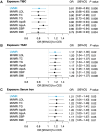
Methods and results: Iron biomarker instrumental variables (total iron-binding capacity [n=208 422], transferrin saturation [n=198 516], serum iron [n=236 612], ferritin [n=257 953]) were selected from a European genome-wide association study meta-analysis. We performed 2-sample univariate MR of each iron trait on CVD outcomes (all-cause ischemic stroke, cardioembolic ischemic stroke, large-artery ischemic stroke, small-vessel ischemic stroke, and coronary heart disease) from MEGASTROKE (n=440 328) and CARDIoGRAMplusC4D (Coronary Artery Disease Genome Wide Replication and Meta-Analysis Plus the Coronary Artery Disease Genetics) (n=183 305). We then implemented multivariate MR conditioning on 7 CVD risk factors from independent European samples to evaluate their potential confounding or mediating effects on the observed iron-CVD associations. With univariate MR analyses, we found higher genetically predicted iron status to be associated with a greater risk of cardioembolic ischemic stroke (transferrin saturation: odds ratio, 1.17 [95% CI, 1.03-1.33]; serum iron: odds ratio, 1.21 [95% CI, 1.02-1.44]; total iron-binding capacity: odds ratio, 0.81 [95% CI, 0.69-0.94]). The detrimental effects of iron status on cardioembolic ischemic stroke risk remained unaffected when adjusting for CVD risk factors (all P<0.05). Additionally, we found diastolic blood pressure to mediate between 7.1 and 8.8% of the total effect of iron status on cardioembolic ischemic stroke incidence. Univariate MR initially suggested a protective effect of iron status on large-artery stroke and coronary heart disease, but controlling for CVD factors using multivariate MR substantially diminished these associations (all P>0.05).
Conclusions: Higher iron status was associated with a greater risk of cardioembolic ischemic stroke independent of CVD risk factors, and this effect was partly mediated by diastolic blood pressure. These findings support a role of iron status as a modifiable risk factor for cardioembolic ischemic stroke.
Keywords: Mendelian randomization analysis; biomarkers; blood pressure; genome‐wide association study; iron; ischemic stroke; risk factors.
Figures


Update of
-
Associations between genetically predicted iron status and cardiovascular disease risk: A Mendelian randomization study.medRxiv [Preprint]. 2024 Feb 6:2024.02.05.24302373. doi: 10.1101/2024.02.05.24302373. medRxiv. 2024. Update in: J Am Heart Assoc. 2024 Jun 4;13(11):e034991. doi: 10.1161/JAHA.124.034991. PMID: 38370765 Free PMC article. Updated. Preprint.
References
-
- Jenkitkasemwong S, Wang CY, Coffey R, Zhang W, Chan A, Biel T, Kim JS, Hojyo S, Fukada T, Knutson MD. SLC39A14 is required for the development of hepatocellular iron overload in murine models of hereditary hemochromatosis. Cell Metab. 2015;22:138–150. doi: 10.1016/j.cmet.2015.05.002 - DOI - PMC - PubMed
MeSH terms
Substances
Grants and funding
LinkOut - more resources
Full Text Sources
Medical

