. 2024 Oct 4;14(10):1860-1878.
doi: 10.1158/2159-8290.CD-23-1092.

CRISPR Dependency Screens in Primary Hematopoietic Stem Cells Identify KDM3B as a Genotype-specific Vulnerability in IDH2- and TET2-mutant Cells

Affiliations

CRISPR Dependency Screens in Primary Hematopoietic Stem Cells Identify KDM3B as a Genotype-specific Vulnerability in IDH2- and TET2-mutant Cells

Michael R Waarts et al. Cancer Discov. .

Abstract

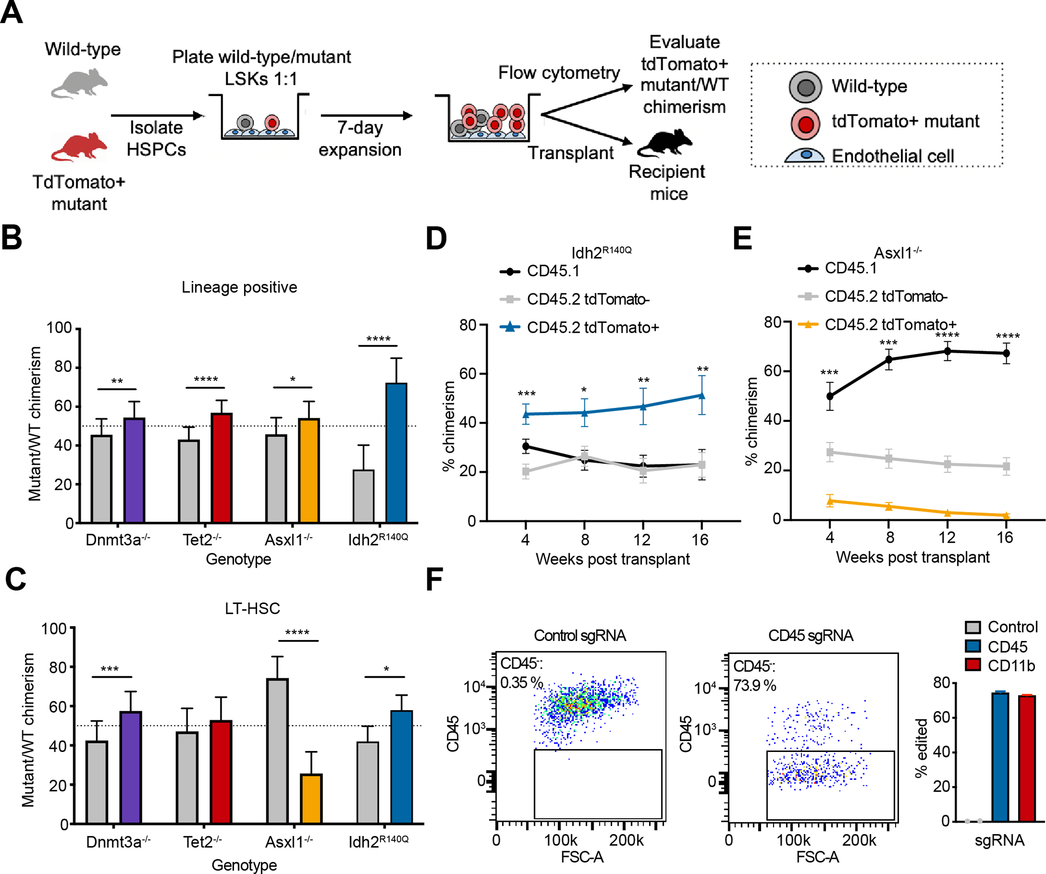
Clonal hematopoiesis (CH) is a common premalignant state in the blood and confers an increased risk of blood cancers and all-cause mortality. Identification of therapeutic targets in CH has been hindered by the lack of an ex vivo platform amenable for studying primary hematopoietic stem and progenitor cells (HSPCs). Here, we utilize an ex vivo co-culture system of HSPCs with bone marrow endothelial cells to perform CRISPR/Cas9 screens in mutant HSPCs. Our data reveal that loss of the histone demethylase family members Kdm3b and Jmjd1c specifically reduces the fitness of Idh2- and Tet2-mutant HSPCs. Kdm3b loss in mutant cells leads to decreased expression of critical cytokine receptors including Mpl, rendering mutant HSPCs preferentially susceptible to inhibition of downstream JAK2 signaling. Our study nominates an epigenetic regulator and an epigenetically regulated receptor signaling pathway as genotype-specific therapeutic targets and provides a scalable platform to identify genetic dependencies in mutant HSPCs. Significance: Given the broad prevalence, comorbidities, and risk of malignant transformation associated with CH, there is an unmet need to identify therapeutic targets. We develop an ex vivo platform to perform CRISPR/Cas9 screens in primary HSPCs. We identify KDM3B and downstream signaling components as genotype-specific dependencies in CH and myeloid malignancies. See related commentary by Khabusheva and Goodell, p. 1768.


Conflict of interest statement

Conflict of interest statement: R.L.L. is on the supervisory board of Qiagen and on the board of directors of Ajax Therapeutics, for which he receives compensation and equity support. He is or has recently been a scientific advisor to Imago, Mission Bio, Syndax. Zentalis, Ajax, Bakx, Auron, Prelude, C4 Therapeutics and Isoplexis for which he receives equity support. He has research support from Ajax and AbbVie, consulted for Janssen, and received honoraria from Astra Zeneca and Kura for invited lectures. L.A.M. has received honoraria from Mission Bio and previously served as a member of the Mission Bio Speakers Bureau. S.F.C. is a consultant for Daiichi-Sankyo and Ursamin. He was previously a consultant for Dava Oncology and held equity interest in Imago Biosciences, none of which are directly related to the content of this paper. R.K. is a co-founder of and consultant for Econic Biosciences. S.A.A. has been a consultant and/or shareholder for Neomorph Inc, Imago Biosciences, Cyteir Therapeutics, C4 Therapeutics, Nimbus Therapeutics and Accent Therapeutics. SAA has received research support from Janssen and Syndax. S.A.A. is named as an inventor on a patent application related to MENIN inhibition WO/2017/132398A1. W.X. received research support from Stemline Therapeutics. No other authors report competing interests.

Figures

Figure 1:
Figure 1:. BMEC/HSPC co-cultures of CH-mutant HSPCs yield rapid ex vivo phenotypes.
A, Schematic representation of BMEC co-culture with HSPCs. LSK cells are isolated from wild-type or tdT+ CH-mutant mice and competitively cultured in a 1:1 ratio over a feeder layer of BMECs. Expanded cells are transplanted into recipient mice or analyzed by flow cytometry after 7 days. B and C, Flow cytometric analysis of tdT+ mutant and wild-type chimerism measured after 7 days of culture within lineage+ cells (B) or long-term hematopoietic stem cells (LT-HSCs) (C). Grey bars represent wild-type chimerism and colored bars represent mutant chimerism. (n = 9–12) D and E, Peripheral blood chimerism of recipient mice (% CD45.1) transplanted with cells from BMEC/HSPC co-cultures of wild-type (% CD45.2 tdT) versus Idh2R140Q (% CD45.2 tdT+) (D) or Asxl1−/− (% CD45.2 tdT+) (E) cells. (n = 10) F, Representative flow cytometry plots and quantification of editing efficiency in HSPCs maintained in BMEC/HSPC co-cultures 7 days after lentiviral infection of sgRNAs. Editing was assessed by flow cytometric analysis of cell surface expression of CD45 or CD11b. Empty control represents Cas9 delivery only and measurement of CD45 negative cells. (n = 3–4). All data are mean ± SEM. *p < 0.05, **p < 0.01, ***p < 0.001, ****p < 0.0001
Figure 2:
Figure 2:. CRISPR screens identify vulnerabilities in HSPCs with different CH mutations.
A, Schematic representation of CRISPR screens with BMEC/HSPC platform. HSPCs are isolated from Cas9-expressing mice, infected with a lentivirus encoding an epigenome sgRNA library, and co-cultured with BMECs. DNA is extracted pre- and post-culture for sgRNA library amplification and sequencing. MAGeCK is used to assess depleted and enriched sgRNAs. B, Correlation of normalized read counts from sgRNA libraries pre- and post-BMEC/HSPC co-culture of wild-type HSPCs without Cas9. C, Genes with depleted sgRNAs in Cas9-expressing HSPCs as assessed by sgRNA abundance post-culture normalized to pre-culture. D, Venn diagram of genes targeted by sgRNAs relatively depleted in mutant HSPCs compared to wild-type HSPCs. Genes that are also depleted in wild-type HSPCs are marked as shared dependencies. E and F, Genes with depleted or enriched sgRNAs in Idh2R140Q (E) or Tet2−/− (F) HSPCs as compared to wild-type HSPCs. G and H, Idh2R140Q tdT+ chimerism as assessed by flow cytometry in lineage (G) or LSK (H) infected cells measured after 14 days of BMEC/HSPC co-culture. Wild-type and Idh2R140Q tdT+ HSPCs were mixed 1:1 and infected with a BFP+ lentivirus encoding an sgRNA targeting Rosa or Kdm3b. (n = 3) I and J, Tet2−/− tdT+ chimerism as assessed by flow cytometry in lineage (I) or LSK (J) infected cells measured after 14 days of BMEC/HSPC co-culture. Wild-type and Tet2−/− tdT+ HSPCs were mixed 1:1 and infected with a BFP+ lentivirus encoding an sgRNA targeting Rosa or Kdm3b. (n = 3). All data are mean ± SEM. **p < 0.01, ***p < 0.001
Figure 3:
Figure 3:. Kdm3b is a dependency in Idh2- and Tet2-mutant HSPCs.
A, Schematic representation of in vivo CRISPR screens. HSPCs from Idh2R140Q or WT mice were infected with a lentivirus encoding a library of sgRNAs targeting dependencies identified in ex vivo screens. HSPCs were transplanted and sgRNA abundance was assessed in recipient mice 16 weeks after transplant. B, Genes with depleted sgRNAs in the bone marrow of recipient mice engrafted with Idh2R140Q cells as compared to wild-type cells. (DNA pooled from n = 10 mice) C and D, Peripheral blood chimerism of Idh2R140Q (C) or Tet2−/− (D) within infected cells engrafted in recipient mice. Wild-type and mutant tdT+ HSPCs were mixed 1:1, infected with a BFP+ lentivirus encoding an sgRNA targeting Rosa or Kdm3b, and transplanted into recipient mice. (n = 5–8) E and F, Idh2R140Q (E) or Tet2−/− (F) chimerism within infected cells in bone marrow compartments of recipient mice. Wild-type and mutant tdT+ HSPCs were mixed 1:1, infected with a BFP+ lentivirus encoding an sgRNA targeting Rosa or Kdm3b, and transplanted into recipient mice. (n = 5–8) G and H, Chimerism within infected myeloid cells in the peripheral blood and LT-HSCs in the bone marrow of recipient mice engrafted with cells from Idh2R140Q (G) or Tet2−/− (H) primary transplants from Fig. 3C–F. (n = 3–5) I, Schematic representation of inducible sgRNA experiments. HSPCs were isolated from wild-type or tdT+ Idh2R140Q-mutant mice, mixed in a 1:1 ratio, infected with a BFP+ lentivirus encoding a doxycycline-inducible sgRNA, and transplanted into recipient mice. Mice were treated with doxycycline (Dox) 8 weeks post-transplant to induce expression of sgRNAs. Chimerism within infected cells in the peripheral blood and bone marrow of recipient mice was assessed 20 weeks post-transplant. J, Peripheral blood chimerism of Idh2R140Q within infected cells engrafted in recipient mice. HSPCs were isolated from wild-type or tdT+ Idh2R140Q-mutant mice, mixed in a 1:1 ratio, infected with a BFP+ lentivirus encoding a doxycycline-inducible sgRNA, and transplanted into recipient mice. Mice were treated with doxycycline 8 weeks post-transplant as indicated. (n = 10–14) K, Idh2R140Q chimerism within infected cells in bone marrow compartments of recipient mice. HSPCs were isolated from wild-type or tdT+ Idh2R140Q-mutant mice, mixed in a 1:1 ratio, infected with a BFP+ lentivirus encoding a doxycycline-inducible sgRNA, and transplanted into recipient mice. Mice were treated with doxycycline 8 weeks post-transplant. (n = 10–14) L, Colony forming assay of wild-type or TET2-mutant iPSC-derived HSPCs electroporated with RNPs targeting AAVS1 or KDM3B. Colonies were scored 10 days after plating. (n = 3) M, Colony forming assay of cord blood HSPCs sequentially electroporated with sgRNAs against AAVS1 or TET2 followed by additional sgRNAs against AAVS1 or KDM3B. Colonies were scored 12 days after plating. (n = 2) N, Rosa or Kdm3b variant allele frequency (VAF) in the peripheral blood of recipient mice engrafted with Tet2−/−/Npm1c/Flt3ITD leukemic cells electroporated with RNPs targeting Rosa or Kdm3b. (n = 3). VAFs were assessed by NGS and normalized to week 2. All data are mean ± SEM. *p < 0.05, **p < 0.01, ***p < 0.001, ****p < 0.0001
Figure 4:
Figure 4:. Kdm3b vulnerability in Idh2-mutant cells is dependent on 2-HG and loss of KDM3B enzymatic activity.
A and B, Flow cytometry measurement of BFP+ infected wild-type lineage (A) or LSK (B) cells measured after 14 days of BMEC/HSPC co-culture. Wild-type HSPCs were infected with a BFP+ lentivirus encoding an sgRNA targeting Rosa or Kdm3b and treated with vehicle or 500 μM 2-HG. (n = 3) C and D, Idh2R140Q tdT+ chimerism as measured by flow cytometry in lineage (C) or LSK (D) infected cells measured after 14 days of BMEC/HSPC co-culture. Wild-type and Idh2R140Q tdT+ HSPCs were mixed 1:1, infected with a BFP+ lentivirus encoding an sgRNA targeting Rosa or Kdm3b, and treated with increasing doses of enasidenib. (n = 3) (E) Depletion of BFP+ shRNAs targeting KDM3B or scrambled (SCR) control in TF-1 cells isogenic for the IDH2 R140Q mutation. Measured as % BFP+ by flow cytometry 14 days after infection and normalized to % BFP+ 2 days after infection. (n = 3) (F) Depletion of BFP+ shRNAs targeting KDM3B or scrambled (SCR) control within TF-1 cells isogenic for the IDH2 R140Q mutation and carrying the indicated NGFR+ cDNA. Measured as % BFP+NGFR+ by flow cytometry 14 days after infection and normalized to % BFP+NGFR+ by flow cytometry 14 days after infection. (n = 3). All data are mean ± SEM. *p < 0.05, **p < 0.01, ***p < 0.001
Figure 5:
Figure 5:. KDM3B regulates the expression of critical cytokine receptors in IDH2-mutant cells.
A, Heatmap showing relative gene expression of differentially expressed genes in Kdm3b KO Idh2R140Q HSPCs as compared to Rosa KO wild-type HSPCs. Denoted groups shown are wild-type or Idh2R140Q HSPCs with Rosa or Kdm3b KO. B, ssGSEA scores of the gene set derived from downregulated genes in Kdm3b KO Idh2R140Q HSPCs. (n = 3) C, Mpl expression in primary HSPCs in the indicated groups by RNA-seq data. (n = 3) D, Mean normalized ATAC-seq signal at genes with decreased expression in KDM3B KD IDH2 R140Q TF-1 cells. E, Normalized ATAC-seq signal in TF-1 cells on surrounding loci at CSF2RB and MPL. F, Idh2R140Q tdT+ chimerism as measured by flow cytometry in LSK infected cells measured after 14 days of BMEC/HSPC co-culture. Wild-type and Idh2R140Q tdT+ HSPCs were mixed 1:1, infected with a BFP+ lentivirus encoding an sgRNA targeting Rosa or Kdm3b, and treated with increasing doses of TPO. (n = 3) G, Median fluorescent intensity (MFI) of phospho-STAT3 (pSTAT3) as measured by flow cytometry in lineageKit+ infected cells measured after 7 days of BMEC/HSPC co-culture. Wild-type and Idh2R140Q HSPCs were infected with a BFP+ lentivirus encoding an sgRNA targeting Rosa or Kdm3b. (n = 3) H, Idh2R140Q tdT+ chimerism as measured by flow cytometry in LSK infected cells measured after 14 days of BMEC/HSPC co-culture. Wild-type and Idh2R140Q tdT+ HSPCs were mixed 1:1, infected with a BFP+ lentivirus encoding an sgRNA targeting Rosa or Kdm3b, and treated with increasing doses of ruxolitinib. (n = 3). All data are mean ± SEM. *p < 0.05, **p < 0.01, ***p < 0.001, ****p < 0.0001, #FDR < 0.1

References

    1. Jaiswal S & Ebert BL Clonal hematopoiesis in human aging and disease. Science 366 (2019). 10.1126/science.aan4673 - DOI - PMC - PubMed
    1. Jaiswal S, Fontanillas P, Flannick J, Manning A, Grauman PV, Mar BG et al. Age-related clonal hematopoiesis associated with adverse outcomes. N Engl J Med 371, 2488–2498 (2014). 10.1056/NEJMoa1408617 - DOI - PMC - PubMed
    1. Xie M, Lu C, Wang J, McLellan MD, Johnson KJ, Wendl MC et al. Age-related mutations associated with clonal hematopoietic expansion and malignancies. Nat Med 20, 1472–1478 (2014). 10.1038/nm.3733 - DOI - PMC - PubMed
    1. Genovese G, Kahler AK, Handsaker RE, Lindberg J, Rose SA, Bakhoum SF et al. Clonal hematopoiesis and blood-cancer risk inferred from blood DNA sequence. N Engl J Med 371, 2477–2487 (2014). 10.1056/NEJMoa1409405 - DOI - PMC - PubMed
    1. Jaiswal S, Natarajan P, Silver AJ, Gibson CJ, Bick AG, Shvartz E et al. Clonal Hematopoiesis and Risk of Atherosclerotic Cardiovascular Disease. N Engl J Med 377, 111–121 (2017). 10.1056/NEJMoa1701719 - DOI - PMC - PubMed

MeSH terms