. 2024 Dec 5;73(6):463-472.
doi: 10.1093/jmicro/dfae023.

Visualization of Bacillus subtilis spore structure and germination using quick-freeze deep-etch electron microscopy

Affiliations

Visualization of Bacillus subtilis spore structure and germination using quick-freeze deep-etch electron microscopy

Kiran Jalil et al. Microscopy (Oxf). .

Abstract

Bacterial spores, known for their complex and resilient structures, have been the focus of visualization using various methodologies. In this study, we applied quick-freeze and replica electron microscopy techniques, allowing observation of Bacillus subtilis spores in high-contrast and three-dimensional detail. This method facilitated visualization of the spore structure with enhanced resolution and provided new insights into the spores and their germination processes. We identified and described five distinct structures: (i) hair-like structures on the spore surface, (ii) spike formation on the surface of lysozyme-treated spores, (iii) the fractured appearance of the spore cortex during germination, (iv) potential connections between small vesicles and the core membrane and (v) the evolving surface structure of nascent vegetative cells during germination.

Keywords: bacteria; cortex; dormant; hairy structure; peptidoglycan; rodlet layer.


Figures

Fig. 1.
Fig. 1.
Schematic illustrations of spore structures. Left: dormant spore. Right: germinating spore. Conventional names are shown with names based on the present study in parentheses.
Fig. 2.
Fig. 2.
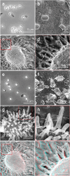
Surface structures. (a) Phase-contrast microscopy of untreated spores. (b–d) QFDE-EM. (d) Magnified image of the boxed area in (c). The diameter of the hair-like filament was measured as shown by white arrows. (e) Phase-contrast microscopy of spores treated by lysozyme. (f–h) QFDE-EM of lysozyme-treated spores. (h) Magnified image of the boxed area in (f). The lengths and anaglyph diameters of the spikes were measured as shown by the white arrows. (i, j) Stereoscopic view of (c, d), which can be observed using a pair of glasses with red filter on the left side and blue on the right side. Abbreviation: QFDE-EM, quick-freeze deep-etch electron microscopy.
Fig. 3.
Fig. 3.
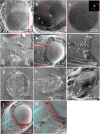
Dormant spores are visualized by QFDE-EM. (a) A slightly fractured spore features a hair-like structure on the surface (blue asterisk) and a rodlet coat layer (brown asterisk). (b) Magnified image of the boxed area in (a). The rodlet coat layers’ overlap, as marked by white arrows. (c) Magnified and FFT images of the circle area in (b). The FFT image is characterized by spots marked by red triangles. (d) Field image of fractured spores without etching. (e) Fractured image of a spore. The rodlet layer (brown asterisk), cortex (pink asterisk) and core surface (red asterisk) are shown. (f) Magnified image of the boxed area in (e). White arrows show dimensions. A sectional view of the cortex is marked by a green asterisk. (g–i) Sectional image of spore. The rodlet layer (brown asterisk, brown triangles), outer vesicle (green triangles), core membrane (yellow triangles) and core membrane vesicles (black triangles) are shown. (j and k) Stereo view of structures shown in (e and f) from different angles. Abbreviation: QFDE-EM, quick-freeze deep-etch electron microscopy.
Fig. 4.
Fig. 4.
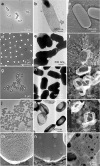
Effect of lysozyme on the isolated cortex. (a) Phase-contrast microscopy of vegetative cells treated with SDS. (b) Negative staining EM of PG sac isolated from vegetative cells. (c) QFDE-EM of PG sac from vegetative cells. (d) Phase-contrast microscopy of untreated spores. (e) Negative staining EM of untreated spores. (f) QFDE-EM of untreated spores. (g) Phase-contrast microscopy of spores treated with SDS, DTT and proteinase K. (h) Negative staining EM of treated spores. (i) QFDE-EM image of treated spores. (j) Phase-contrast microscopy of spores treated with SDS, DTT and proteinase K and then with lysozyme. (k) Negative staining EM of spores additionally treated with lysozyme. (l) QFDE-EM of spores additionally treated with lysozyme. (m) Magnified QFDE-EM image of PG sac isolated from vegetative cells. (n) Magnified QFDE-EM image of spores treated with SDS, DTT and proteinase K. (o) Magnified QFDE-EM image of spores additionally treated by lysozyme. Abbreviation: QFDE-EM, quick-freeze deep-etch electron microscopy.
Fig. 5.
Fig. 5.
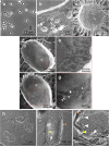
Germinating spores after 45-min incubation in germination medium. (a) Phase-contrast microscopy. (b) Field image using quick-freeze deep-etch replica EM. (c) Quick-freeze and deep-etch replica EM of germinating spore. (d and f) The rodlet layer is marked by a brown asterisk. (e and g) Magnified images of boxed areas in the left panels. Cracks in the rodlet layer (white star) and micro-holes (white triangles) are shown. (h) Field image of quick-freeze and fracture EM. (i and j) Sectioned cell images. Disintegrated core membrane vesicles (white triangles), rodlet layer (brown triangles) and extending core membrane (yellow triangles) are shown. Abbreviation: EM, electron microscopy.
Fig. 6.
Fig. 6.
Germinating spores at 4.5-h incubation in germination medium. (a) Phase-contrast microscopy. (b) Field image using quick-freeze deep-etch replica EM. (c–f) Quick-freeze deep-etch replica EM. (d and f) Magnified images of boxed areas in the left panels. (e) Germ cell surface observed in the crack of the spore. Spore (brown asterisk) and germ cell (purple asterisk) surfaces are shown. (g and h) Partially hatched cells. The remaining spore (brown asterisk) and germinating cell (purple asterisk) are shown. The boxed region is magnified as an inset. Small protrusions are marked by red triangles. (i and j) Stereo view of (c and d). Abbreviation: EM, electron microscopy.

Similar articles

Cited by

References

    1. Carlin F (2011) Origin of bacterial spores contaminating foods. Food Microbiol. 28: 177–182. - PubMed
    1. Galperin M Y, Mekhedov S L, Puigbo P, Smirnov S, Wolf Y I, and Rigden D J (2012) Genomic determinants of sporulation in Bacilli and Clostridia: towards the minimal set of sporulation-specific genes. Environ. Microbiol. 14: 2870–2890. - PMC - PubMed
    1. de Hoon M J, Eichenberger P, and Vitkup D (2010) Hierarchical evolution of the bacterial sporulation network. Curr. Biol. 20: R735–45. - PMC - PubMed
    1. Nicholson W L, Munakata N, Horneck G, Melosh H J, and Setlow P (2000) Resistance of Bacillus endospores to extreme terrestrial and extraterrestrial environments. Microbiol. Mol. Biol. Rev. 64: 548–572. - PMC - PubMed
    1. Setlow P (2006) Spores of Bacillus subtilis: their resistance to and killing by radiation, heat and chemicals. J. Appl. Microbiol. 101: 514–525. - PubMed

MeSH terms

LinkOut - more resources