. 2024 Aug;49(8):2087-2104.
doi: 10.1007/s11064-024-04173-1. Epub 2024 May 31.

ACKR3 Antagonism Enhances the Repair of Demyelinated Lesions Through Both Immunomodulatory and Remyelinating Effects

Affiliations

ACKR3 Antagonism Enhances the Repair of Demyelinated Lesions Through Both Immunomodulatory and Remyelinating Effects

Laetitia Pouzol et al. Neurochem Res. 2024 Aug.

Abstract

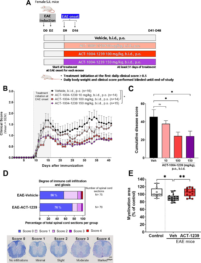
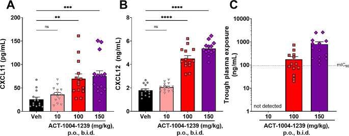
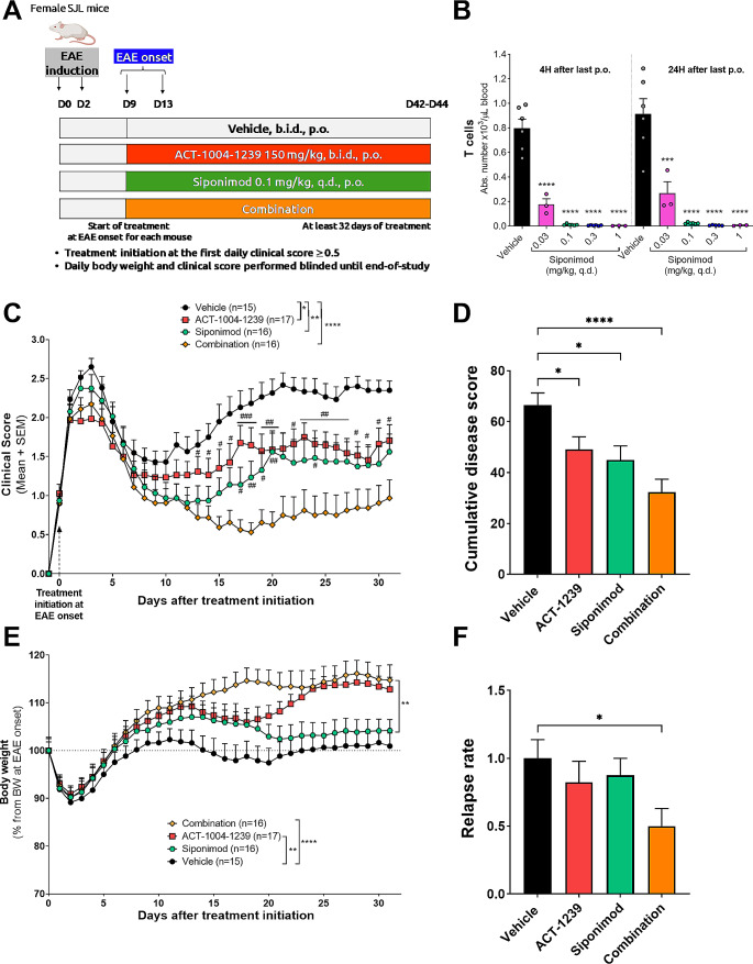
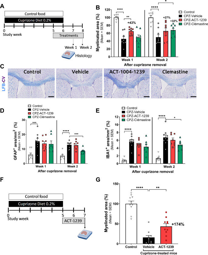
Addressing inflammation, demyelination, and associated neurodegeneration in inflammatory demyelinating diseases like multiple sclerosis (MS) remains challenging. ACT-1004-1239, a first-in-class and potent ACKR3 antagonist, currently undergoing clinical development, showed promise in preclinical MS models, reducing neuroinflammation and demyelination. However, its effectiveness in treating established disease and impact on remyelination after the occurrence of demyelinated lesions remain unexplored. This study assessed the therapeutic effect of ACT-1004-1239 in two demyelinating disease models. In the proteolipid protein (PLP)-induced experimental autoimmune encephalomyelitis (EAE) model, ACT-1004-1239 administered upon the detection of the first signs of paralysis, resulted in a dose-dependent reduction in EAE disease severity, concomitant with diminished immune cell infiltrates in the CNS and reduced demyelination. Notably, efficacy correlated with elevated plasma concentrations of CXCL11 and CXCL12, two pharmacodynamic biomarkers of ACKR3 antagonism. Combining ACT-1004-1239 with siponimod, an approved immunomodulatory treatment for MS, synergistically reduced EAE severity. In the cuprizone-induced demyelination model, ACT-1004-1239 administered after 5 weeks of cuprizone exposure, significantly accelerated remyelination, already quantifiable one week after cuprizone withdrawal. Additionally, ACT-1004-1239 penetrated the CNS, elevating brain CXCL12 concentrations. These results demonstrate that ACKR3 antagonism significantly reduces the severity of experimental demyelinating diseases, even when treatment is initiated therapeutically, after the occurrence of lesions. It confirms the dual mode of action of ACT-1004-1239, exhibiting both immunomodulatory effects by reducing neuroinflammation and promyelinating effects by accelerating myelin repair. The results further strengthen the rationale for evaluating ACT-1004-1239 in clinical trials for patients with demyelinating diseases.

Keywords: ACKR3/CXCR7; CXCL11; CXCL12; Immunomodulation; Multiple sclerosis; Remyelination.


Conflict of interest statement

All the authors acknowledge that the work was performed as employees of Idorsia Pharmaceuticals Ltd. The authors declare no other financial interests.

Figures

Fig. 1
Fig. 1
Therapeutic ACT-1004-1239 treatment dose-dependently reduces disease severity in the PLP-induced EAE model. EAE was induced by immunization of female SJL mice with PLP139 − 151/CFA and pertussis toxin. Vehicle or ACT-1004-1239 (10, 100, or 150 mg/kg) was given orally (p.o.), twice daily (b.i.d.), starting at disease onset for each mouse (therapeutic mode). (A) Study design, created with Biorender.com. Mice were assigned to a treatment group at EAE onset. The main readout was the daily clinical score assessing paralysis, performed in a blinded manner. (B) Mean clinical score of EAE mice for each treatment group after PLP139 − 151/CFA immunization. Results are expressed as mean + SEM (n = 14–16 per group). A two-way ANOVA was performed to analyze the effect of time and treatments on clinical scores. There was a significant interaction between the effects of time and treatment (F120,2200=1.4, p = 0.003). Simple main effects analysis showed that both independent variables, the time and treatment, had a statistically significant effect on clinical score (p < 0.0001 and p = 0.024, respectively). Multiple comparisons uncorrected Fisher’s test was then performed, *p < 0.05 compared to vehicle control (C) Cumulative disease score, defined as the sum of the clinical scores for each mouse over the 41-days study, in each treatment group. Results are expressed as mean + SEM, n = 14–16 per group. *p < 0.05 vs. vehicle-treated EAE mice using a Kruskal-Wallis followed by uncorrected Dunn’s multiple comparisons test. (D) At the end of the study, spinal cords from vehicle-treated mice and from mice treated with 100 mg/kg ACT-1004-1239, b.i.d., were scored for degree of inflammation and gliosis as indicated for ≥ 70 sections for each treatment group. Data are shown as percentage of total sections evaluated. Representative images of spinal cord sections stained with Cresyl Violet and Luxol Fast Blue are displayed, as well as criteria used to define histopathological scores. Scale: 200 μm. (E) Quantification of the LFB stain (% myelination area) in the spinal cord sections from vehicle-treated mice and from mice treated with 100 mg/kg ACT-1004-1239, b.i.d. Values were normalized to the median value of the non-immunized mice (control) which was set to 100%. *p < 0.05, **p < 0.01 vs. vehicle-treated EAE mice using a Kruskal-Wallis followed by uncorrected Dunn’s multiple comparisons test
Fig. 2
Fig. 2
ACT-1004-1239 dose-dependently increases plasma CXCL11 and CXCL12 concentrations in PLP-EAE mice. EAE was induced by immunization of female SJL mice with PLP139 − 151/CFA and pertussis toxin. Vehicle or ACT-1004-1239 (10, 100, or 150 mg/kg) was given orally (p.o.), twice daily (b.i.d.), starting at disease onset for each mouse (therapeutic mode). Plasma samples were prepared at sacrifice, after 31–33 days of treatment for each mouse, 14 h after last dosing (at trough). (A) CXCL11 and (B) CXCL12 plasma concentrations at trough. Results are expressed as mean + SEM (n = 13–14 group). One-way ANOVA was performed to compare the effect of treatment on chemokines plasma concentrations. There was a statistically significant difference in both CXCL11 and CXCL12 levels between at least two groups (F3,52=9.134, p < 0.0001 and F3,51=112.4, p < 0.0001, respectively). Dunnett’s multiple comparisons test was then performed ** p < 0.01, ***p < 0.001, ****p < 0.0001 versus vehicle-treated EAE mice (C) ACT-1004-1239 plasma concentrations measured at trough, expressed as mean + SEM (n = 13–14 per group). The dotted line represents the concentration inhibiting 90% of ACKR3 molecules (mouse IC90) based on an in vitro β-arrestin recruitment assay
Fig. 3
Fig. 3
ACT-1004-1239 dose-dependently increases blood lymphocyte counts in the PLP-induced EAE model EAE was induced by immunization of female SJL mice with PLP139 − 151/CFA and pertussis toxin. Vehicle or ACT-1004-1239 (10, 100, or 150 mg/kg) was given orally (p.o.), twice daily (b.i.d.), starting at disease onset for each mouse (therapeutic mode). Blood samples were collected at sacrifice, after 31 to 33 days of treatment for each mouse, 14 h after the last oral gavage, at trough. Blood cell subpopulations were analyzed by flow cytometry. (A) Lymphocyte subtype counts are expressed as mean + SEM (n = 14–16 per group). One-way ANOVA was performed to compare the effect of treatment on lymphocyte counts. There was a statistically significant difference in both overall T and B cell counts between at least two treatment groups (F3,52=3.683, p = 0.018 and F3,52=6.139, p = 0.001, respectively). *p < 0.05, **p < 0.01, using Dunnett’s multiple comparisons test. (B) Representative flow cytometry plots from vehicle-treated and ACT-1004-1239 100 mg/kg, b.i.d.-treated EAE mice depicting the gating strategy among T cells (CD3+) for naïve T cells (CD62L+CD44), central memory (CM) T cells (CD62L+CD44+), and effector memory/effector (EM/Eff) T cells (CD62LCD44+)
Fig. 4
Fig. 4
ACT-1004-1239 reduces CNS immune cells infiltration without affecting T cell activation in draining lymph nodes EAE was induced by immunization of female SJL mice with PLP139 − 151/CFA and pertussis toxin. Vehicle or ACT-1004-1239 (100 mg/kg) was given orally (p.o.), twice daily (b.i.d.), starting at PLP139 − 151/CFA immunization (n = 10/group). Negative control mice were injected with PBS/IFA. Mice were sacrificed before appearance of clinical signs, 9 days after immunization. (A) Study design (B) Right brain hemisphere and spinal cord of each mouse were collected and processed as neural single cell suspensions for flow cytometry analysis. ACT-1004-1239 reduces immune cell infiltrates into the CNS. MdCs: monocyte-derived cells. Results are expressed as mean + SEM. * p < 0.05, **p < 0.01 using one-way ANOVA followed by uncorrected Fisher’s test. (C) Cervical lymph nodes were collected and processed as single cell suspensions, counted, and plated at 2*106 cells/condition with or without PMA/ionomycine stimulation overnight and analyzed by flow cytometry. Absolute counts of CD69+ T cells in cervical lymph nodes from vehicle- or ACT-1004-1239 treated mice after stimulation. Representative plots of unstimulated and stimulated conditions. (D) ACT-1004-1239 does not affect the proportion of TNF-α, IFN-γ, IL-17 A and IL-10-secreting T cells in the draining lymph nodes. Results are expressed as mean + SEM
Fig. 5
Fig. 5
ACT-1004-1239 does not affect directly CXCL12-induced chemotaxis in vitro. EAE was induced by immunization of female SJL mice with PLP139 − 151/CFA and pertussis toxin. (A) Spleens were collected, processed, and assessed in an in vitro CXCL12 migration assay. Results are expressed as mean + SD, (triplicates/condition). (B) CXCR4 antagonism (CXCR4 ant) with AMD3100 reduces dose-dependently CXCL12 (100 ng/mL)-induced chemotaxis of splenocytes extracted from EAE mice. ACKR3 or CXCR3 antagonism (ACT-1004-1239 or AMG287, respectively) did not affect splenocyte migration. Results are expressed as mean + SD. (Each condition was performed in triplicates, which were averaged for each mouse; n = 3 mice). ****p < 0.0001 using one way ANOVA followed by Dunnett’s multiple comparisons test
Fig. 6
Fig. 6
Combination of ACT-1004-1239 with siponimod further reduces EAE disease severity compared to each monotherapy. EAE was induced by immunization of female SJL mice with PLP139 − 151/CFA and pertussis toxin. Vehicle, ACT-1004-1239 (150 mg/kg, twice daily (b.i.d.)), siponimod (0.1 mg/kg, once daily (q.d.)), or the combination of ACT-1004-1239 with siponimod was given orally (p.o.), starting at disease onset for each mouse (therapeutic mode). (A) Study design, created with Biorender.com (B) Naive SJL mice were treated for 3 days with vehicle or Siponimod (0.03, 0.1, 0.3, or 1 mg/kg). Blood was collected 4–24 h after the last oral dose and processed for flow cytometry. Absolute counts of T cells are expressed as mean + SEM (n = 3–6/group). ****p < 0.0001 using one way ANOVA followed by Dunnett’s multiple comparisons test versus vehicle-treated mice. (C) Mean clinical score of EAE mice for each treatment group after treatment initiation. Results are expressed as mean + SEM (n = 15–17 per group). A two-way ANOVA was performed to analyze the effect of time and treatments on clinical scores. There was a significant interaction between the effects of time and treatment (F96,1920=3.953, p < 0.0001). Simple main effects analysis showed that both independent variables, the time and treatment, had a statistically significant effect on clinical score (p < 0.0001 and p = 0.0001, respectively). Multiple comparisons uncorrected Fisher’s test was then performed, *p < 0.05, **p < 0.01, ***p < 0.001 compared to vehicle control. #p < 0.05, ##p < 0.01, ###p < 0.001 using t-test versus combination-treated group. (D) Cumulative disease index, defined as the sum of the clinical scores for each mouse over the 42-days study, in each treatment group. Results are expressed as mean + SEM, n = 15–17 per group. *p < 0.05, ****p < 0.0001 vs. vehicle-treated EAE mice using a Kruskal-Wallis followed by uncorrected Dunn’s multiple comparisons test. (E) Body weight evolution after treatment initiation. Results are expressed as the mean percentage of the body weight of each mouse at the time of treatment initiation, which was set to 100%. A mixed-effects analysis was performed to analyze the effect of time and treatments on body weight. There was a significant interaction between the effects of time and treatment (F93,1849=5.864, p < 0.0001). Simple main effects analysis showed that both independent variables, the time and treatment, had a statistically significant effect on body weight (p < 0.0001 and p < 0.0001, respectively). **p < 0.01, ****p < 0.0001 using Tukey’s multiple comparisons test. (F) Relapse rate. Results are expressed as mean + SEM (n = 15–17/group). *p < 0.05 vs. vehicle-treated EAE mice using a Kruskal-Wallis followed by uncorrected Dunn’s multiple comparisons test
Fig. 7
Fig. 7
ACT-1004-1239 enhances remyelination in the cuprizone model. Male C57BL/6 mice were exposed to control food (control) or cuprizone diet 0.2% (CPZ) for 6 weeks and then to normal chow for subsequent 2 weeks. Vehicle, ACT-1004-1239 (100 mg/kg, twice daily), or clemastine (10 mg/kg, once daily) were given orally, after 5 weeks of CPZ exposure for 2 or 3 weeks. (A) Study design, created with BioRender.com. Representatives of each group (n = 8–9/group) were euthanized one or two weeks after CPZ removal. (B) Quantitative analysis of myelinated areas in the corpus callosum, 1 or 2 weeks after cuprizone removal. Results are expressed as percentage of the mean positive myelinated area normalized to the mean myelinated area in control, set to 100% for each independent time point; n = 8–9 mice in each group. For each time point, a one-way ANOVA was performed to compare the effect of the different treatments on the myelinated area. There was a significant difference in myelinated area between at least two treatment groups (F3,28=27.08, p < 0.0001, at week 1 and F3,29=12.03, p < 0.0001 at week 2). Multiple comparisons uncorrected Fisher’s test was then performed, *p < 0.05, **p < 0.01, ****p < 0.0001 compared to vehicle control. (C) Representative Luxol Fast Blue-Cresyl Violet (LFB-CV) images of the corpus callosum sections from each treatment group, 1 week after CPZ removal; scale bar 380 μm. ACT-1004-1239 did not impact cuprizone-induced astrogliosis (D) and microgliosis (E) in the corpus callosum. Quantitative analysis of GFAP (D) and IBA1 (E) are expressed as the percentage of the mean positive area, normalized by the selected region of interest in mm2; n = 7–9 mice in each group. A One-way ANOVA was performed to compare the effect of the different treatments on astrogliosis and microgliosis. There was a significant difference in GFAP+ area and IBA1+ area between at least two treatment groups (F3,29=6.33, p = 0.002 and F3,29=22.50, p < 0.0001 at week 1, respectively; F3,29=20.76, p < 0.0001 and F3,28=22.2, p < 0.0001 at week 2). Multiple comparisons uncorrected Fisher’s test was then performed *p < 0.05, ***p < 0.001, ****p < 0.0001 versus vehicle-treated CPZ mice. In a second independent experiment, mice were treated orally with vehicle or ACT-1004-1239 (100 mg/kg), twice daily for two weeks, after 5 weeks of CPZ exposure. (F) Study design (G) Quantitative analysis of myelinated areas in the corpus callosum, 2 weeks after cuprizone removal. Results are expressed as percentage of the mean positive LFB+ area normalized to the mean % in mice fed with control food, set to 100% for each study week; n = 10 mice/group. A One-way ANOVA was performed to compare the effect of the different treatments on the myelinated area. There was a significant difference in myelinated area between at least two treatment groups (F3,27=46.19, p < 0.0001). **p < 0.01, ****p < 0.0001 using Dunnett’s multiple comparisons test versus CPZ mice
Fig. 8
Fig. 8
ACT-1004-1239 increases the levels of CXCL12 in both plasma and brain tissue. Healthy female C57BL/6 mice were orally treated with vehicle or ACT-1004-1239 (100 mg/kg), twice daily for 3 days, for a total of 5 doses. Blood and brain samples were collected 1 h after the last oral administration and analyzed for CXCL12 concentrations. ACT-1004-1239 significantly increased (A) plasma and (B) brain tissue CXCL12 concentrations. Results are expressed as mean + SEM, n = 5/group. ***p < 0.001, ****p < 0.0001 using t-test versus vehicle-treated mice

Similar articles

References

    1. Plemel JR, Liu W-Q, Yong VW. Remyelination therapies: a new direction and challenge in multiple sclerosis. Nat Rev Drug Discovery. 2017;16(9):617–634. - PubMed
    1. Cunniffe N, Coles A. Promoting remyelination in multiple sclerosis. J Neurol. 2021;268(1):30–44. - PMC - PubMed
    1. Melchor GS, Khan T, Reger JF, Huang JK. Remyelination Pharmacotherapy investigations Highlight Diverse mechanisms underlying multiple sclerosis progression. ACS Pharmacol Translational Sci. 2019;2(6):372–386. - PMC - PubMed
    1. Cruz-Orengo L, Holman DW, Dorsey D, Zhou L, Zhang P, Wright M, et al. CXCR7 influences leukocyte entry into the CNS parenchyma by controlling abluminal CXCL12 abundance during autoimmunity. J Exp Med. 2011;208(2):327–339. - PMC - PubMed
    1. Chu T, Shields LBE, Zhang YP, Feng SQ, Shields CB, Cai J. CXCL12/CXCR4/CXCR7 Chemokine Axis in the Central Nervous System: therapeutic targets for Remyelination in Demyelinating diseases. Neuroscientist. 2017;23(6):627–648. - PubMed

MeSH terms

LinkOut - more resources