. 2024 May 30;16(11):9498-9517.
doi: 10.18632/aging.205880. Epub 2024 May 30.

THY1 is a prognostic-related biomarker via mediating immune infiltration in lung squamous cell carcinoma (LUSC)

Affiliations

THY1 is a prognostic-related biomarker via mediating immune infiltration in lung squamous cell carcinoma (LUSC)

Changsheng Yi et al. Aging (Albany NY). .

Abstract

Thymus cell antigen 1 (THY1) has been proven to play pivotal roles in many diseases. However, we do not fully understand its functional mechanism, especially in lung squamous cell carcinoma (LUSC). Here, we aimed to perform a comprehensive analysis to explore the expression and prognostic values of THY1 in LUSC using bioinformatic technology. Some online public databases (e.g., ONCOMINE, PrognoScan, TIMER, Kaplan-Meier plotter, STRING, LinkedOmics, and GEPIA) were used to explore the expression, prognostic significance, and potential molecular mechanism of THY1. The analysis indicated that THY1 was significantly up-regulated and closely correlated with poor prognosis in many malignant tumors, including LUSC. Further analysis revealed that over-expression of THY1 was significantly correlated with clinicopathological parameters (e.g., individual cancer stage, age, smoking habits, nodal metastasis status, and TP53 mutation status) in LUSC. The CpG islands methylation of THY1 was negatively correlated with THY1 mRNA expression in The Cancer Genome Atlas Program (TCGA). Further enrichment analysis of THY1 correlated genes revealed that they were mainly correlated with the formation of extracellular matrix (ECM), and got involved in the pathway of epithelial mesenchymal transition (EMT). Furthermore, differentially expressed THY1 was significantly correlated with immune cell infiltrations and poor prognosis in LUSC. In summary, bioinformatic analysis demonstrated that THY1 was significantly over-expressed and closely correlated with unfavorable prognosis in LUSC, which may apply as a promising diagnostic and therapeutic biomarker for LUSC in the future.

Keywords: LUSC; THY1; bioinformatics; biomarker; immune infiltration.


Conflict of interest statement

CONFLICTS OF INTEREST: The authors declare no conflicts of interest related to this study.

Figures

Figure 1
Figure 1
Differentially expressed THY1 in a variety of cancers and normal tissues. (A) The interactive body-map revealed the median expression of THY1 in tumor (red) and normal samples (green) using GEPIA (scale: Log2 (TPM+1)). (B) The expression levels of THY1 in different tumors compared to normal tissues from ONCOMINE database. (C) The expression levels of THY1 in 33 types of cancers compared to normal tissues from TCGA in TIMER database.
Figure 2
Figure 2
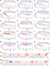
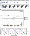
The prognostic significance of up-regulated THY1 in many types of cancers. (AO) Relationships between higher expression of THY1 and prognosis in different types of cancers in PrognoScan database. (P) The prognostic value (OS) of differentially expressed THY1 in 33 types of cancer form TCGA in GEPIA database. (Q) The prognostic value (DFS) of differentially expressed THY1 in 33 types of cancer from TCGA in GEPIA database. Abbreviations: OS: overall survival; DFS: disease-free survival; DMFS: disease-metastasis free survival; DSS: disease specific survival; RFS: relapse free survival.
Figure 3
Figure 3
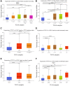
Relationships between THY1 expression and clinicopathological parameters in LUSC. (AF) Relationships between THY1 expression and patient’s smoking habits, age, TP53 mutation status, nodal metastasis status, gender, and individual cancer stage in LUSC from UALCAN database. *P < 0.05; **P < 0.01; ***P < 0.001; ****P < 0.0001.
Figure 4
Figure 4
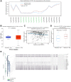
The methylation level of THY1 in LUSC and normal tissues. (A) Mean methylation level of THY1 in LUSC and normal tissues regarding different probes using the TCGA Wander. (B) Promoter methylation level of THY1 in LUSC using UALCAN. (C) Relationships between THY1 methylation and mRNA expression in LUSC using GSCA. (D) Expression level of THY1 in LUSC and normal tissues using TCGA Wander. (E) Relationships between different THY1 CpG islands methylation and THY1 expression using MEXPRESS. *P < 0.05; **P < 0.01; ***P < 0.001.
Figure 5
Figure 5
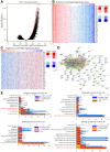
Functional enrichment analysis of correlated genes of THY1 in LUSC. (A) Genes that positively or negatively correlated with THY1 in LUSC measured by Pearson test in LinkedOmics. (B) Heatmap of positively correlated genes of THY1 in LUSC. (C) Heatmap of negatively correlated genes of THY1 in LUSC. (D) Protein-protein interactive network of top correlated genes constructed by STRING. (E) Functional enrichment analysis of these top correlated genes regarding cellular component, molecular function, biological process, and biological pathway using FunRich 3.v.13.
Figure 6
Figure 6
Relationships between THY1 expression and immune infiltration levels in lung squamous cell carcinoma (LUSC) and cholangiocarcinoma (CHOL). (A) Differentially expressed THY1 was significantly correlated with lymphocytes infiltration levels in LUSC, but not in CHOL. (B) Copy number alteration of different immune lymphocytes was significantly correlated with immune infiltration levels in LUSC, but not in CHOL. (C) Higher THY1 expression and dendritic cell infiltration level significantly correlated with poorer prognosis in LUSC.
Figure 7
Figure 7
Associations between THY1 expression and gene markers of dendritic cells. (AG) The relationships between THY1 expression and HLA-DPB1, HLA-DQB1, HLA-DRA, HLA-DPA1, CD1C, NRP1, and ITGAX in LUSC from TIMER database.

Similar articles

References

    1. Reck M, Rabe KF. Precision Diagnosis and Treatment for Advanced Non-Small-Cell Lung Cancer. N Engl J Med. 2017; 377:849–61. 10.1056/NEJMra1703413 - DOI - PubMed
    1. Ettinger DS, Akerley W, Borghaei H, Chang AC, Cheney RT, Chirieac LR, D'Amico TA, Demmy TL, Govindan R, Grannis FW Jr, Grant SC, Horn L, Jahan TM, et al., and National comprehensive cancer network. Non-small cell lung cancer, version 2.2013. J Natl Compr Canc Netw. 2013; 11:645–53. 10.6004/jnccn.2013.0084 - DOI - PubMed
    1. Hirsch FR, Scagliotti GV, Mulshine JL, Kwon R, Curran WJ Jr, Wu YL, Paz-Ares L. Lung cancer: current therapies and new targeted treatments. Lancet. 2017; 389:299–311. 10.1016/S0140-6736(16)30958-8 - DOI - PubMed
    1. Kulasingam V, Diamandis EP. Strategies for discovering novel cancer biomarkers through utilization of emerging technologies. Nat Clin Pract Oncol. 2008; 5:588–99. 10.1038/ncponc1187 - DOI - PubMed
    1. Lung HL, Cheung AK, Cheng Y, Kwong FM, Lo PH, Law EW, Chua D, Zabarovsky ER, Wang N, Tsao SW, Stanbridge EJ, Lung ML. Functional characterization of THY1 as a tumor suppressor gene with antiinvasive activity in nasopharyngeal carcinoma. Int J Cancer. 2010; 127:304–12. 10.1002/ijc.25047 - DOI - PubMed

MeSH terms