

Differences in ankle and knee muscle architecture and plantar pressure distribution among women with knee osteoarthritis
- PMID: 38820170
- PMCID: PMC11296719
- DOI: 10.1002/jfa2.12028
Differences in ankle and knee muscle architecture and plantar pressure distribution among women with knee osteoarthritis
Abstract
Background: The aim of this study was to compare the plantar pressure distribution and knee and ankle muscle architecture in women with and without knee osteoarthritis (OA).
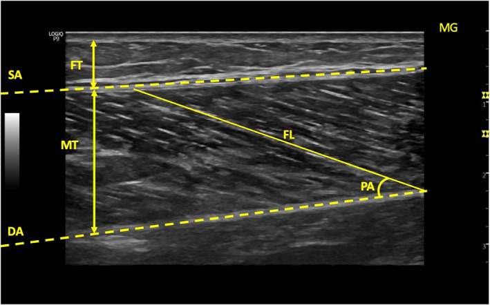
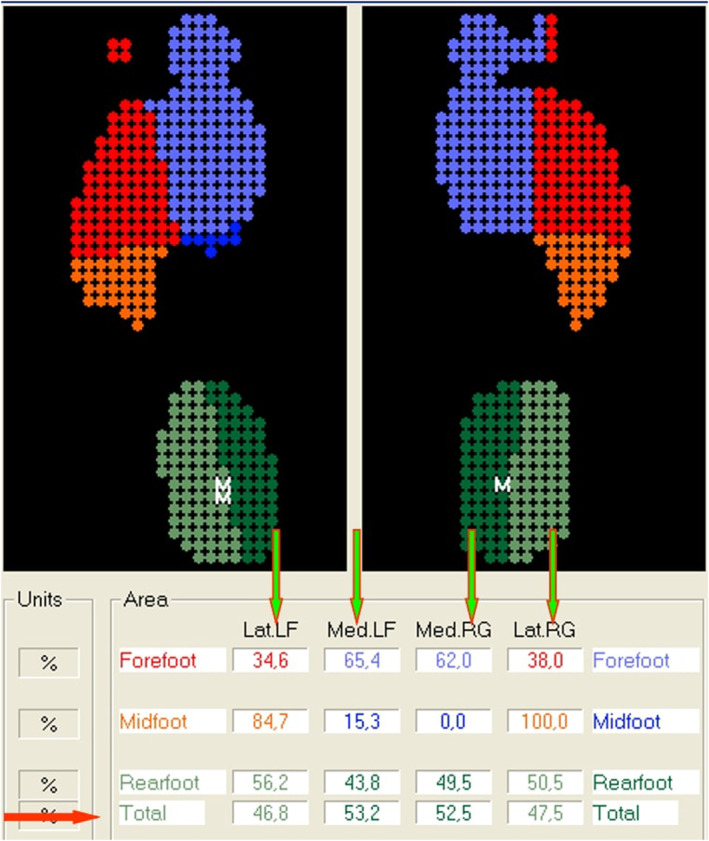
Methods: Fifty women with knee OA (mean age = 52.11 ± 4.96 years, mean Body mass index (BMI) = 30.94 ± 4.23 kg/m2) and 50 healthy women as a control group (mean age = 50.93 ± 3.78 years, mean BMI = 29.06 ± 4.82 kg/m2) were included in the study. Ultrasonography was used to evaluate knee and ankle muscles architecture and femoral cartilage thickness. The plantar pressure distribution was evaluated using the Digital Biometry Scanning System and Milleri software (DIASU, Italy). Static foot posture was evaluated using the Foot Posture Index (FPI), and pain severity was assessed using the Visual Analog Scale.
Results: The OA group exhibited lower muscle thickness in Rectus Femoris (RF) (p = 0.003), Vastus Medialis (VM) (p = 0.004), Vastus Lateralis (p = 0.023), and Peroneus Longus (p = 0.002), as well as lower Medial Gastrocnemius pennation angle (p = 0.049) and higher Fat thickness (FT) in RF (p = 0.033) and VM (p = 0.037) compared to the control group. The OA group showed thinner femoral cartilage thickness (p = 0.001) and higher pain severity (p = 0.001) than the control groups. FPI scores were higher (p = 0.001) in OA group compared to the control group. The plantar pressure distribution results indicated an increase in total surface (p = 0.027), total load (p = 0.002), medial load (p = 0.005), and lateral load (p = 0.002) on dominant side in OA group compared to the control group.
Conclusions: Knee and ankle muscle architecture, knee extensor muscle FT, and plantar pressure distribution in the dominant foot differed in individuals with knee OA compared to the control group.
Keywords: foot; knee osteoarthritis; muscle architecture; muscles; ultrasonography.
© 2024 The Author(s). Journal of Foot and Ankle Research published by John Wiley & Sons Australia, Ltd on behalf of Australian Podiatry Association and The Royal College of Podiatry.
Conflict of interest statement
Not applicable.
Figures
References
-
- Fujita, R. , Matsui Y., Harada A., Takemura M., Kondo I., Nemoto T., and Ota S.. 2014. “Relationship between Muscle Strength and Knee Pain in Knee Osteoarthritis Patients.” Osteoarthritis and Cartilage 22: S415. 10.1016/j.joca.2014.02.782. - DOI
-
- Aily, J. B. , de Noronha M., de Almeida A. C., Pedroso M. G., Maciel J. G., Mattiello‐Sverzut A. C., and Mattiello Stela Marcia. 2019. “Evaluation of Vastus Lateralis Architecture and Strength of Knee Extensors in Middle‐Aged and Older Individuals with Knee Osteoarthritis.” Clinical Rheumatology 38(9): 2603–2611. 10.1007/s10067-019-04539-9. - DOI - PubMed
MeSH terms
LinkOut - more resources
Full Text Sources
Medical
