. 2024 May 30;134(14):e173586.
doi: 10.1172/JCI173586.

Loss-of-function mutations of the TIE1 receptor tyrosine kinase cause late-onset primary lymphedema

Affiliations

Loss-of-function mutations of the TIE1 receptor tyrosine kinase cause late-onset primary lymphedema

Pascal Brouillard et al. J Clin Invest. .

Abstract

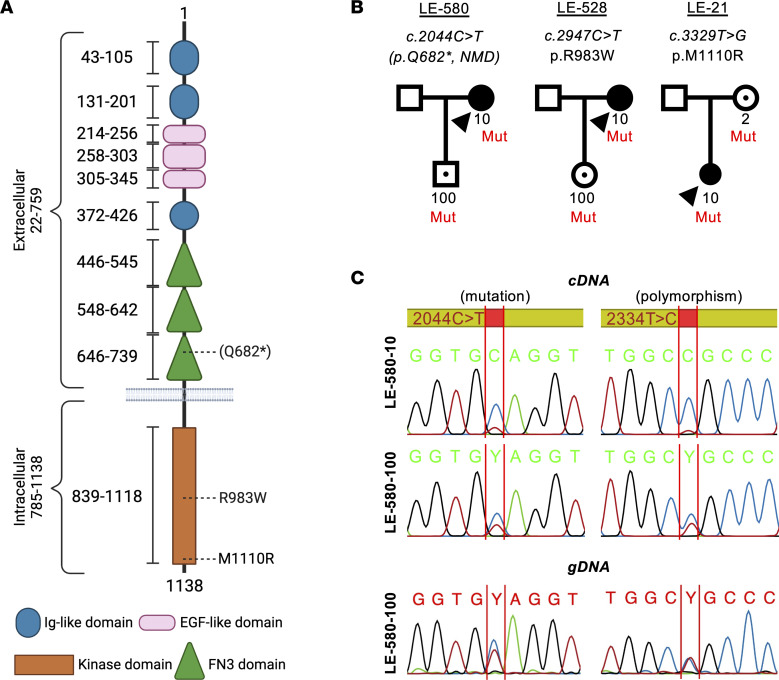
Primary lymphedema (PL), characterized by tissue swelling, fat accumulation, and fibrosis, results from defects in lymphatic vessels or valves caused by mutations in genes involved in development, maturation, and function of the lymphatic vascular system. Pathogenic variants in various genes have been identified in about 30% of PL cases. By screening of a cohort of 755 individuals with PL, we identified two TIE1 (tyrosine kinase with immunoglobulin- and epidermal growth factor-like domains 1) missense variants and one truncating variant, all predicted to be pathogenic by bioinformatic algorithms. The TIE1 receptor, in complex with TIE2, binds angiopoietins to regulate the formation and remodeling of blood and lymphatic vessels. The premature stop codon mutant encoded an inactive truncated extracellular TIE1 fragment with decreased mRNA stability, and the amino acid substitutions led to decreased TIE1 signaling activity. By reproducing the two missense variants in mouse Tie1 via CRISPR/Cas9, we showed that both cause edema and are lethal in homozygous mice. Thus, our results indicate that TIE1 loss-of-function variants can cause lymphatic dysfunction in patients. Together with our earlier demonstration that ANGPT2 loss-of-function mutations can also cause PL, our results emphasize the important role of the ANGPT2/TIE1 pathway in lymphatic function.

Keywords: Genetic diseases; Genetics; Lymph; Mouse models; Vascular biology.


Figures

Figure 1
Figure 1. Identification of three TIE1 variants in primary lymphedema patients.
(A) Schematic structure of TIE1 shown with its domains and the variants, created with BioRender (biorender.com). (B) Pedigrees of the 3 families and cosegregation of the variants. Arrowheads, index patients; black symbols, affected individuals; black dots within symbols, unaffected variant carriers. Only numbered individuals were tested. (C) Reverse transcriptase PCR showing degradation of the alternative TIE1 allele in LE-580-10 and partial degradation in LE-580-100, also confirmed by use of the c.2334T>C;p.Ala778= heterozygous polymorphism. LE-580-100 genomic DNA (gDNA) sequences show heterozygous levels of the variant peaks.
Figure 2
Figure 2. Clinical images of 2 patients with TIE1 variants.
(A) Patient LE-580-10, with premature stop codon Q682*, has a stable clinical presentation of mild distal stage 2a lower-limb edema, according to the ISL classification. Lymphoscintigraphy (middle) shows bilateral insufficiency of lymph drainage and fewer lymph nodes, at 4 hours after injection, as well as the presence of a deep popliteal lymph node only on the right leg. The MRI image (right) shows edema (bright signal, red arrows) in the ankles, more marked on the left foot. (B) Photograph of patient LE-21-10 (M1110R missense variant). Lymphoscintigraphy taken 30 minutes after tracer injection reveals defective lymphatic function in the left leg.
Figure 3
Figure 3. Analysis of TIE1 protein in HEK293T expressing the different TIE1 variants.
(A and B) Western blotting of WT-TIE1 and the 3 variants in HEK293T cells transfected using a lentiviral vector. The truncated TIE1-Q682* variant is detected in cell lysate (A) only after 5 minutes of exposure, and is weak in the supernatant (B) after a 20-second exposure. Asterisks indicate the truncated Q682* polypeptide. The lanes were run on the same gel but were noncontiguous. (C) Relative TIE1 protein signals in the lysates and culture supernatants from n = 3 experiments like in A and B. (D) Flow cytometry analysis of TIE1 in transfected HEK293T cells. (E) Quantification of D from n = 3 experiments. Statistical significance in C and E was determined with Brown-Forsythe ANOVA with Dunnett’s post hoc test for multiple comparisons. Data shown as mean ± SEM. **P < 0.01, ***P < 0.001, ****P < 0.0001.
Figure 4
Figure 4. Analysis of TIE1 protein baseline phosphorylation in LECs expressing the different TIE1 variants.
(A) Western blotting analysis of TIE1 baseline phosphorylation using the 4G10 anti-pY antibody and total TIE1 protein in LECs transduced with WT-, R983W-, or M1110R-TIE1 variant. (B) Quantification of phosphorylated TIE1 relative to total TIE1, from n = 3 independent experiments like in A. (C) Immunofluorescence staining for DAPI and TIE1 in non-permeabilized and permeabilized LECs transduced with WT-, R983W-, or M1110R-TIE1 variant. Scale bars: 100 μm. (D) Quantification of surface TIE1 relative to total TIE1 from 3–7 images like in C per group. Statistical significance in B and D was determined with Brown-Forsythe ANOVA with Dunnett’s post hoc test for multiple comparisons. Data shown as mean ± SEM. ***P < 0.001.
Figure 5
Figure 5. Effect of TIE1 variants R983W and M1110R on TIE1 activation and downstream signaling in endothelial cells.
(A) Western blot analysis of TIE1 and TIE2 phosphorylation at Y1007 or Y992 residue (both pY1007 and pY992 detected by anti-pY992 antibody AF2720, R&D Systems) in Comp-ANGPT1–stimulated (cANG1-stimulated) LECs transduced with the WT-, R983W-, or M1110R-TIE1 variant. (B) Quantification of the pY1007/total TIE1 ratios in the samples from experiments like in A. Mean ± SEM, n = 3–5. (C) Western blot analysis of cANG1-stimulated TIE2-PAE cells transduced with the WT-, R983W-, or M1110R-TIE1 variant. (D) Quantification of phospho-AKT-S475/total AKT and phospho-ERK/total ERK ratios from experiments like in C. Data shown as mean ± SEM, n = 4 experiments. Statistical significance in B and D was analyzed by 1-way ANOVA with Tukey’s post hoc test for multiple comparisons. *P < 0.05, **P < 0.01, ***P < 0.001.
Figure 6
Figure 6. Embryos homozygous for the Tie1R979W allele display lymphatic defects and altered cell-surface/intracellular TIE1 ratio.
(A) Macroscopic images of Tie1WT and homozygous Tie1R979W/R979W embryos at E15.5. Scale bars: 2 mm. (B) VEGFR3 staining of dorsal skin from WT and Tie1R979W/R979W embryos at E18.5. Scale bars: 100 μm. (C) Analysis of VEGFR3 polypeptides from the indicated skin lysates at E18.5. (D) Western blot analysis of TIE1 polypeptides from indicated lung lysates at E18.5. (E) Quantification of the proportions of cell-surface 135 kDa and intracellular 125 kDa TIE1 polypeptides relative to total TIE1 from the Western blots in D. The graph “Both bands” is the total TIE1 normalized to HSC70. n = 3 experiments. Statistical analysis by 1-way ANOVA with Tukey’s post hoc test for multiple comparisons. Data shown as mean ± SEM. *P < 0.05, ***P < 0.001, ****P < 0.0001. (F) Percentages of the indicated genotypes among pups born from heterozygous TIE1-WT/R979W matings. n = 8 litters (observed WT/WT n = 17, WT/R979W n = 25, R979W/R979W n = 0).

References

    1. Karkkainen MJ, et al. A model for gene therapy of human hereditary lymphedema. Proc Natl Acad Sci U S A. 2001;98(22):12677–12682. doi: 10.1073/pnas.221449198. - DOI - PMC - PubMed
    1. Brouillard P, et al. Primary lymphoedema. Nat Rev Dis Primers. 2021;7(1):77. doi: 10.1038/s41572-021-00309-7. - DOI - PubMed
    1. Potente M, Makinen T. Vascular heterogeneity and specialization in development and disease. Nat Rev Mol Cell Biol. 2017;18(8):477–494. doi: 10.1038/nrm.2017.36. - DOI - PubMed
    1. Petrova TV, Koh GY. Organ-specific lymphatic vasculature: from development to pathophysiology. J Exp Med. 2018;215(1):35–49. doi: 10.1084/jem.20171868. - DOI - PMC - PubMed
    1. Petrova TV, Koh GY. Biological functions of lymphatic vessels. Science. 2020;369(6500):eaax4063. doi: 10.1126/science.aax4063. - DOI - PubMed