

Achillea fragrantissima (Forssk.) Sch.Bip instigates the ROS/FADD/c-PARP expression that triggers apoptosis in breast cancer cell (MCF-7)
- PMID: 38820323
- PMCID: PMC11142488
- DOI: 10.1371/journal.pone.0304072
Achillea fragrantissima (Forssk.) Sch.Bip instigates the ROS/FADD/c-PARP expression that triggers apoptosis in breast cancer cell (MCF-7)
Abstract
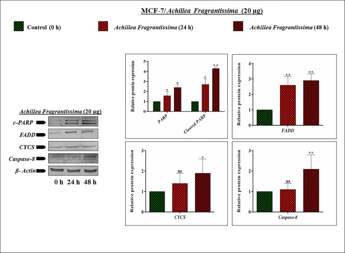
Achillea fragrantissima is a shrub plant that belongs to the Asteraceae family in Arabia and Egypt. It is used as folk medicine and is a good source of phenolic acids, flavonoids, and some active compounds. To investigate the anti-cancer effect of A.fragrantissima on breast cancer MCF-7 cells and find the critical mechanism involved in apoptosis. The toxicity and pharmacokinetic studies of ethanolic extract of A.fragrantissima was examined for anti-breast cancer properties. In turn, cytotoxicity and cell viability were achieved by the MTT method. Furthermore, the trypan blue exclusion and microscopy examination proved the presence of apoptotic cells. Again, fluorescent staining such as AO/EtBr, DCFH-DA, Rho-123, and Hoechst-33342 reveals the cellular cytoplasmic disciplines upon A. fragrantissima effect. Moreover, cellular functioning tests like wound healing, colony formation, and Transwell invasion assay were demonstrated. In addition, the qRT-PCR technique authenticates the A. fragrantissima -induced apoptotic network genes (Caspase-3, Caspase-8, Caspase-9, Cytochrome c, BCL-2, BID, BAX, PARP, PTEN, PI3K, and Akt) expression were evaluated. Mainly, the Immunoblot technique proved the expressed level of apoptotic proteins such as cleaved PARP, CYCS, and FADD. This study confirmed that the A. fragrantissima exerts cytotoxicity at 20 μg/mL for 24 hrs in MCF-7 cells. Also, decreases cellular viability, producing apoptotic cells and damaged cellular surfaces with dead matter. Consequently, it creates ROS species accumulation, loss of mitochondrial membrane potential, and fragmentation of DNA in MCF-7 cells. Furthermore, it arrests cell migration, induces colony-forming ability loss, and suppresses cell invasion. In addition, A. fragrantissima significantly upregulates genes such as caspase-3, 9, cytochrome c, BID, BAX, and PTEN while downregulating the Pi3K/ Akt signaling. Nonetheless, A.fragrantissima induced cleaved PARP, CYCS, and FADD proteins in MCF-7 cells to avail apoptosis.
Copyright: © 2024 Abdulrahman Alasmari. This is an open access article distributed under the terms of the Creative Commons Attribution License, which permits unrestricted use, distribution, and reproduction in any medium, provided the original author and source are credited.
Conflict of interest statement
The authors have declared that no competing interests exist.
Figures


Similar articles
-
[Mechanism of ergosterol peroxide on MCF-7 breast cancer cells based on network pharmacology and in vitro experiments].Zhongguo Zhong Yao Za Zhi. 2024 Jul;49(13):3627-3635. doi: 10.19540/j.cnki.cjcmm.20240202.705. Zhongguo Zhong Yao Za Zhi. 2024. PMID: 39041135 Chinese.
-
Oldenlandia diffusa suppresses metastatic potential through inhibiting matrix metalloproteinase-9 and intercellular adhesion molecule-1 expression via p38 and ERK1/2 MAPK pathways and induces apoptosis in human breast cancer MCF-7 cells.J Ethnopharmacol. 2017 Jan 4;195:309-317. doi: 10.1016/j.jep.2016.11.036. Epub 2016 Nov 20. J Ethnopharmacol. 2017. PMID: 27876502
-
Cytosolic and mitochondrial ROS production resulted in apoptosis induction in breast cancer cells treated with Crocin: The role of FOXO3a, PTEN and AKT signaling.Biochem Pharmacol. 2020 Jul;177:113999. doi: 10.1016/j.bcp.2020.113999. Epub 2020 Apr 28. Biochem Pharmacol. 2020. PMID: 32353423
-
Achillea fragrantissima (Forssk.) Sch.Bip. methanolic extract exerts potent antimicrobial activity and causes cancer cell death via induction of caspase-dependent apoptosis and S-phase arrest.Nat Prod Res. 2022 Sep;36(18):4645-4650. doi: 10.1080/14786419.2021.2010074. Epub 2021 Nov 30. Nat Prod Res. 2022. PMID: 34847782
-
Promising impacts of Achillea spp., beyond A medicinal plant, against toxins, toxicities, and injuries: In vivo and in vitro mechanisms.Biochem Biophys Rep. 2025 Apr 22;42:102023. doi: 10.1016/j.bbrep.2025.102023. eCollection 2025 Jun. Biochem Biophys Rep. 2025. PMID: 40330076 Free PMC article. Review.
Cited by
-
Effect of bispecific recombinant oncolytic adenovirus carrying apoptin on apoptosis of MCF-7 cells.Front Immunol. 2025 May 16;16:1530583. doi: 10.3389/fimmu.2025.1530583. eCollection 2025. Front Immunol. 2025. PMID: 40453070 Free PMC article.
-
Silver nanoparticles synthesized using aerial part of Achillea fragrantissima and evaluation of their bioactivities.Sci Rep. 2024 Oct 21;14(1):24703. doi: 10.1038/s41598-024-75558-z. Sci Rep. 2024. PMID: 39433875 Free PMC article.
-
Antineoplastic potential of Kushneria avicenniae pigments via modulation of the BAX/BCL-2 axis and CASP-9 pathway in inducing G2/M arrest and apoptosis in liver and breast cancer.Med Oncol. 2025 Aug 2;42(9):400. doi: 10.1007/s12032-025-02949-1. Med Oncol. 2025. PMID: 40753129
References
MeSH terms
Substances
LinkOut - more resources
Full Text Sources
Medical
Research Materials
Miscellaneous
