. 2024 May 31;18(5):e0012106.
doi: 10.1371/journal.pntd.0012106. eCollection 2024 May.

A panel of phenotypically and genotypically diverse bioluminescent:fluorescent Trypanosoma cruzi strains as a resource for Chagas disease research

Affiliations

A panel of phenotypically and genotypically diverse bioluminescent:fluorescent Trypanosoma cruzi strains as a resource for Chagas disease research

Francisco Olmo et al. PLoS Negl Trop Dis. .

Abstract

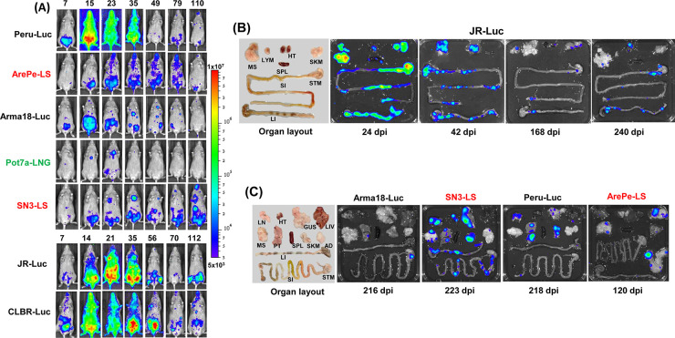
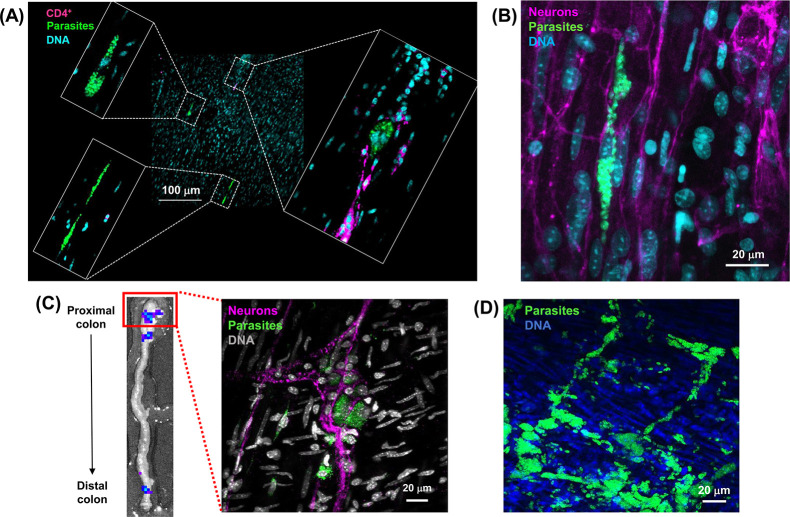
Chagas disease is caused by Trypanosoma cruzi, a protozoan parasite that displays considerable genetic diversity. Infections result in a range of pathological outcomes, and different strains can exhibit a wide spectrum of anti-parasitic drug tolerance. The genetic determinants of infectivity, virulence and therapeutic susceptibility remain largely unknown. As experimental tools to address these issues, we have generated a panel of bioluminescent:fluorescent parasite strains that cover the diversity of the T. cruzi species. These reporters allow spatio-temporal infection dynamics in murine models to be monitored in a non-invasive manner by in vivo imaging, provide a capability to detect rare infection foci at single-cell resolution, and represent a valuable resource for investigating virulence and host:parasite interactions at a mechanistic level. Importantly, these parasite reporter strains can also contribute to the Chagas disease drug screening cascade by ensuring that candidate compounds have pan-species in vivo activity prior to being advanced into clinical testing. The parasite strains described in this paper are available on request.


Conflict of interest statement

The authors have declared that no competing interests exist.

Figures

Fig 1
Fig 1. Growth profile of selected T. cruzi reporter strains in severe combined immunodeficient (SCID) mice.
CB17 SCID mice were infected with a range of T. cruzi strains that expressed red-shifted luciferase (L), or as indicated, fusion proteins also containing mNeonGreen (LNG) or mScarlet (LS) fluorescent components (Table 2) (Materials and Methods). Inoculations were by the i.p. route, with the number of tissue culture trypomastigotes, as indicated. The mice were monitored regularly by bioluminescence imaging and the total flux (p/s) from ventral view images recorded (Materials and Methods). The number of days post-infection is indicated. Mice were euthanized at humane end-points by exsanguination under terminal anaesthesia.
Fig 2
Fig 2. Bioluminescence imaging of BALB/c mice infected with a range of T. cruzi strains.
(A) Dynamics of infection. BALB/c mice were infected with T. cruzi reporter strains by the i.p. route and monitored regularly by in vivo imaging until they had transitioned to the chronic stage (Materials and Methods). Ventral images are shown and the number of days post-infection indicated. -Luc; strains expressing the red-shifted luciferase PpyRE9h. -LS, -LNG; strains expressing luciferase:mScarlet and luciferase:mNeonGreen fusion proteins, respectively. Further details on parasite strains are provided in Tables 1 and 2. (B) Assessment of the parasite burden in tissues and organs of T. cruzi CLBR-Luc infected mice during the acute (17 days post-infection; dpi) and chronic (168 dpi) stages. Mice were injected with D-luciferin prior to necropsy, during which the organs and tissues were harvested, arranged as shown (inset), and examined by ex vivo imaging (Materials and Methods). (C) Assessment of parasite burden in tissues and organs of BALB/c mice during chronic stage infections with a range of strains as indicated. Organs from a T. cruzi CLBR-Luc infected mouse during the acute stage (19 dpi) are shown for comparison. The heat-maps are on a log10 scale and indicate intensity of bioluminescence from low (blue) to high (red); the minimum and maximum radiances for the pseudocolour scale are indicated. This scale was used for both in vivo and ex vivo images.
Fig 3
Fig 3. Bioluminescence imaging of C3H/HeN mice infected with a variety of T. cruzi reporter strains.
(A) Dynamics of infection. C3H/HeN mice were infected as described (Materials and Methods), and monitored by in vivo imaging (ventral view shown) at the days post-infection, as indicated. The bioluminescence scale and abbreviated strain names are as in Fig 1 (see Table 2 for further details). (B) Ex vivo imaging of organs and tissues harvested from T. cruzi JR-Luc infected mice over the course of infection (Materials and Methods). (C) Ex vivo images of organs and tissues from mice chronically infected with a range of reporter strains. Days post-infection (dpi) are indicated. The same heat-map scale indicating intensity of bioluminescence was used for both in vivo and ex vivo images. Organ layout is shown (left) (some images do not contain the full set of organs) using the following abbreviations: lymph nodes–LYM, Lungs—LN, gut mesenteric tissue—MS, heart—HT, spleen—SPL, skeletal muscle—SKM, stomach—STM, small intestine—SI, large intestine–LI, genito-urinary system–GUS, liver–LIV, peritoneum–PT, and adipose tissue–AD.
Fig 4
Fig 4. The use of bioluminescent:fluorescent T. cruzi reporter strains to monitor tissue microenvironment in the colon during infection.
(A) Differential recruitment of T cells to closely localised T. cruzi CLBR-Luc:NeonGreen chronic stage (121 dpi) infection foci in a C3H/HeN mouse colon. Smooth muscle wall sections were prepared from colonic whole mounts, following bioluminescence-guided excision, and screened using a Zeiss LSM880 confocal microscope to localise fluorescent infection foci [–21] (Materials and Methods). The image expansions show three closely localised infection foci, only one of which has triggered recruitment of T cells. Parasites, green (fluorescence); DNA, cyan (Hoechst 33342); CD4+ T cells, magenta. (B) Chronic stage (230 dpi) T. cruzi CLBR-Luc:NeonGreen infections of colonic smooth muscle in the context of local enteric neurons. C3H/HeN tissue sections were prepared as above. Neurons, magenta (TuJ1+). (C) A whole mount colon tissue section from a C3H/HeN mouse infected with T. cruzi JR-Luc:NeonGreen imaged by fluorescence microscopy, showing parasites (green) in the myenteric nerve plexus (magenta) (139 dpi). The inset (left) shows the proximal colon bioluminescence image that was used to guide tissue excision and fluorescent imaging of infection foci at single cell resolution (Materials and Methods). (D) Whole mount colonic tissue obtained from an immunocompromised SCID mouse during a fulminant infection with T. cruzi CLBR-Luc:NeonGreen (21 dpi) at the level of the myenteric nerve plexus (unlabelled).

Similar articles

Cited by

References

    1. WHO (2023) https://www.who.int/news-room/fact-sheets/detail/chagas-disease-(america....
    1. Ribeiro AL, Nunes MP, Teixeira MM, Rocha MO. Diagnosis and management of Chagas disease and cardiomyopathy. Nat Rev Cardiol. 2012; 9: 576–589. doi: 10.1038/nrcardio.2012.109 - DOI - PubMed
    1. Cunha-Neto E, Chevillard C. Chagas disease cardiomyopathy: immunopathology and genetics. Mediat Inflamm. 2014; 2014: 683230. doi: 10.1155/2014/683230 - DOI - PMC - PubMed
    1. Wilkinson SR, Kelly JM. Trypanocidal drugs: mechanisms, resistance and new targets. Exp Rev Molec Med. 2009; 11: e31, pp1-24. doi: 10.1017/S1462399409001252 - DOI - PubMed
    1. Gaspar L, Moraes CB, Freitas-Junior LH, Ferrari S, Costantino L, Costi MP, et al.. Current and future chemotherapy for Chagas disease. Curr Med Chem. 2015; 22: 4293–4312. doi: 10.2174/0929867322666151015120804 - DOI - PubMed