Review
. 2024 Jun;33(3):151426.
doi: 10.1016/j.sempedsurg.2024.151426. Epub 2024 May 23.

Lymphatic failure and lymphatic interventions: Knowledge gaps and future directions for a new frontier in congenital heart disease

Affiliations
Review

Lymphatic failure and lymphatic interventions: Knowledge gaps and future directions for a new frontier in congenital heart disease

Benjamin Smood et al. Semin Pediatr Surg. 2024 Jun.

Abstract

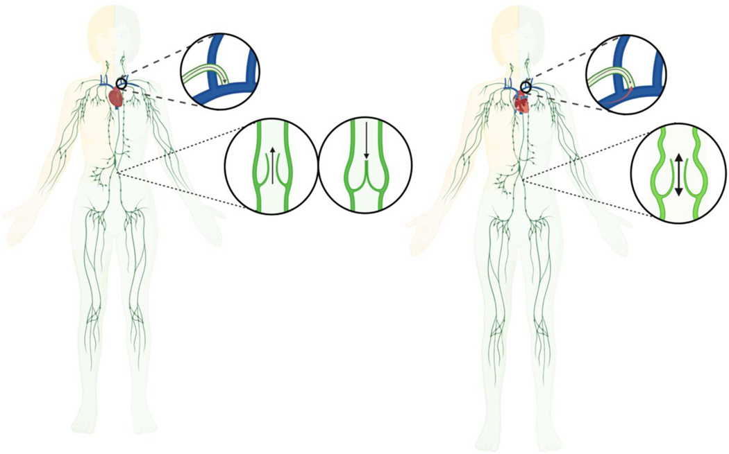
Lymphatic failure is a broad term that describes the lymphatic circulation's inability to adequately transport fluid and solutes out of the interstitium and into the systemic venous circulation, which can result in dysfunction and dysregulation of immune responses, dietary fat absorption, and fluid balance maintenance. Several investigations have recently elucidated the nexus between lymphatic failure and congenital heart disease, and the associated morbidity and mortality is now well-recognized. However, the precise pathophysiology and pathogenesis of lymphatic failure remains poorly understood and relatively understudied, and there are no targeted therapeutics or interventions to reliably prevent its development and progression. Thus, there is growing enthusiasm towards the development and application of novel percutaneous and surgical lymphatic interventions. Moreover, there is consensus that further investigations are needed to delineate the underlying mechanisms of lymphatic failure, which could help identify novel therapeutic targets and develop innovative procedures to improve the overall quality of life and survival of these patients. With these considerations, this review aims to provide an overview of the lymphatic circulation and its vasculature as it relates to current understandings into the pathophysiology and pathogenesis of lymphatic failure in patients with congenital heart disease, while also summarizing strategies for evaluating and managing lymphatic complications, as well as specific areas of interest for future translational and clinical research efforts.


Figures

Figure 1:
Figure 1:
Overview of the Systemic and Lymphatic Circulation in Biventricular and Single Ventricle Hearts. Unlike the systemic circulation, the lymphatic circulation lacks a central pump. Left: Lymphatic fluid is normally driven from the capillaries to the systemic venous circulation by intrinsic and extrinsic forces. Right: The Fontan procedure provides surgical palliation for single ventricle congenital heart defects by creating a nonphysiological shunt between the systemic venous circulation and the pulmonary artery to bypass the subpulmonic ventricle. As a result, central venous pressures are often chronically and/or obligatorily elevated, which can lead to progressive overload and congestion of the lymphatic circulation that ultimately results in lymphatic failure. Individuals with Fontan circulation may develop dilated and tortuous lymphatic vessels with multiple collaterals and channels, which likely reflect early or failed adaptive responses and pathologic remodeling to reroute abnormal lymphatic fluid flow. Illustration reproduced with permission from Kelly, et al. (34).
Figure 2:
Figure 2:
Lymphatic fluid produced by the right side of the head, neck, thorax, and the right upper extremity normally drains into the systemic venous circulation via the right thoracic duct, whereas lymphatic fluid produced by the rest of the body is typically returned through the left lymphatic duct (i.e., thoracic duct), which is the largest lymphatic vessel in the body. Left: Lymphatic fluid normally passes through the thoracic duct before emptying into the systemic venous circulation via an ostial valve near the lymphovenous junction at the confluence between the left subclavian and jugular veins, which helps maintain unidirectional lymphatic fluid flow and prevent blood from regurgitating into the lymphatic circulation. Right: Chronically elevated central venous pressures excessively increase hepatosplanchnic lymphatic fluid production and afterload on the thoracic duct, which impedes lymphatic fluid drainage into the systemic venous circulation. These imbalances in lymphatic fluid overproduction and impaired drainage cause the lymphatic circulation to become overloaded and congested. As a result, lymphatic vessels and valves may become dilated and incompetent, which further impairs unidirectional, antegrade lymphatic fluid flow and exacerbates lymphatic dysfunction and dysregulation. Illustration adapted and modified with permission from Fudim, et al. (1).
Figure 3:
Figure 3:
As blood passes through the systemic microcirculation, interstitial fluid is produced by transvascular capillary filtration that drives plasma fluid into the interstitium. Left: Under normal physiologic circumstances, the rate of lymphatic fluid production, transportation, and flow through the lymphatic circulation is autoregulated to maintain a relatively constant volume of interstitial fluid. The removal of interstitial fluid and its contents (i.e., immune cells, proteins and macromolecules, lipids and chylomicrons, etc.) is essential for regulating immune responses, the absorption of dietary fats, and maintaining fluid balance. Right: When lymphatic failure occurs, the lymphatic circulation and its vasculature are unable to adequately transport interstitial fluid and its contents into and through the lymphatic circulation, which may result from pathologic imbalances in homeostatic hydrostatic and osmotic pressures, disruptions in normal capillary permeability and excessive transvascular capillary filtration, as well as impairments in lymphatic reuptake and lymphatic fluid transportation. Progressive overload and congestion of the lymphatic circulation ultimately gets transmitted to smaller and more peripheral lymphatic vessels, which prevents lymphatic capillaries from resorbing fluid and other contents from the interstitium of tissues. Illustration adapted with permission from Rossitto, et al. (9).
Figure 4:
Figure 4:
The lymphatic vasculature is organized into lymphatic capillaries, pre-collector and collector vessels, and larger lymphatic trunks and central collecting ducts that ultimately empty into the systemic venous circulation via a lymphovenous junction near the internal jugular and subclavian veins. The structural and functional integrity of the lymphatic circulation and its vasculature are essential for homeostatic fluid balance regulation and the removal of excess interstitial fluid. Lymphatic capillaries are comprised of highly-permeable lymphatic capillary endothelial cells that have a discontinuous or entirely lacking basement membrane, and are joined together by ‘button-like’ intercellular junctions that form overlapping flaps between cell borders. Anchoring filaments prevent the collapse of lymphatic capillaries when interstitial fluid production increases and interstitial pressures rise, which drives interstitial fluid and its contents into the lymphatic microvasculature. Lymphatic fluid is transported into larger lymphatic pre-collector and collector vessels that are comprised of relatively impermeable lymphatic endothelial cells that have a solid, continuous basement membrane, as well as tight, zipper-like intercellular junctions. Precollector and collector vessels are functionally separated by two unidirectional valves, and each individual segment is collectively referred to as a lymphangion, which are surrounded by smooth muscle cells that autonomously. LEC: lymphatic endothelial cell; LMC: lymphatic muscle cell. Illustration reproduced with permission from Brakenhielm, et al. (55).
Figure 5:
Figure 5:
Plastic bronchitis and protein-losing enteropathy are among the most devastating complications associated with lymphatic failure. Top: In normal lungs, pulmonary lymphatics drain fluid away from small airways and alveoli, whereas lymphatic vessels in individuals with lymphatic failure are often dilated and have multiple collaterals. Increased transvascular capillary filtration and impaired lymphatic reuptake can cause lymphatic fluid to leak into the pleural cavity and airways, leading to pleural effusions, inflammation, and plastic bronchitis with the production of thick proteinaceous casts. Bottom: Intestinal lymphatics (lacteals) normally transport lipids and fat-soluble nutrients into the lymphatic circulation, while also helping to clear fluid, proteins, and lymphocytes within the interstitium. These functions are critical for nutrient absorption, regulating immune responses, and maintaining homeostasis in the enteric tract. In patients with lymphatic failure, impaired lymphatic drainage can cause retrograde lymphatic fluid flow and protein-losing enteropathy. In patients with protein-losing enteropathy, lacteals are often dilated and engorged, and the intestinal epithelium may become compromised. Failure to collect proteins extravasated into the interstitium and the leakage of interstitial fluid into the lumen of the bowel can result in uncompensated plasma protein loss, lipid depletion, lymphopenia, among other complications. Illustrations adapted and modified with permission from Kelly, et al. and Ozen, et al. (34,68).

Similar articles

References

    1. Fudim M, Salah HM, Sathananthan J, et al. Lymphatic Dysregulation in Patients With Heart Failure: JACC Review Topic of the Week. J Am Coll Cardiol. 2021;78(1):66–76. doi:10.1016/j.jacc.2021.04.090 - DOI - PubMed
    1. RochéRodríguez M, DiNardo JA. The Lymphatic System in the Fontan Patient-Pathophysiology, Imaging, and Interventions: What the Anesthesiologist Should Know. J Cardiothorac Vasc Anesth. 2022;36(8 Pt A):2669–2678. doi:10.1053/j.jvca.2021.07.049 - DOI - PubMed
    1. Udink Ten Cate FEA, Tjwa ETTL. Imaging the Lymphatic System in Fontan Patients. Circ Cardiovasc Imaging. 2019;12(4):e008972. doi:10.1161/CIRCIMAGING.119.008972 - DOI - PubMed
    1. Kreutzer C, Kreutzer G. The Lymphatic System: The Achilles Heel of the Fontan-Kreutzer Circulation. World J Pediatr Congenit Heart Surg. 2017;8(5):613–623. doi:10.1177/2150135117720685 - DOI - PubMed
    1. Kreutzer J, Kreutzer C. Lymphodynamics in Congenital Heart Disease: The Forgotten Circulation. J Am Coll Cardiol. 2017;69(19):2423–2427. doi:10.1016/j.jacc.2017.03.559 - DOI - PubMed

MeSH terms