Review
. 2024 May 31;12(5):e008025.
doi: 10.1136/jitc-2023-008025.

Cytokine-armed oncolytic herpes simplex viruses: a game-changer in cancer immunotherapy?

Affiliations
Review

Cytokine-armed oncolytic herpes simplex viruses: a game-changer in cancer immunotherapy?

Hongbin Wang et al. J Immunother Cancer. .

Abstract

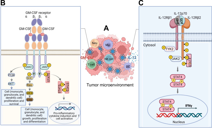
Cytokines are small proteins that regulate the growth and functional activity of immune cells, and several have been approved for cancer therapy. Oncolytic viruses are agents that mediate antitumor activity by directly killing tumor cells and inducing immune responses. Talimogene laherparepvec is an oncolytic herpes simplex virus type 1 (oHSV), approved for the treatment of recurrent melanoma, and the virus encodes the human cytokine, granulocyte-macrophage colony-stimulating factor (GM-CSF). A significant advantage of oncolytic viruses is the ability to deliver therapeutic payloads to the tumor site that can help drive antitumor immunity. While cytokines are especially interesting as payloads, the optimal cytokine(s) used in oncolytic viruses remains controversial. In this review, we highlight preliminary data with several cytokines and chemokines, including GM-CSF, interleukin 12, FMS-like tyrosine kinase 3 ligand, tumor necrosis factor α, interleukin 2, interleukin 15, interleukin 18, chemokine (C-C motif) ligand 2, chemokine (C-C motif) ligand 5, chemokine (C-X-C motif) ligand 4, or their combinations, and show how these payloads can further enhance the antitumor immunity of oHSV. A better understanding of cytokine delivery by oHSV can help improve clinical benefit from oncolytic virus immunotherapy in patients with cancer.

Keywords: Cytokine; Immune modulatory; Oncolytic virus.


Conflict of interest statement

Competing interests: SDR is a co-inventor on patents relating to oncolytic herpes simplex viruses, owned, and managed by Georgetown University and Massachusetts General Hospital, which have received royalties from Amgen and Acti\Vec Inc., and acted as a consultant and received honoraria from Replimune, Cellinta, and Greenfire Bio, and honoraria and equity from EG 427. HLK is an employee of Ankyra Therapeutics and has received honoraria for participating on advisory boards for Castle Biosciences, Midatech Pharma, Marengo Therapeutics, and Virogin. The remaining authors declare that the research was conducted in the absence of any commercial or financial relationships that could be construed as a potential conflict of interest.

Figures

Figure 1
Figure 1
(A) Mechanism of action of cytokine/chemokine-armed oHSVs. Armed oHSVs can enter healthy or normal host cells. However, the deletion or mutation of viral genes (such as ICP34.5, ICP6) and the presence of cellular antiviral defense mechanisms in healthy cells (such as the antiviral PKR-eIF2α pathway) prevent oHSV replication. As a result, healthy cells remain non-permissive to oHSV replication and infection (upper panel). In contrast, in cancer cells, antiviral mechanisms like the PKR-eIF2α pathway are often defective, allowing viral replication (ie, permissiveness). This leads to the killing of cancer cells and the release of tumor (and viral) antigens. These antigens provoke an antitumor immune response. Through oncolysis, oHSVs spread to surrounding cancer cells, a process known as in-situ amplification and spread. This further augment the immune response, often referred to as the in-situ vaccine effect (lower panel). The virus-induced in-situ vaccine effect can be enhanced by the viral expression of cytokines/chemokines from the armed oHSVs in the tumor microenvironment. The cytokine/chemokine expression results in the further orchestration of the immune response, including the recruitment of various cells into the tumor. These recruited cells contribute to the production of a cancer-killing effect. This figure was created with BioRender.com. (B) Cytokine/chemokine-expressing oHSVs in the tumor microenvironment and their antitumor immune effects. (1) GM-CSF-armed oHSVs demonstrated significant antitumor efficacy not only in the injected tumor but also in non-injected tumors in preclinical and clinical studies. GM-CSF stimulated granulocyte and monocyte differentiation, enhanced T-cell recruitment at the tumor site, locally reduced regulatory T cells (Tregs) and myeloid-derived suppressor cells (MDSCs), and induced TH9 cell expansion and NK cell activation, along with dendritic cell (DC) maturation. (2) IL-15-armed oHSVs significantly enhance infiltration and activation of NK and CD8+ T cells in tumor and inhibit tumor growth and prolong survival. (3) IL-12-armed oHSVs significantly boosted antitumor immune responses, promoting intratumoral CD8+ T-cell infiltration, inducing Th1 (T-bet+) cell differentiation, reducing CD4+FoxP3+ regulatory T cells (Tregs) and Th2 cell activity, polarizing macrophages towards antitumor M1 phenotypes, increasing release of IFN-γ, TNF-α, GM-CSF, and IL-2, and decreasing CD31+ tumor vascularity. (4) IL-2-armed oHSVs greatly improved antitumor efficacy compared with unarmed oHSV. Elevated IL-2 levels drove differentiation and expansion of activated T cells toward Th1/CTLs, enhancing TNF-α and IFN-γ release, while potentially increasing Treg cells and suppressing IL-17A. (5) IL-18-armed oHSVs did not exhibit improved antitumor efficacy compared with unarmed oHSVs. However, when combined with vHsv-IL-12, vHsv-IL-18 significantly enhanced antitumor immunity synergistically with IL-12 expression, increasing IFN-γ and GM-CSF release, and promoting the infiltration of CD4+ and CD8+ T lymphocytes and NK cells. (6) FLT3L-armed oHSVs significantly improved long-term survival rates in glioma-bearing mice by enhancing IFN-α-expressing plasmacytoid and conventional DC infiltration in the tumor microenvironment. (7) CCL-5-armed oHSVs enhance antitumor immune responses by recruiting CD8+, CD4+, and NK cells into tumor microenvironment. (8) TNF-α-armed oHSVs effectively inhibited injected and non-injected tumors, stimulating T-cell mediated antitumor immunity, promoting M1 polarization of tumor-associated macrophages, inhibiting Treg cells and facilitating the recruitment of antigen-presenting cells and neutrophils. (9) CCL2-armed oHSVs demonstrated inconsistent antitumor efficacy, but the combination therapy of CCL2 expression with IL-12 expression showed superior antitumor effects characterized by recruiting CD4+ and CD8+ T lymphocytes to the tumor site. CCL2 expression also activated monocytes, macrophages, memory T lymphocytes, and NK cells, leading to the release of pro-inflammatory cytokines, such as IL-1, IL-6, and TNF-α. (10) CXCL4-armed oHSVs significantly outperformed unarmed viruses in controlling tumor growth, resulting in a substantial extension of median survival. CXCL4 expression activated DCs, leading to CD4+ T-cell proliferation/activation and IL-17 production. This figure was created with BioRender.com. APC, antigen-presenting cells; CCL2, chemokine (C-C motif) ligand 2; CCL5, chemokine (C-C motif) ligand 5; CTLs, cytotoxic T lymphocytes; CXCL4, chemokine (C-X-C motif) ligand 4; DC, dendritic cell; ECM, extracellular matrix; Fib, fibroblasts; FLT3L, FMS-like tyrosine kinase 3 ligand; GM-CSF, granulocyte-macrophage colony-stimulating factor; IFN, interferon; IL, interleukin; MΦ, macrophage; NK, natural killer; PKR, protein kinase R; oHSV, oncolytic herpes simplex virus; TNF, tumor necrosis factor.
Figure 2
Figure 2
(A) oHSV-infected tumor microenvironment. GM-CSF-armed or IL-12-armed oHSVs infect and lyse cancer cells, resulting in the release of GM-CSF or IL-12 in the tumor microenvironment (TME), respectively, for induction of antitumor immunity. (B) GM-CSF expression and its associated signaling pathways in antitumor immunity. GM-CSF released from oHSVs binds to its receptor, which is a heterodimer, consisting of α chains that specifically bind GM-CSF and βc chains that are responsible for signaling. Neither the α chains nor the βc chains of the GM-CSF receptor contains a tyrosine (Y) kinase catalytic domain. The βc chain of the GM-CSF receptor links to tyrosine kinase JAK2. Once the α chain binds to GM-CSF, it subsequently binds to the βc chain to form a multimer. The polymerization of the receptor leads to the activation of JAK2, and the activated JAK2 consequently phosphorylates the tyrosine residues on the βc chain of the receptor. The phosphorylated tyrosine on the βc chain recruits STAT-5 by binding to the SH2 domain of STAT5. JAK2 further phosphorylates STAT-5 and activates the JAK-STAT pathway, which induces pro-inflammatory cytokines that activate T lymphocytes within the TME. Furthermore, JAK2 activates PI3K and initiates the PI3K-Akt signaling pathway, which promotes the proliferation and survival of monocytes, macrophages, and granulocytes within the TME. The phosphorylated tyrosine on the βc chain of the GM-CSF receptor also recruits the adaptor protein SHC, which in turn activates RAS/MEK/ERK signaling pathway to induce signaling in the nucleus, enhancing the differentiation and proliferation of macrophages, neutrophils, and dendritic cells within the TME. (C) IL-12 expression and its associated signaling pathways in antitumor immunity. IL-12 (IL-12p70) is composed of two covalently linked subunits, p35 and p40. The released IL-12 from oHSVs binds to IL-12 receptor, which is composed of two subunits, IL-12Rβ1 and IL-12Rβ2. JAK2 and TYK2 become trans-phosphorylated after the association of IL-12p40 and IL-12p35 with IL-12Rβ1 and IL-12Rβ2, respectively. Phosphorylated IL-12Rβ2 binds to STAT4, which then dimerizes with another STAT4 molecule. STAT4 homodimers translocate to the nucleus and promote transcription of the IFN-γ gene. The IL-12 and IFN-γ induce the activity and proliferation of macrophages, NK cells, and T lymphocytes in TME. This figure was created with BioRender.com. DC, dendritic cell; GM-CSF, granulocyte-macrophage colony-stimulating factor; IFN, interferon; IL, interleukin; Mφ, macrophage; Neu, neutrophil; NK, natural killer cell; oHSV, oncolytic herpes simplex virus.
Figure 3
Figure 3
Immunological alterations in the tumor microenvironment following oHSV-IL-12 treatment as monotherapy and combination therapy. (1) oHSV-12 monotherapy enhances antitumor immune responses characterized by increased intratumoral infiltration of CD8+ T cells, enhanced Th1 (T-bet+) cell differentiation, decreased CD4+FoxP3+ regulatory T cells (Tregs), increased polarization of macrophages toward an antitumor M1 phenotype, elevated levels of IFN-γ and IL-12, and reduced CD31+ tumor vascularity. (2) Immune checkpoint inhibitors significantly enhance the effectiveness of oHSV-IL-12 monotherapy. The combination therapy markedly increases infiltration of CD3+ and CD8+ T cells, reduces infiltration of CD4+FoxP3+ Tregs, enhances Th1 differentiation (T-bet+), and augments influx of M1-like antitumoral macrophages. (3) oHSV-IL-12 in combination with angiogenic inhibitors leads to enhanced tumor oncolysis, decreased tumor vascularity (CD34+ vessels), and greater infiltration of macrophages (CD68+). (4) However, oHSV-IL-12 in combination with a chemotherapy agent, temozolomide (TMZ), yields no significant survival benefit in the combination group when compared with oHSV-IL-12 monotherapy, rather TMZ treatment abrogates the antitumor efficacy of oHSV-IL-12. This is due to TMZ treatment, which substantially impairs immune responses, characterized by the reduction of T cells (CD4+ and CD8+) and macrophages (CD68+) in the tumor microenvironment, along with CD4+ T-cell reduction systemically in the spleen. This figure was created with BioRender.com. IFN, interferon; IL, interleukin; oHSV, oncolytic herpes simplex virus; TAM, tumor-associated macrophage.

Similar articles

Cited by

References

    1. Tian Y, Xie D, Yang L. Engineering strategies to enhance Oncolytic viruses in cancer Immunotherapy. Signal Transduct Target Ther 2022;7:117. 10.1038/s41392-022-00951-x - DOI - PMC - PubMed
    1. Pol JG, Workenhe ST, Konda P, et al. . Cytokines in Oncolytic Virotherapy. Cytokine Growth Factor Rev 2020;56:4–27. 10.1016/j.cytogfr.2020.10.007 - DOI - PubMed
    1. Jahan N, Ghouse SM, Martuza RL, et al. . In situ cancer vaccination and Immunovirotherapy using Oncolytic HSV. Viruses 2021;13:1740. 10.3390/v13091740 - DOI - PMC - PubMed
    1. Ma R, Li Z, Chiocca EA, et al. . The emerging field of Oncolytic virus-based cancer Immunotherapy. Trends Cancer 2023;9:122–39. 10.1016/j.trecan.2022.10.003 - DOI - PMC - PubMed
    1. Hemminki O, Dos Santos JM, Hemminki A. Oncolytic viruses for cancer Immunotherapy. J Hematol Oncol 2020;13:84. 10.1186/s13045-020-00922-1 - DOI - PMC - PubMed