. 2024 May 31;15(1):4644.
doi: 10.1038/s41467-024-48991-x.

Nanoscale cellular organization of viral RNA and proteins in SARS-CoV-2 replication organelles

Affiliations

Nanoscale cellular organization of viral RNA and proteins in SARS-CoV-2 replication organelles

Leonid Andronov et al. Nat Commun. .

Abstract

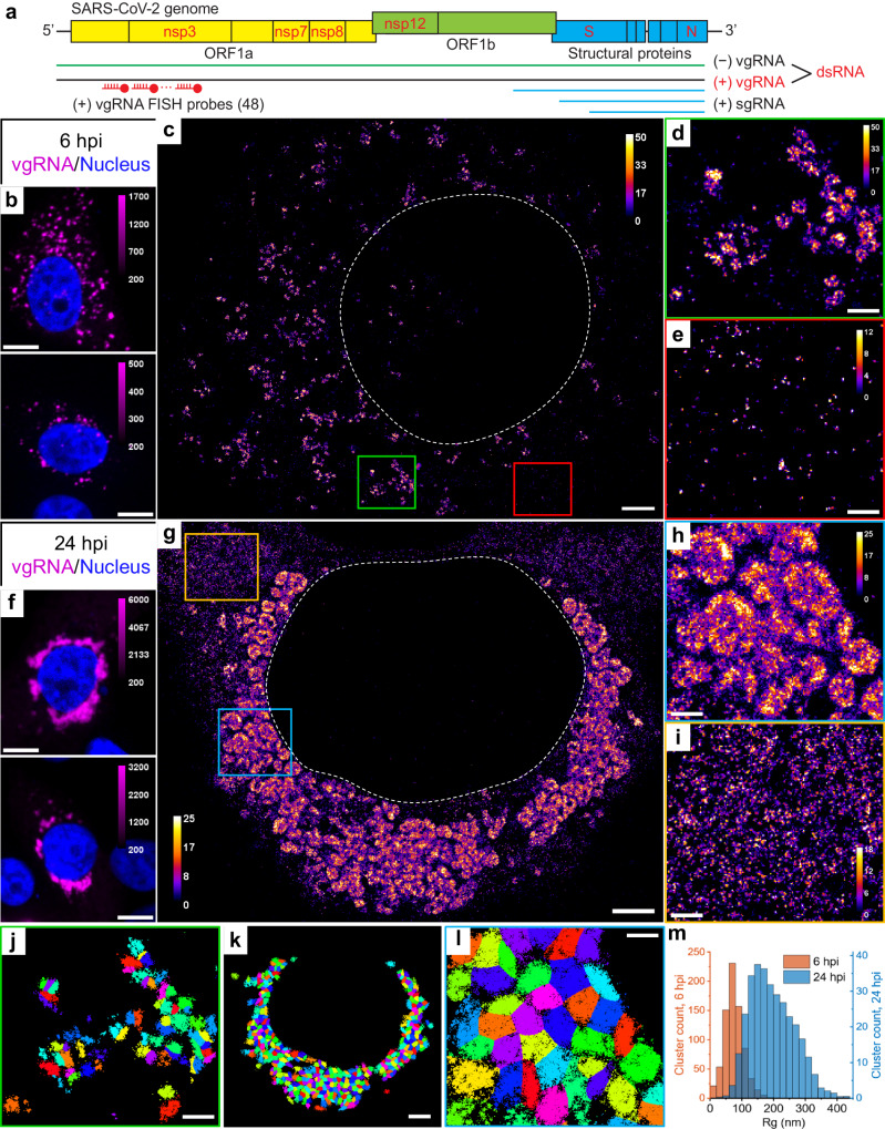
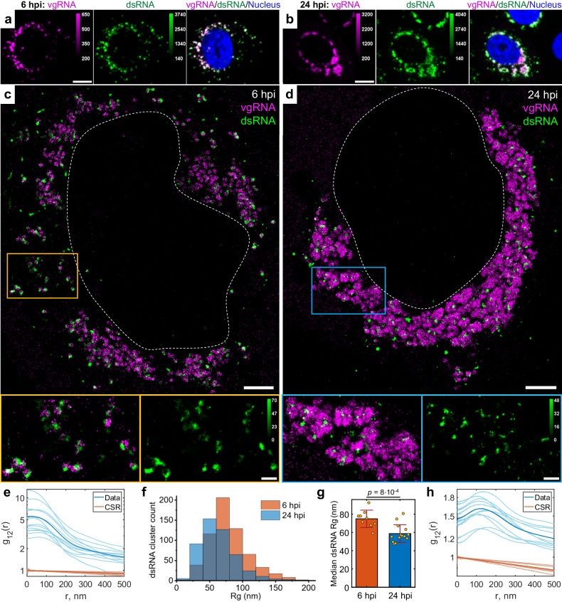
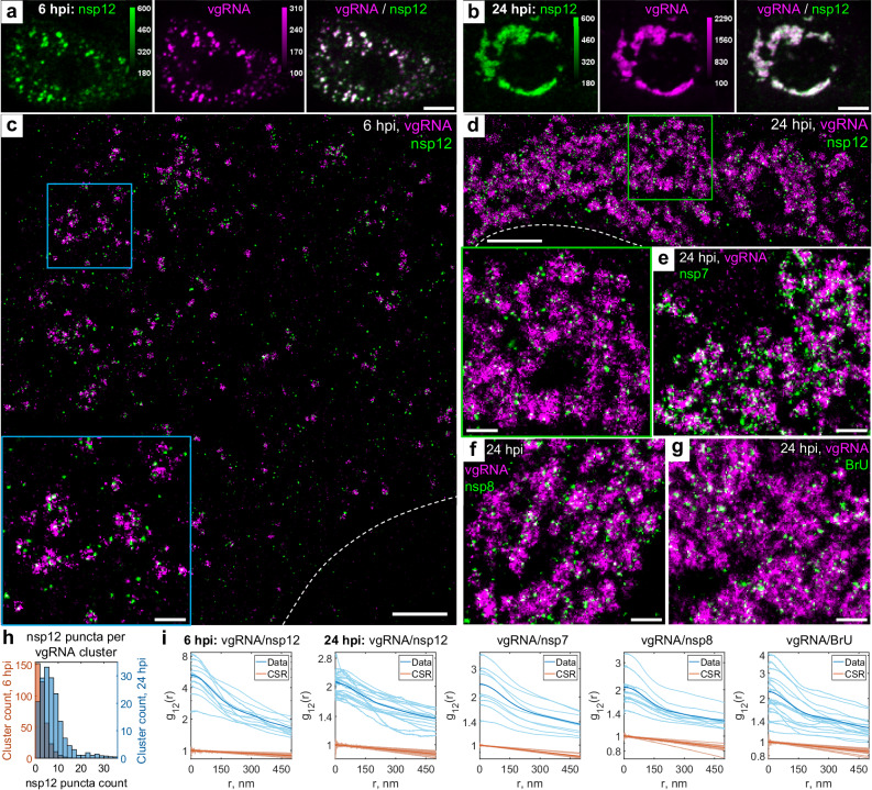
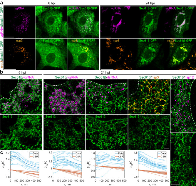
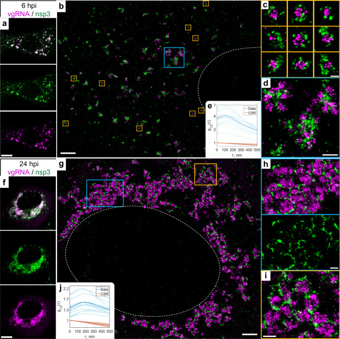
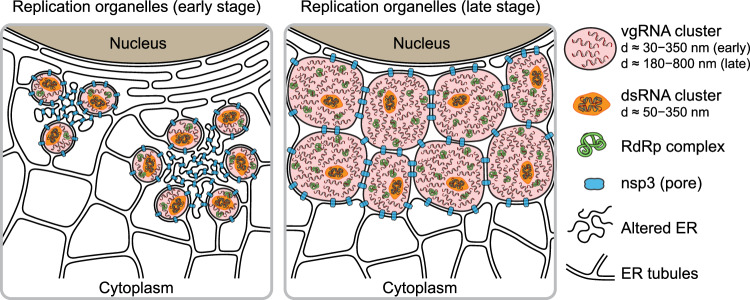
The SARS-CoV-2 viral infection transforms host cells and produces special organelles in many ways, and we focus on the replication organelles, the sites of replication of viral genomic RNA (vgRNA). To date, the precise cellular localization of key RNA molecules and replication intermediates has been elusive in electron microscopy studies. We use super-resolution fluorescence microscopy and specific labeling to reveal the nanoscopic organization of replication organelles that contain numerous vgRNA molecules along with the replication enzymes and clusters of viral double-stranded RNA (dsRNA). We show that the replication organelles are organized differently at early and late stages of infection. Surprisingly, vgRNA accumulates into distinct globular clusters in the cytoplasmic perinuclear region, which grow and accommodate more vgRNA molecules as infection time increases. The localization of endoplasmic reticulum (ER) markers and nsp3 (a component of the double-membrane vesicle, DMV) at the periphery of the vgRNA clusters suggests that replication organelles are encapsulated into DMVs, which have membranes derived from the host ER. These organelles merge into larger vesicle packets as infection advances. Precise co-imaging of the nanoscale cellular organization of vgRNA, dsRNA, and viral proteins in replication organelles of SARS-CoV-2 may inform therapeutic approaches that target viral replication and associated processes.


Conflict of interest statement

The authors declare no competing interests.

Figures

Fig. 1
Fig. 1. Clustering of vgRNA in the cytoplasm of infected cells.
a Scheme of SARS-CoV-2 genome with constructs used for its detection in infected cells. 48 antisense DNA oligonucleotide probes were used to target the ORF1a-coding region of vgRNA that is exclusive to the positive-sense vgRNA and does not occur in the sgRNAs. The RNA FISH probes are conjugated with AF647 or CF568. b Representative confocal images of vgRNA in infected Vero E6 cells at 6 hpi display scattered diffraction-limited (DL) puncta. c Representative SR image of an infected cell at 6 hpi reveals distinct vgRNA clusters in the cytoplasm. d Zoomed-in region of the SR image (green frame in c) displays an agglomeration of vgRNA clusters. e Zoomed-in region of the SR image (red frame in c) shows nanoscale puncta of individual vgRNA molecules. f Representative confocal images of vgRNA in infected Vero E6 cells at 24 hpi display large DL foci in the perinuclear region of the cytoplasm. g Representative SR image of an infected cell at 24 hpi reveals large perinuclear vgRNA clusters. h Zoomed-in region of the SR image (blue frame in g) displays dense vgRNA clusters. i Zoomed-in region of the SR image (yellow frame in g) displays nanoscale puncta of vgRNA molecules. j BIC-GMM cluster analysis of the region shown in d. k BIC-GMM cluster analysis of the cell shown in g. l BIC-GMM cluster analysis of the region shown in h. m Histogram of the radii of gyration (Rg) of the vgRNA clusters indicate their size increase between 6 hpi (tan) and 24 hpi (blue). Data for the histograms are obtained from 10 cells (6 hpi) and 16 cells (24 hpi) and 2 independent experiments for each time point. Scale bars, 10 µm (b, f), 2 µm (c, g, k), 500 nm (d, e, h, i, j, l). Dashed lines in c and g indicate the boundary of the cell nucleus (large dark region). Localizations that belong to the same cluster in j, k, l are depicted with the same color, but colors are reused. Color bars in ce, gi show the number of single-molecule localizations within each SR pixel (20 × 20 nm2).
Fig. 2
Fig. 2. Association of dsRNA with vgRNA clusters.
Representative confocal images of SARS-CoV-2 infected cells display DL colocalization between dsRNA (green) and vgRNA (magenta) at both 6 hpi (a) and 24 hpi (b). Representative SR images of SARS-CoV-2 infected cells indicate association between dsRNA and vgRNA at 6 hpi (c) and short-range anti-correlation often with concentric localization at 24 hpi (d). Bottom panels, zoomed-in images of corresponding colored boxes. e Bivariate pair-correlation functions g12(r) calculated between the localizations of dsRNA and vgRNA indicate their close association at 6 hpi. f Histogram of Rg of dsRNA clusters as determined by the BIC-GMM cluster analysis. g Median Rg of dsRNA clusters per cell significantly decreases between 6 hpi and 24 hpi. Data points denote median Rg values of dsRNA clusters in individual analyzed cells; error bars represent mean ± standard deviation of these values. Data for the histograms (f, g) are obtained from 9 cells (6 hpi) and 14 cells (24 hpi). h Bivariate pair-correlation functions g12(r) reveal nanoscale anti-correlation between dsRNA and vgRNA at 24 hpi. CSR, complete spatial randomness. Thin lines correspond to g12(r) of individual cells and bold lines represent the mean values of g12(r) from all analyzed cells. Scale bars, 10 µm (a, b), 2 µm (c, d), 500 nm (c, d, bottom panels). Dashed lines in c and d indicate the boundary of the cell nucleus.
Fig. 3
Fig. 3. Association of SARS-CoV-2 replication enzyme with vgRNA clusters.
Representative confocal images of SARS-CoV-2 infected cells display DL colocalization between nsp12, the catalytic subunit of RdRp (green) and vgRNA (magenta) at both 6 hpi (a) and 24 hpi (b). Representative SR images of SARS-CoV−2 infected cells indicate nanoscale association between nsp12 and vgRNA at both 6 hpi (c) and 24 hpi (d). Insets show magnified images of corresponding regions in the colored boxes. Representative SR images of vgRNA with nsp7 (e) or nsp8 (f) in the perinuclear regions of SARS-CoV-2 infected cells indicate association of nsp7 and nsp8 with vgRNA clusters. g Representative SR image of vgRNA with newly synthesized viral RNAs labeled by BrU in a SARS-CoV-2 infected cell indicates localization of newly synthesized viral RNAs within the perinuclear clusters of vgRNA. h Number of nanoscale puncta of nsp12 per vgRNA cluster. Data for the histograms are obtained from 5 cells (6 hpi) and 9 cells (24 hpi). i Bivariate pair-correlation functions for vgRNA and nsp12, nsp7, nsp8 and newly transcribed viral RNA labeled with BrU peak at r = 0 nm indicating association between these target pairs. Scale bars, 10 µm (a, b), 2 µm (c, d), 500 nm (eg and insets in c and d). Dashed lines in c and d indicate the edge of the cell nucleus.
Fig. 4
Fig. 4. vgRNA clusters are encapsulated in membranes of remodeled ER.
a Representative confocal images of SARS-CoV-2 infected cells indicate an appearance of dense perinuclear foci of Sec61β ER labeling (green) at 24 hpi that colocalizes with vgRNA and nsp3. b SR images reveal concentric organization of Sec61β around vgRNA, dsRNA and nsp12, and colocalization of Sec61β with nsp3. c Bivariate pair-correlation functions indicate anti-correlation of Sec61β with vgRNA and dsRNA and association of Sec61β with nsp3. Scale bars, 20 µm (a) and 1 µm (b). Dashed lines in b indicate the boundary of the cell nucleus.
Fig. 5
Fig. 5. Nsp3 localizes at the surface of vgRNA clusters.
a Representative confocal images of a SARS-CoV-2 infected cell display DL colocalization between punctate vgRNA (magenta) and nsp3 (green) labeling at 6 hpi. b Representative SR image of a SARS-CoV-2 infected cell at 6 hpi. c Zoomed-in images of selected vgRNA particles (yellow boxes in b) indicate the localization of nsp3 at the surface of the vgRNA clusters. d Magnified region with aggregates of vgRNA clusters (blue box in b) displays dense nsp3 localization in the core of these aggregates. e Bivariate pair-correlation functions calculated between the SM localizations of vgRNA and nsp3 indicate nanoscale anti-correlation of these targets at 6 hpi. f Confocal images show that vgRNA and nsp3 occupy approximately the same regions in a SARS-CoV-2 infected cell at 24 hpi. g Representative SR image of a SARS-CoV-2 infected cell at 24 hpi. h, i Magnified regions of the SR image (colored boxes in g) reveal that nsp3 localizes in interstitial regions or encapsulates vgRNA clusters. j Bivariate pair-correlation functions indicate nanoscale anti-correlation between vgRNA and nsp3 at 24hpi. Scale bars, 10 µm (a, f), 2 µm (b, g), 500 nm (d, h, i), 200 nm (c). Dashed lines in b and g indicate the boundary of the cell nucleus.
Fig. 6
Fig. 6. Proposed model for SARS-CoV-2 replication organelles.
Replication organelles (ROs) comprise various RNA species (vgRNA, dsRNA) and proteins (RdRp, nsp3) organized by remodeled cellular ER membranes. Our data support a model which specifies the spatial organization of ROs at early (left panel) and late (right panel) stages of infection. Specific RNA and protein molecules are shown to be distinctly organized within the ROs. The replication and transcription of SARS-CoV-2 RNA occur within the double-membrane vesicles (DMVs) and vesicle packets (VPs) in proximity to RdRp.

Update of

References

    1. Schmidt N, et al. The SARS-CoV-2 RNA–protein interactome in infected human cells. Nat. Microbiol. 2021;6:339–353. doi: 10.1038/s41564-020-00846-z. - DOI - PMC - PubMed
    1. Flynn RA, et al. Discovery and functional interrogation of SARS-CoV-2 RNA-host protein interactions. Cell. 2021;184:2394–2411.e2316. doi: 10.1016/j.cell.2021.03.012. - DOI - PMC - PubMed
    1. Knoops K, et al. SARS-coronavirus replication is supported by a reticulovesicular network of modified endoplasmic reticulum. PLoS Biol. 2008;6:e226. doi: 10.1371/journal.pbio.0060226. - DOI - PMC - PubMed
    1. Snijder EJ, et al. A unifying structural and functional model of the coronavirus replication organelle: Tracking down RNA synthesis. PLoS Biol. 2020;18:e3000715. doi: 10.1371/journal.pbio.3000715. - DOI - PMC - PubMed
    1. Klein S, et al. SARS-CoV-2 structure and replication characterized by in situ cryo-electron tomography. Nat. Commun. 2020;11:5885. doi: 10.1038/s41467-020-19619-7. - DOI - PMC - PubMed

MeSH terms