. 2024 May 31;15(1):4629.
doi: 10.1038/s41467-024-48059-w.

Universal paramyxovirus vaccine design by stabilizing regions involved in structural transformation of the fusion protein

Affiliations

Universal paramyxovirus vaccine design by stabilizing regions involved in structural transformation of the fusion protein

Johannes P M Langedijk et al. Nat Commun. .

Abstract

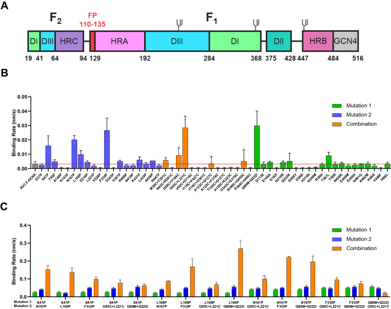
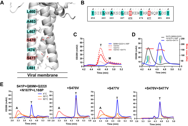
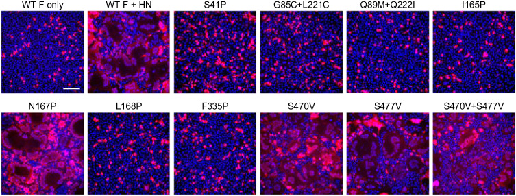
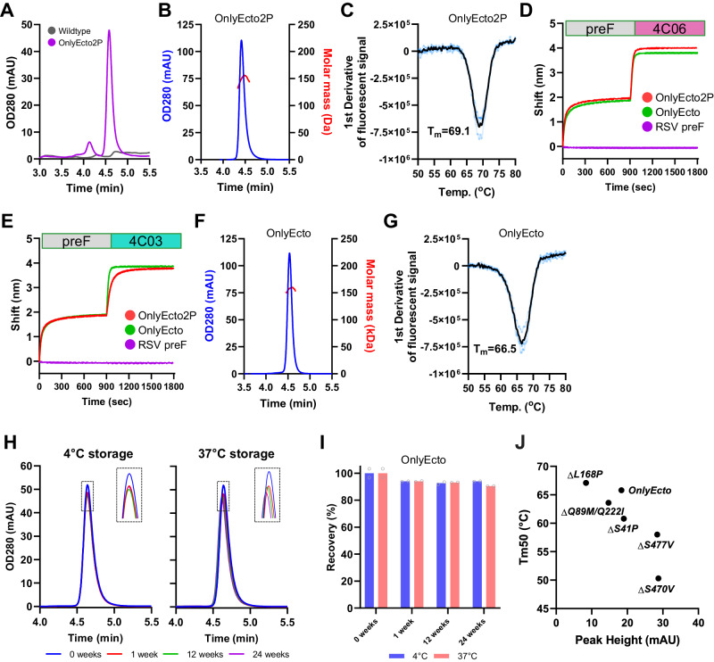
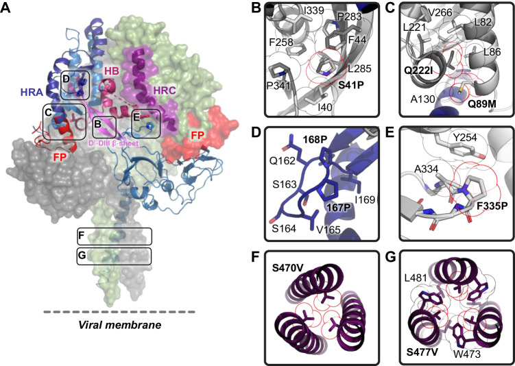
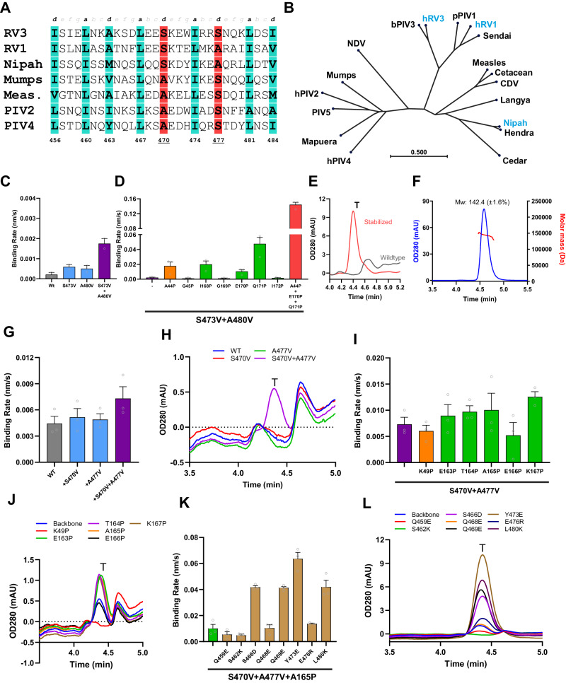
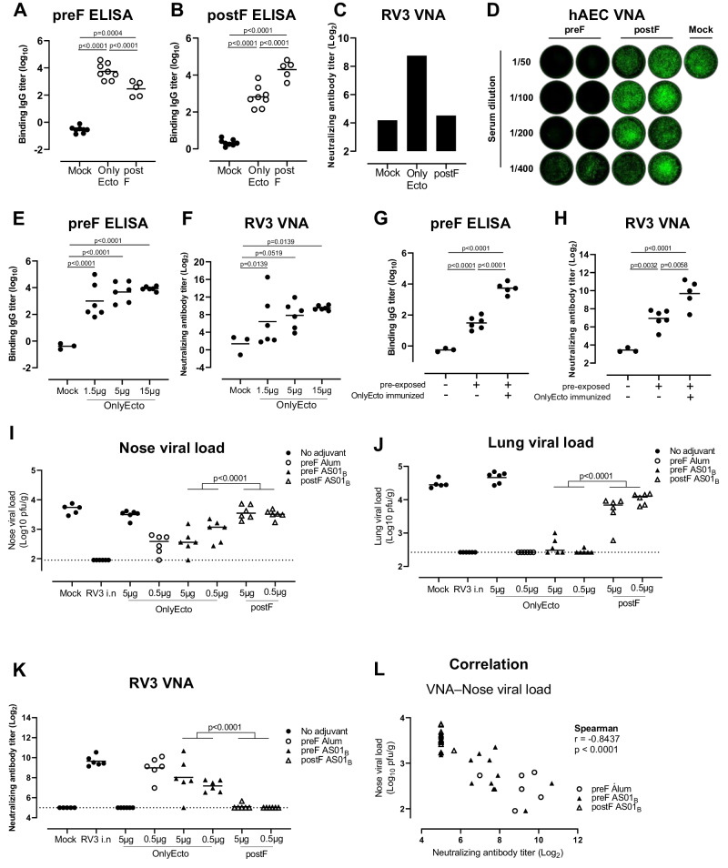
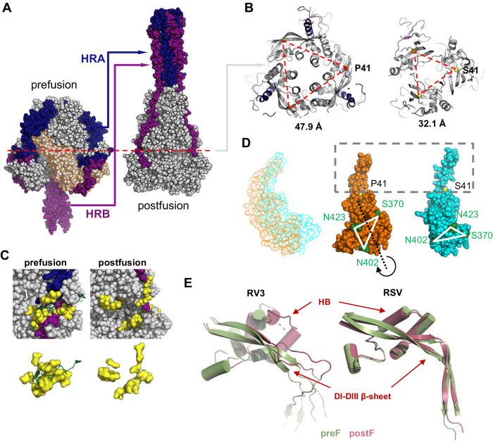
The Paramyxoviridae family encompasses medically significant RNA viruses, including human respiroviruses 1 and 3 (RV1, RV3), and zoonotic pathogens like Nipah virus (NiV). RV3, previously known as parainfluenza type 3, for which no vaccines or antivirals have been approved, causes respiratory tract infections in vulnerable populations. The RV3 fusion (F) protein is inherently metastable and will likely require prefusion (preF) stabilization for vaccine effectiveness. Here we used structure-based design to stabilize regions involved in structural transformation to generate a preF protein vaccine antigen with high expression and stability, and which, by stabilizing the coiled-coil stem region, does not require a heterologous trimerization domain. The preF candidate induces strong neutralizing antibody responses in both female naïve and pre-exposed mice and provides protection in a cotton rat challenge model (female). Despite the evolutionary distance of paramyxovirus F proteins, their structural transformation and local regions of instability are conserved, which allows successful transfer of stabilizing substitutions to the distant preF proteins of RV1 and NiV. This work presents a successful vaccine antigen design for RV3 and provides a toolbox for future paramyxovirus vaccine design and pandemic preparedness.


Conflict of interest statement

J.P.M.L., J.J., and M.J.G.B. are co-inventors on related vaccine patents. J.P.M.L., F.C., D.v.O., L.L., W.v.d.H., R.V., R.Z., L.v.d.F., J.J. and M.J.G.B. are employees of Janssen Vaccines & Prevention BV and may hold stock of Johnson and Johnson.

Figures

Fig. 1
Fig. 1. Identification of RV3 preF-stabilizing substitutions.
A Schematic of the RV3 F ectodomain used to screen for stabilizing substitutions in the head domain. The domain organization is color-coded, with start and end residue numbers indicated. Location of N-linked glycosylation sites are shown. B, C Expression of preF protein variants in supernatant as measured by BioLayer Interferometry (BLI) using immobilized PIA174. The initial slope, V0, at the start of binding is plotted as the average of three independent transfections; shown is the mean with error bars representing the standard deviation (SD). B Individual substitutions were tested and are colored by design feature. The dotted red line indicates the binding rate of the backbone construct RV3 F-GCN4. C Combinations (orange) of promising stabilizing substitutions of (B) were tested and compared to the individual substitutions (green and blue).
Fig. 2
Fig. 2. Stabilization of the RV3 HRB region.
A Cartoon representation of HRB from PDB ID 6MJZ. The heptad register is shown in sticks, with the serines at positions 470 and 477 highlighted in red. B The RV3 F HRB heptad register indicating the suboptimal residues at positions 470 and 477. C Analytical SEC trace of F variants with a minimally stabilized head domain (Q89M, Q222I and L168P) and with or without HRB stabilization in supernatant. The trimer (T) and monomer (M) peaks are indicated. D Analytical SEC-MALS trace of F variants with a stabilized head domain with or without HRB stabilization in supernatant. The molar mass as determined by MALS at peak max of the trimer are indicated. A cartoon to visualize the presumed opening of HRB is shown. E Stability of indicated F variants using analytical SEC of supernatants after 15 min incubation at 4 °C (blue line), 50 °C (orange line), or 60 °C (red line). The trimer (T) and aggregate (A) peaks are indicated.
Fig. 3
Fig. 3. Fusogenicity of full-length RV3 F variants.
Cell-cell fusion assay using HEK293T cells transiently transfected with the F protein of the RV3 JS strain, or variants thereof, in a 15:1 ratio to HN carrying a H552Q substitution, to allow fusion activation, and mScarlet to mark the cytosol of transfected cells in red. Cell nuclei were visualized with Hoechst in blue. Syncytia can be recognized by the dilution of the mScarlet signal after fusion with non-transfected cells, and the clustering of nuclei. Scale bar represents 100 μm. Experiment was performed twice with similar outcome, data shown are from the same, representative experiment.
Fig. 4
Fig. 4. Purification and characterization of RV3 preF lead candidates.
A Analytical SEC trace of wildtype (gray) and stabilized OnlyEcto2P (purple) in supernatant. B SEC-MALS trace of purified OnlyEcto2P. C Melting temperature as determined by DSF of purified OnlyEcto2P of (B). The first derivative of the fluorescence signal is plotted. BLI using kinetic Octet with PIA174 immobilized to anti-human Fc sensors (not shown), followed subsequently by capture of purified preF variants (0−900 s phase) and association of sdAb’s 4C06 (D) and 4C03 (E) (900−1800 s phase). F SEC-MALS trace of purified OnlyEcto. G Melting temperature as determined of OnlyEcto as in (C). H Analytical SEC trace of purified OnlyEcto after storage at either 4 °C (left panel) or 37 °C (right panel) for up to 24 weeks. I Percentage trimer recovery of (H). Shown is the average of two replicates. J Expression and melting temperature of OnlyEcto and back-substitutions as measured by analytical SEC and DSF on crude cell supernatant.
Fig. 5
Fig. 5. Structure of stabilized RV3 preF.
A Structure of OnlyEcto2P. Two monomers are shown as surface representation, and one as cartoon. Different structural elements are color-coded and indicated. Boxes refer to stabilizing substitutions and refer to Fig. 5B−G. (HRA heptad repeat A (dark blue), HRC: heptad repeat C (purple), HB: helical-bundle (maroon), FP: fusion peptide (red), DI-DIII β-sheet (pink)) (B) P41 buried in the hydrophobic pocket forming hydrophobic contacts involving sequence distant residues. C Space filling double substitution Q89M + Q222I in its aliphatic pocket formed by three helices. D The N167P and L168P substitutions in the DI β-hairpin. E Turn stabilizing F335P substitution. F S470V on the threefold axis of the stalk. G S477V forming an unusual coiled-coil structure with neighboring W473 and L481 and a central water channel.
Fig. 6
Fig. 6. Transfer of stabilizing substitutions to other paramyxovirus F proteins.
A Sequence alignment of the HRB heptad register of selected F proteins. The a and d positions are highlighted in cyan, with the suboptimal residues at positions 470 and 477 highlighted in red (numbering according to RV3). B Neighbor joining phylogenetic tree of representative paramyxovirus F proteins. C, D Expression of RV1 F protein variants in supernatant as measured by BLI using immobilized 3 × 1 antibody. The initial slope, V0, at the start of binding is plotted as the average of three independent transfections; error bars represent the SD. RV1 F substitutions A44P, E170P, Q171P, S473V, and A480V substitutions are the equivalents of the following substitutions in RV3, respectively: S41P, N167P, L168P, S470V, and S477V. E Analytical SEC trace of wildtype and stabilized (A44P + E170P + Q171P + S473V + A480V) RV1 F in cell supernatant. The trimer (T) peak is indicated. F Analytical SEC-MALS of purified stabilized RV1 preF. Expression of NiV F protein variants in supernatant as measured by BLI using immobilized 5B3 antibody (G, I, K), or by analytical SEC (H, J, L). For BLI, the initial slope, V0, at the start of binding is plotted as the average of three independent transfections; error bars represent the SD. NiV F substitutions K49P, K167P, S470V, and A477V are the equivalents of the following substitutions in RV3, respectively: S41P, N167P, S470V, and S477V.
Fig. 7
Fig. 7. Preclinical evaluation of RV3 preF (OnlyEcto).
AD Mice (n = 5 or 8) were immunized with 15 µg adjuvanted preF or postF protein or formulation buffer (Mock) at week 0 and 4. Two weeks later, serum samples were taken and preF (A) and postF (B) binding antibody titers were measured by ELISA or virus neutralization titers (VNT) in preF, postF or Mock pooled serum samples were determined with an VNA (virus neutralization assay) on Vero cells (C) or on differentiated human airway epithelial cell cultures (D) using the RV3 JS strain equipped with a GFP reporter gene. E, F Mice (n = 5 or 8) were immunized with a dose range of 1.5, 5 or 15 µg non-adjuvanted OnlyEcto or formulation buffer (Mock) at week 0 and 4. Two weeks later serum samples were taken and preF binding antibody titers were measured by ELISA (E) or RV3 neutralizing antibody titers by RV3-GFP VNA on Vero cells were determined (F). G, H Mice (n = 3 or 6) were intranasally exposed to RV3 or formulation buffer as control and immunized 19 weeks later with OnlyEcto or formulation buffer. Six weeks later, serum samples were taken and preF binding antibody titers were measured by ELISA (G) or RV3 neutralizing antibody titers by RV3-GFP VNA on Vero cells were determined (H). Analysis of variance (ANOVA; 2-sided t-test) was used for statistical comparisons between groups. Tukey-Kramer (A, B and G, H) or Dunnett adjustments. E, F for multiple comparisons were applied. IL Cotton rats (n = 5 or n = 6) were immunized with OnlyEcto with or without adjuvant or postF with adjuvant or formulation buffer (Mock) at week 0 and 4. Three weeks after the final immunization, cotton rats were challenged intranasally with RV3. Four days later viral loads in nose and lung tissue were determined (I, J). RV3 neutralizing antibody titers by plaque reduction neutralization test (PRNT) were determined in pre-challenge sera (K) and the correlation between nose viral loads and VNA titers were calculated with a spearman correlation analysis (L). Mean responses per group are indicated with horizontal lines. Statistical comparisons were performed across dose levels using a Tobit model (IL). P < 0.05 were considered statistically significant.
Fig. 8
Fig. 8. Structural transformation of the RV3 preF conformation.
A The prefusion structure (OnlyEcto2P) compared with the postfusion structure of RV3 F (PDB ID 1ZTM). HRA and HRB, including the preceding loop, are colored in blue and purple respectively. DI and DII in the prefusion structure are indicated with an orange outline. B Cross-section indicated in (A) for the prefusion (left) and postfusion (right) structures. C-alpha of residue 41 has been indicated as yellow sphere at the corner of the triangle and the distances between the C-alpha’s of the three monomers are indicated below. C Fusion peptide (green) pocket (yellow) for the preF and postF structures were plotted in the context of the full protein (upper panels) and without it (lower panels). D left panel Translation of DI and DII during the conformational change, with the preF structure indicated in orange and postF in cyan. Middle and right panel Aligned on the beta sheet around residue 41, with three residues in green to indicate the rotation of the domain during the refolding. E Comparison of the conformational change of the DI-DIII β-sheet structure and helical bundle (HB) between RV3 F and RSV F, with the preF structure indicated in green and postF in red. The structures were aligned based on the top part of the DI-DIII β-sheet.

Similar articles

Cited by

References

    1. Rima B, et al. ICTV virus taxonomy profile: paramyxoviridae. J. Gen. Virol. 2019;100:1593–1594. doi: 10.1099/jgv.0.001328. - DOI - PMC - PubMed
    1. Moscona A. Entry of parainfluenza virus into cells as a target for interrupting childhood respiratory disease. J. Clin. Investig. 2005;115:1688–1698. doi: 10.1172/JCI25669. - DOI - PMC - PubMed
    1. Lam E, Rosen JB, Zucker JR. Mumps: an update on outbreaks, vaccine efficacy, and genomic diversity. Clin. Microbiol. Rev. 2020;33:e00151–19. doi: 10.1128/CMR.00151-19. - DOI - PMC - PubMed
    1. Hübschen JM, Gouandjika-Vasilache I, Dina J. Measles. Lancet. 2022;399:678–690. doi: 10.1016/S0140-6736(21)02004-3. - DOI - PubMed
    1. Bellini WJ, Harcourt BH, Bowden N, Rota PA. Nipah virus: an emergent paramyxovirus causing severe encephalitis in humans. J. Neurovirol. 2005;11:481–487. doi: 10.1080/13550280500187435. - DOI - PubMed

MeSH terms