. 2024 May 31;15(1):4627.
doi: 10.1038/s41467-024-48361-7.

The peptidase DA1 cleaves and destabilizes WUSCHEL to control shoot apical meristem size

Affiliations

The peptidase DA1 cleaves and destabilizes WUSCHEL to control shoot apical meristem size

Guicai Cui et al. Nat Commun. .

Abstract

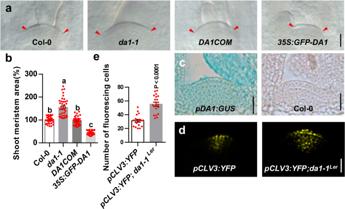
Stem cells in plants and animals are the source of new tissues and organs. In plants, stem cells are maintained in the central zone (CZ) of multicellular meristems, and large shoot meristems with an increased stem cell population hold promise for enhancing yield. The mobile homeodomain transcription factor WUSCHEL (WUS) is a central regulator of stem cell function in plant shoot meristems. Despite its central importance, the factors that directly modulate WUS protein stability have been a long-standing question. Here, we show that the peptidase DA1 physically interacts with and cleaves the WUS protein, leading to its destabilization. Furthermore, our results reveal that cytokinin signaling represses the level of DA1 protein in the shoot apical meristem, thereby increasing the accumulation of WUS protein. Consistent with these observations, loss of DA1 function results in larger shoot apical meristems with an increased stem cell population and also influences cytokinin-induced enlargement of shoot apical meristem. Collectively, our findings uncover a previously unrecognized mechanism by which the repression of DA1 by cytokinin signaling stabilizes WUS, resulting in the enlarged shoot apical meristems with the increased stem cell number during plant growth and development.


Conflict of interest statement

The authors declare no competing interests.

Figures

Fig. 1
Fig. 1. The da1-1 forms large shoot apical meristem with increased stem cell population.
a SAMs of Col-0, da1-1, DA1COM and 35S:GFP-DA1 (n = 38). DA1COM represents that the da1-1 mutant was transformed with the DA1 genomic DNA. b The average SAM area of Col-0, da1-1, DA1COM and 35S:GFP-DA1 (n = 38). The measured region of SAM was shown in Supplementary Fig. 3. Data are mean ± s.e.m. relative to the wild-type value (100%). One-way ANOVA with Tukey’s multiple comparison test was used for statistical analyses(P  <  0.05). c Images of SAMs from pDA1:GUS plants(n = 15). d Images of SAMs from pCLV3:YFP (n = 19)(left) and pCLV3:YFP; da1-1Ler (n = 20)(right) plants. YFP (yellow) fluorescence was shown. e The number of fluorescing cells in SAMs of the pCLV3:YFP (n = 19) and pCLV3:YFP; da1-1Ler (n = 20) plants. Data are mean ± s.e.m. relative to the pCLV3:YFP value. P values are from two-sided Student’s t tests. Scale bars, 20 μm (a, c). 10 μm (d). All the plants were grown for 6 days in long-day conditions. The experiments were done with similar results in at least two independent replicates. Source data are provided as a Source Data file.
Fig. 2
Fig. 2. DA1 acts genetically with WUS to control SAM size.
a SAMs of Ler (n =  55), da1-1Ler(n =  56), clv1-1(n = 63), da1-1Ler clv1-1(n = 62), clv2-1(n = 47), da1-1Ler clv2-1(n = 58), clv3-2 (n = 63), and da1-1Ler clv3-2(n = 21). b The average SAM area of Ler (n =  55), da1-1Ler(n =  56), clv1-1(n = 63), da1-1Ler clv1-1(n = 62), clv2-1(n = 47), da1-1Ler clv2-1(n = 58), clv3-2 (n = 63), and da1-1Ler clv3-2(n = 21). c SAMs of Ler, da1-1Ler, wus-1 and da1-1Ler wus-1(n = 20). d SAM of Ler, da1-1Ler, wus-7 and da1-1Ler wus-7(n = 46). e The average area of Ler, da1-1Ler, wus-7, and da1-1Ler wus-7(n  =  46) SAMs. f The relative expression levels of a set of genes, which have been reported to be upregulated or downregulated by the inducible activation of WUS. SAMs of 6-day-old Col-0 and da1-1 were used to perform quantitative RT-PCR assay. Data was normalized with ACTIN2. Data are mean ± S.D (n = 3 for three biological repeats) relative to the wild-type value. P values are from two-sided Student’s t tests. **P < 0.01 compared with the wild type (Col-0). Data in b and e are presented as mean values ± s.e.m. One-way ANOVA with Tukey’s multiple comparison test was used for statistical analyses (P < 0.05). Scale bars, 50 μm (a, c, d). All the plants were grown for 6 days in long-day conditions. The experiments in a, c, d were done with similar results in at least two independent replicates. Source data are provided as a Source Data file.
Fig. 3
Fig. 3. DA1 physically interacts with and cleaves WUS.
a The indicated construct pairs were co-transformed into yeast strain Y2H Gold (Clontech). Interactions were examined on the control media DDO (SD/-Leu/-Trp) and selective media QDO (SD/-Ade/-His/-Leu/-Trp). b DA1 directly interacts with WUS in vitro. GST-DA1 was pulled down (PD) by MBP-WUS immobilized on amylose resin and analyzed by immunoblotting (IB) with an anti-GST antibody. c WUS interacts with DA1 in vivo. N. benthamiana leaves were transformed by injection of Agrobacterium tumefaciens GV3101 cells harboring 35S:GFP-WUS and 35S:MYC-DA1 plasmids. MYC-DA1 was detected in the immunoprecipitated GFP-WUS complex, indicating that there is a physical association between DA1 and WUS in vivo. d Co-immunoprecipitation analyses showing the interactions between WUS and DA1 in Arabidopsis. Total protein extracts of pWUS:WUS-HA; 35S:GFP and pWUS:WUS-HA; 35S:GFP-DA1 plants were incubated with GFP-Trap agarose beads. Precipitates were detected by western blot with anti-GFP and anti-HA antibody. e, f Arabidopsis da1-ko1 dar1-1 mesophyll protoplasts were cotransformed with plasmids expressing WUS-FLAG or FLAG-WUS with HA-DA1 and HA-DA1pep, respectively. The specific cleavage products of WUS-FLAG and FLAG-WUS were indicated by red arrowheads. IP immunoprecipitation; IN input; IB immunoblot. Experiments in bf were repeated independently at least twice with similar results. Source data are provided as a Source Data file.
Fig. 4
Fig. 4. DA1 destabilizes WUS.
a, b SAMs from pWUS:WUS-GFP (a)(n = 14) and pWUS:WUS-GFP; da1-1 (b)(n = 10) transgenic plants. Plants were grown for 9 days in long-day conditions. c, d IMs from pWUS:WUS-GFP (c) (n = 19) and pWUS:WUS-GFP; da1-1 (d) (n = 14) transgenic plants. Plants were grown for 35 days in long-day conditions. e The fluorescing cell number of SAMs from pWUS:WUS-GFP (n = 14) and pWUS:WUS-GFP; da1-1 (n = 10) plants. f The fluorescing cell number of IMs in pWUS:WUS-GFP (n = 19) and pWUS:WUS-GFP; da1-1 (n = 14) plants. g, h SAMs from pWUS:WUS-GFP; pER8:MYC-DA1 plants treated without β -estradiol for 12 h (mock) (n = 8) and with β -estradiol for 12 h (treated) (n = 10). i, j SAMs from pWUS:WUS-GFP treated without β -estradiol for 12 h (mock) (n = 12) and with β -estradiol for 12 h (treated) (n = 12). k The protein levels of MYC-DA1 in pWUS:WUS-GFP; pER8:MYC-DA1 transgenic plants treated with or without (mock) β -estradiol for 12 h. Total protein extracts were subjected to immunoblot assays using anti-MYC, and anti-ACTIN (as loading control) antibodies. l The fluorescing cell number of SAMs from pWUS:WUS-GFP; pER8:MYC-DA1 plants treated without β-estradiol for 12 h (mock) (n = 8) and with β-estradiol for 12 h (n = 10). m The fluorescing cell number of SAMs from pWUS:WUS-GFP plants treated without β -estradiol for 12 h (mock) (n = 12) and with β -estradiol 12 h (treated) (n = 12). n Quantification of WUS-GFP mRNA levels in pWUS:WUS-GFP; pER8:MYC-DA1 plants without β -estradiol for 12 h (mock) and with β -estradiol for 12 h (treated). Data are mean ± s.e.m with three biological replicates. o Quantification of WUS-GFP mRNA levels in pWUS:WUS-GFP plants treated without β -estradiol for 12 h (mock) and with β -estradiol for 12 h (treated). Expression level of WUS-GFP at mock was set at 1. Data are mean ± s.e.m with three biological replicates. Data in e, f, l, and m are presented as mean values ± s.e.m. P values are from two-sided Student’s t tests. GFP (green) fluorescence were shown in ad, gj. Scale bars, 10 μm (ad, gj). Experiments in ad, gk were repeated independently at least twice with similar results. Source data are provided as a Source Data file.
Fig. 5
Fig. 5. Specific expression of DA1 driven by the WUS promoter represses the large SAM phenotype of da1-1.
a Morphologies of da1-1 SAMs. bd Morphologies of pWUS:DA1; da1-1 SAMs. e, f Morphologies of abnormal SAMs in pWUS:DA1; da1-1. g, h Morphologies of pWUS:DA1pep; da1-1 SAMs(n = 18). i The average SAM area of da1-1, pWUS:DA1; da1-1 and pWUS:DA1pep; da1-1 (n = 18). Data are mean ± s.e.m relative to the da1-1 value (100%). One-way ANOVA with Dunnett’s multiple comparison test was used for statistical analyses (P  <  0.05). Scale bars, 20 μm (ah). All plants were grown for 6 days in long-day conditions. The experiments in ah were done with similar results in at least two independent replicates. Source data are provided as a Source Data file.
Fig. 6
Fig. 6. DA1 is involved in cytokinin-induced accumulation of WUS protein and enlargement of the SAM.
a, b 6-BA treatment decreases the level of DA1 protein. a Total protein extracts were detected by anti-GFP and anti-ACTIN (loading control) antibodies b Quantification of GFP-DA1 protein levels in a. c, d Quantification of mRNA levels in 35S:GFP-DA1 or Col-0 treated with or without 6-BA. e SAMs from pWUS:WUS-GFP transgenic plants (n = 14). f SAMs from pWUS:WUS-GFP transgenic plants treated with 6-BA (n = 13). g SAMs from pWUS:WUS-GFP; da1-1 transgenic plants(n = 9). h SAMs from pWUS:WUS-GFP; da1-1 transgenic plants treated with 6-BA (n = 13). i SAMs of 6-day-old Col-0 plants. j SAMs of 6-day-old Col-0 plants treated with 6-BA. k SAMs of 6-day-old da1-1 plants. l SAMs of 6-day-old da1-1 plants treated with 6-BA (n = 30). m The fluorescing cell number of SAMs in pWUS:WUS-GFP (n = 14), pWUS:WUS-GFP + 6-BA (n = 13), pWUS:WUS-GFP; da1-1 (n = 9), and pWUS:WUS-GFP; da1-1+6-BA (n = 13) plants. n Quantification of WUS-GFP mRNA levels. pWUS:WUS-GFP (C), pWUS:WUS-GFP+6-BA (C+6), pWUS:WUS-GFP; da1-1 (d) and pWUS:WUS-GFP; da1-1 + 6-BA (d+6). o The average area of SAMs of Col-0, Col-0 + 6-BA, da1-1 and da1-1 + 6-BA SAMs (n = 30). p A model for cytokinin-DA1-WUS module in controlling the shoot stem cell function and SAM size. DA1 represses WUS to regulate shoot stem cell function and SAM size, while Cytokinin represses DA1 level in the SAM. Cytokinin promotes the WUS accumulation partially through the repression of DA1. The da1-1 mutation caused the WUS accumulation, resulting in the large SAMs with increased stem cell population. Cytokinin also promotes WUS expression. Data in b, c, d and n are mean ± s.e.m with three biological repeats. P values are from two-sided Student’s t tests(bd) and one-way ANOVA with Tukey’s multiple comparison test (n). Data in m and o are presented as mean values ± s.e.m relative to the mock value (100%). P values are from one-way ANOVA with Tukey’s multiple comparison test (P  <  0.05). GFP (green) fluorescence were shown. Scale bars, 20 μm (el). The experiments in em and o were repeated independently at least twice with similar results. Source data are provided as a Source Data file.

References

    1. Scheres B. Stem-cell niches: nursery rhymes across kingdoms. Nat. Rev. Mol. Cell Biol. 2007;8:345–354. doi: 10.1038/nrm2164. - DOI - PubMed
    1. Dinneny JR, Benfey PN. Plant stem cell niches: standing the test of time. Cell. 2008;132:553–557. doi: 10.1016/j.cell.2008.02.001. - DOI - PubMed
    1. Weigel D, Jurgens G. Stem cells that make stems. Nature. 2002;415:751–754. doi: 10.1038/415751a. - DOI - PubMed
    1. Jurgens G. Apical-basal pattern formation in Arabidopsis embryogenesis. EMBO J. 2001;20:3609–3616. doi: 10.1093/emboj/20.14.3609. - DOI - PMC - PubMed
    1. Satina S, Blakeslee AF, Avery AG. Demonstration of the three germ layers in the shoot apex of Datura by means of induced polyploidy in periclinal chimeras. Am. J. Bot. 1940;27:895–905. doi: 10.1002/j.1537-2197.1940.tb13952.x. - DOI

MeSH terms