. 2024 May 31;14(1):12559.
doi: 10.1038/s41598-024-63337-9.

Construction of Vero cell-adapted rabies vaccine strain by five amino acid substitutions in HEP-Flury strain

Affiliations

Construction of Vero cell-adapted rabies vaccine strain by five amino acid substitutions in HEP-Flury strain

Michiko Harada et al. Sci Rep. .

Abstract

Rabies virus (RABV) causes fatal neurological disease. Pre-exposure prophylaxis (PrEP) and post-exposure prophylaxis (PEP) using inactivated-virus vaccines are the most effective measures to prevent rabies. In Japan, HEP-Flury, the viral strain, used as a human rabies vaccine, has historically been propagated in primary fibroblast cells derived from chicken embryos. In the present study, to reduce the cost and labor of vaccine production, we sought to adapt the original HEP-Flury (HEP) to Vero cells. HEP was repeatedly passaged in Vero cells to generate ten- (HEP-10V) and thirty-passaged (HEP-30V) strains. Both HEP-10V and HEP-30V grew significantly better than HEP in Vero cells, with virulence and antigenicity similar to HEP. Comparison of the complete genomes with HEP revealed three non-synonymous mutations in HEP-10V and four additional non-synonymous mutations in HEP-30V. Comparison among 18 recombinant HEP strains constructed by reverse genetics and vesicular stomatitis viruses pseudotyped with RABV glycoproteins indicated that the substitution P(L115H) in the phosphoprotein and G(S15R) in the glycoprotein improved viral propagation in HEP-10V, while in HEP-30V, G(V164E), G(L183P), and G(A286V) in the glycoprotein enhanced entry into Vero cells. The obtained recombinant RABV strain, rHEP-PG4 strain, with these five substitutions, is a strong candidate for production of human rabies vaccine.


Conflict of interest statement

The authors declare no competing interests.

Figures
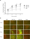
Figure 1
Figure 1
Viral growth and spread of HEP-Flury (HEP) and Vero-adapted viruses (HEP-10V and -30V) in Vero cells. Vero cells were inoculated with HEP, HEV-10V, or HEV-30V at a multiplicity of infection (M.O.I.) of 0.05. Supernatants were collected once daily through 4 days post infection (d.p.i.). (a) Viral titers were determined using MNA cells. Antigen-positive foci were counted under a fluorescence microscope and quantified as focus forming unit (FFU) per milliliter. Viral titers are plotted as the mean and standard deviation (S.D.) from three independent experiments. Significant differences are indicated (*: p < 0.05, **: p < 0.01, ***: p < 0.001) between HEP and HEP-10V or HEP-30V after application of two-way analysis of variance (ANOVA) followed by Tukey. HEP-10V vs HEP-30V had no significant difference at all time points (p > 0.05). (b) At the indicated time points, virus- and mock-infected cells were fixed with 80% cold acetone. Fixed cells were stained with the fluorescein isothiocyanate (FITC) anti-rabies monoclonal globulin (FUJIREBIO, Tokyo, Japan) and examined under a fluorescence microscope. The stained cells were observed using NIS-Elements D version 5.20.00 imaging software (Nikon, Tokyo, Japan). Cells are stained red with Evans Blue (FUJIFILM Wako Pure Chemical Corporation, Osaka, Japan). Scale bars, 100 μm; magnification, ×40.
Figure 2
Figure 2
Comparison of expression of viral proteins among HEP, HEP-10V and HEP-30V in Vero cells. Vero cells were inoculated with each virus at an M.O.I. of 5 and harvested every day. (a) Rabies virus (RABV) glycoprotein (G protein) and nucleoprotein (N proteins) were visualized using polyclonal rabbit anti-serum against each protein. Glyceraldehyde-3-phosphate dehydrogenase (GAPDH), a housekeeping protein, was detected by monoclonal mouse antibody for use as a loading control. (b) Virus-specific bands were quantified using ImageJ software (National Institutes of Health, Bethesda, MD, USA) and levels of RABV G and N proteins were normalized to those of GAPDH. The means and S.D. were calculated from two independent experiments. Significant differences are indicated (*: p < 0.05, **: p < 0.01, ***: p < 0.001) after application of two-way ANOVA followed by Tukey.
Figure 3
Figure 3
Comparison of virus-neutralizing antibody titers against HEP, HEP-10V and HEP-30V using vaccinated rabbit sera. Sera were collected from each of four rabbits (H1, H2, A1, A2). H1 and H2 were inoculated with the human-RABV vaccine (Rabipur, GSK Biologicals, Wavre, Belgium) and A1 and A2 were inoculated with the animal-RABV vaccine (KMB, KM Biologics, Kumamoto, Japan). Virus-neutralizing antibody (VNA) titers were determined by rapid fluorescent focus inhibition tests (RFFITs) and quantified using the World Health Organization (WHO) international units (IU/ml). Means and S.D. were calculated from four independent experiments. Significant difference is indicated (*: p < 0.05) in the neutralizing activities against HEP and HEP-30V in the H1 rabbit serum after application of two-way ANOVA followed by Tukey.
Figure 4
Figure 4
Comparison of pathogenicity among HEP, HEP-10V and HEP-30V in suckling and 6-week-old mice, and VNA titers against HEP-Flury using sera pooled from each infected-adult mice group. Suckling (n = 10/group) and 6-week-old mice (n = 8/group) were inoculated by intracerebral injection with 105 FFU per mouse of the respective virus, or with an equivalent volume of medium (mock). Plots show the survival rate of suckling mice using a Kaplan–Meier plot (a) and relative body weights (normalized to baseline) of 6-week-old mice (b). Body weight data are presented as mean and error bars represent the S.D. of each group. (c) The mouse sera were collected from surviving 6-week-old mice which were infected by intracerebral route with HEP, HEP-10V or HEP-30V. VNA titers were determined by RFFIT and VNA titers are quantified using IU/ml. Means and S.D. was calculated from three independent experiments. Error bars represent the S.D. of each group. Significant difference is indicated (*: p < 0.05) in the neutralizing activity between HEP vs HEP-10V on the inoculated HEP mice group after application of two-way ANOVA followed by Tukey.
Figure 5
Figure 5
Comparison of amino acid sequences between HEP, Vero-adapted viruses. (a) Schematic of the rabies genome and rectangles indicate the open reading frames for the indicated proteins. Change of amino acids and their positions are shown by arrowheads. (b) Sequence details of the adapted viruses that are described in the present study. Amino acids are indicated by the standard one-letter abbreviations. *: Amino acid sequence identical with HEP. **: Amino acid positions are numbered based on the mature G protein without signal peptide.
Figure 6
Figure 6
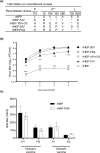
Efficiency of viral growth, entry, and expression of G protein of each recombinant viruses in Vero cells. (a) Information on recombinant viruses used in this figure. *: Amino acid sequence identical with rHEP. **: Amino acid positions are numbered based on the mature G protein without signal peptide. (b) Vero cells were inoculated with the indicated recombinant viruses (a) at a M.O.I. of 0.05 and supernatants were collected every day until 4 d.p.i. Viral titers were determined in MNA cells. (c) The total number of focuses was determined by counting the number of focuses stained with the FITC anti-rabies monoclonal globulin in Vero or MNA cells under a fluorescence microscope. The ratio of the number of focuses in Vero to that in MNA was compared. (d) The particle titer of each secreted alkaline phosphatase (SEAP)-expressing VSVp stock was determined in MNA cells. Vero cells then were inoculated with two-fold serial dilutions (starting from 150 particles) of VSVp pseudotyped with the HEP or HEP-10V G protein SEAP activity was assessed in culture supernatants and detected by optical density (OD). (e) Vero cells were inoculated with each recombinant virus at an M.O.I. of 5 and harvested at 2 d.p.i. The cells were stained with anti-rabies G protein monoclonal antibody (#7-1-9) and FITC-conjugated anti-mouse secondary. After that, cells were fixed with 4% paraformaldehyde, and finally analyzed by BD FACS Canto II flow cytometer (Becton Dickinson and Company; BD, Franklin Lakes, NJ, USA) and Kaluza analysis software Version 2.1 (Beckman Coulter Life Sciences, Indianapolis, IN, USA). Data are presented as the mean and S.D. from three (b, d, e) or four (c) independent experiments. Significant differences are indicated in the comparison between rHEP and each recombinant virus after application of two-way ANOVA followed by Tukey (b), or each virus after application of one-way ANOVA followed by Turkey (c, e) or two-way ANOVA followed by Sidaks (d) (*: p < 0.05, **: p < 0.01, ***: p < 0.001).
Figure 7
Figure 7
Comparison of viral growth in Vero cells among recombinant HEP strains, and entry of VSVp pseudotyped with the G protein of HEP, HEP-10V or HEP-30V. (a) Information on recombinant viruses used in this figure. *: Amino acid sequence identical with rHEP. **: Amino acid positions are numbered based on the mature G protein without signal peptide. Four recombinant viruses were infected to Vero cells at an M.O.I. of 0.05 (b), while nine recombinant viruses were inoculated at an M.O.I. of 0.01 (c). Viral titers were determined in MNA cells. The means and S.D. of the log10 of the viral titers are calculated from three independent experiments. Significant differences are indicated in the comparison between rHEP-10V and recombinant viruses after application of two-way ANOVA followed by Tukey (*: p < 0.05, **: p < 0.01, ***: p < 0.001). (d) Vero and MNA cells then were inoculated with 150 particles of VSVp pseudotyped with the G protein of HEP, HEP-10V, or HEP-30V. Following growth, SEAP activity was assessed in culture supernatants and detected by OD value, and the ratio of values in Vero and MNA cells was calculated. Means and S.D. were calculated from four independent experiments. Significant differences are indicated (*: p < 0.05, **: p < 0.01) after application of one-way ANOVA followed by Tukey.
Figure 8
Figure 8
Viral growth in Vero cells of recombinant HEP with 5 amino acid substitutions identified in Vero-adapted strains, named rHEP-PG4. (a) Information on recombinant viruses used in this figure. *: Amino acid sequence identical with rHEP. **: Amino acid positions are numbered based on the mature G protein without signal peptide. (b) Vero cells were inoculated with the recombinant strains at an M.O.I. of 0.05. Viral titers were determined in MNA cells. The means and S.D. of the log10 of the viral titer calculated from three independent experiments. Significant differences are indicated in the comparison between rHEP-30V and each strain after application of two-way ANOVA followed by Tukey (**: p < 0.01, ***: p < 0.001). rHEP-30Vvs rHEP-10V+G3, rHEP-PG4 had no significant difference at all time points after application of two-way ANOVA followed by Tukey (p > 0.05). (c) Virus-neutralizing antibody (VNA) titers were determined by RFFITs using rabbit serum and quantified using IU/mL. Means and S.D. were calculated from three independent experiments. No significant difference is indicated (p > 0.05) in the neutralizing activities against rHEP and rHEP-PG4 after application of two-way ANOVA followed by Tukey.

Similar articles

References

    1. World Health Organization . WHO Expert Consultation on Rabies: Third Report. World Health Organization; 2018.
    1. Hampson K, et al. Estimating the global burden of endemic canine rabies. PLoS Negl. Trop. Dis. 2015;9:e0003709. doi: 10.1371/journal.pntd.0003709. - DOI - PMC - PubMed
    1. Fooks AR, et al. Rabies. Nat. Rev. Dis. Primer. 2017;3:17091. doi: 10.1038/nrdp.2017.91. - DOI - PubMed
    1. World Health Organization, Food and Agriculture Organization of the United Nations, & World Organisation for Animal Health. In Zero by 30: The Global Strategic Plan to End Human Deaths from Dog-Mediated Rabies by 2030 (World Health Organization, 2018).
    1. Banyard AC, McElhinney LM, Johnson N, Fooks AR. Introduction History of rabies control by vaccination. Rev. Sci. Tech. 2018;37:305–322. doi: 10.20506/rst.37.2.2804. - DOI - PubMed

Substances