. 2024 May 31;7(1):672.
doi: 10.1038/s42003-024-06377-1.

Cryo-EM structure of cadmium-bound human ABCB6

Affiliations

Cryo-EM structure of cadmium-bound human ABCB6

Seung Hun Choi et al. Commun Biol. .

Abstract

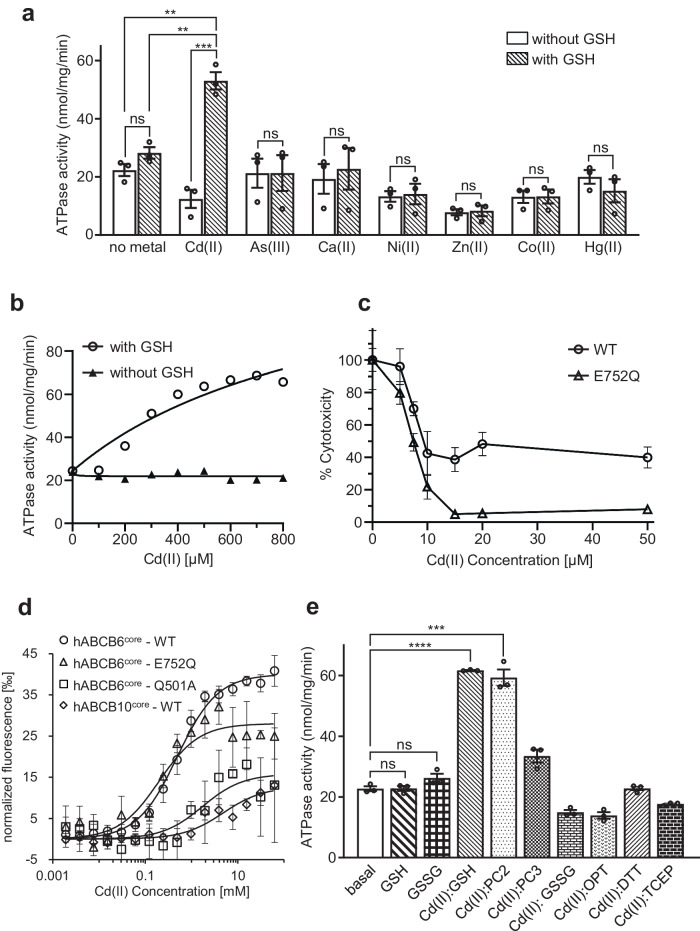
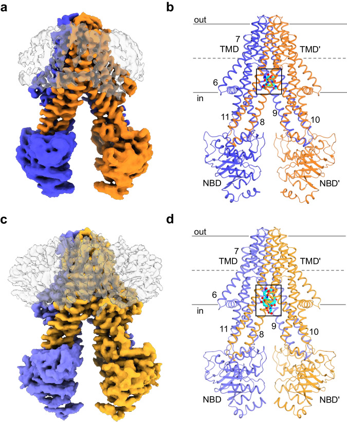
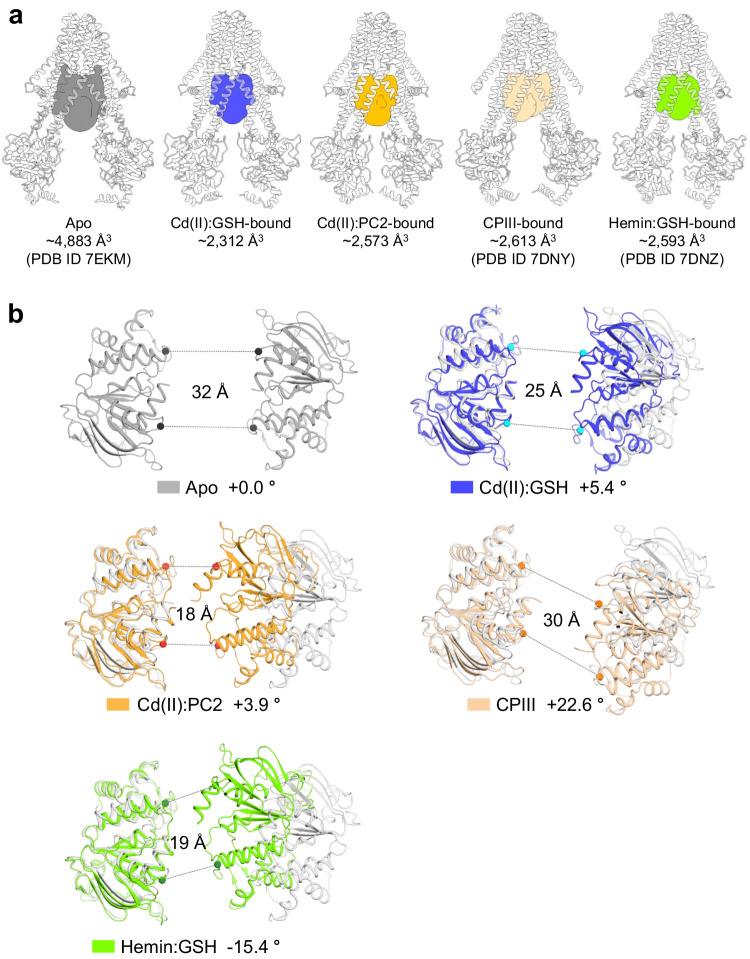
ATP-binding cassette transporter B6 (ABCB6), a protein essential for heme biosynthesis in mitochondria, also functions as a heavy metal efflux pump. Here, we present cryo-electron microscopy structures of human ABCB6 bound to a cadmium Cd(II) ion in the presence of antioxidant thiol peptides glutathione (GSH) and phytochelatin 2 (PC2) at resolutions of 3.2 and 3.1 Å, respectively. The overall folding of the two structures resembles the inward-facing apo state but with less separation between the two halves of the transporter. Two GSH molecules are symmetrically bound to the Cd(II) ion in a bent conformation, with the central cysteine protruding towards the metal. The N-terminal glutamate and C-terminal glycine of GSH do not directly interact with Cd(II) but contribute to neutralizing positive charges of the binding cavity by forming hydrogen bonds and van der Waals interactions with nearby residues. In the presence of PC2, Cd(II) binding to ABCB6 is similar to that observed with GSH, except that two cysteine residues of each PC2 molecule participate in Cd(II) coordination to form a tetrathiolate. Structural comparison of human ABCB6 and its homologous Atm-type transporters indicate that their distinct substrate specificity might be attributed to variations in the capping residues situated at the top of the substrate-binding cavity.


Conflict of interest statement

The authors declare no competing interests.

Figures

Fig. 1
Fig. 1. Functional characterization of ABCB6 in mediating cadmium detoxification.
a The ATPase activities of hABCB6core were measured against various metal species at a concentration of 800 μM, in the presence and absence of 1 mM GSH. Values represent the means ± standard error of the mean (SEM) of at least triplicate measurements using two different batches of nanodisc-purified protein. b ATPase activity of hABCB6core as a function of Cd(II) concentration with and without 1 mM GSH. c Cytotoxicity assay and cell viability profile. Sf9 cells expressing hABCB6core WT or E752Q mutant were cultured with various concentrations of CdCl2 for 2 h. The percentage cell viability was calculated as the ratio of the number of live cells in the presence of Cd(II) to that without Cd(II). d Normalized fluorescence changes (ΔFnorm = Fhot/Fcold) were measured to yield binding curves of Cd(II) to hABCB6core (or its variants) in the presence of GSH. hABCB10core served as a negative control. e Cd(II)-stimulated ATPase activity of hABCB6core in the presence of 1 mM GSH or its derivatives. The derivatives tested in the study included phytochelatin 2 (PC2), PC3, oxidized glutathione (GSSG), ophthalmic acid (OPT), 1,4-dithiothreitol (DTT), and Tris(2-carboxyethyl)phosphine (TCEP). GSH (or GSSG) alone-stimulated ATPase activities of hABCB6core were used as negative controls. The symbols **, ***, ****, and ns denote significant differences at p < 0.01, p < 0.001, p < 0.0001, and not statistically significant, respectively, with p-values calculated using a two-sided unpaired t-test and adjusted by the Welch’s correction method.
Fig. 2
Fig. 2. Overall levels of hABCB6core in complex with Cd(II) and GSH (or PC2).
a Cryo-EM map of Cd(II):GSH-bound hABCB6core. The two monomers are colored blue and orange, respectively. The nanodisc is depicted in a gray surface representation. b Molecular structure of Cd(II):GSH-bound hABCB6core. Cd(II) ion and GSH are shown in green and cyan sphere representation, respectively. The area enlarged in Fig. 4a is boxed. Single primes are used for the residues (or helices) of one monomer to differentiate them from those of the other monomer. c Cryo-EM map of Cd(II):PC2-bound hABCB6core. The two monomers are colored light blue and light orange, respectively. d Molecular structure of Cd(II):PC2-bound hABCB6core. The area enlarged in Fig. 4c is boxed.
Fig. 3
Fig. 3. Comparison of the substrate-binding cavities and NBDs of hABCB6core in the presence and absence of bound substrate.
a Overall structures and cavities of hABCB6core in the apo state (PDB ID 7EKM), Cd(II):GSH-bound state, Cd(II):PC2-bound state, CPIII-bound state (PDB ID 7DNY), and hemin:GSH-bound state (PDB ID 7DNZ). The substrate-binding cavities are colored gray, blue, orange, wheat, and green, respectively. b Cartoon representations of the NBDs in apo afnd substrate-bound states. The Cα distances between the conserved G626 of the Walker A motif and S728 of the signature motif are indicated. The tilt angle of one NBD in the substrate-bound state with respect to the other NBD in the apo state is indicated. Positive tilt angles represent a clockwise rotation, while negative tilt angles indicate a counterclockwise rotation.
Fig. 4
Fig. 4. Close-up view of the substrate-binding site.
a Zoom-in view of the Cd(II):GSH-binding site. Bound Cd(II) ion and GSHs are represented by green sphere and cyan stick models, respectively. Cryo-EM maps (gray mesh) of Cd(II) and GSHs are contoured at the 9 and 4 σ level, respectively. The W546 residue is omitted for simplicity (see also Supplementary Fig. 13). The view is rotated by 90 degrees along the vertical axis from Fig. 2b. b Interactions between hABCB6core and the Cd(II):GSH complex analyzed by LigPlot+ software. Residues involved in nonpolar and van der Waals interactions within 4 Å are depicted as red semicircles. c Zoom-in view of bound Cd(II) ion and PC2s. d Interactions between hABCB6core and the Cd(II):PC2 complex analyzed by LigPlot+ software. e ATPase activities of mutants affecting GSH binding. Activity was measured in the presence and absence of 800 μM Cd(II) and 1 mM GSH. Data points represent mean ± standard error of the mean (SEM) of at least three measurements using two different batches of purified protein. The symbol *** and ns denote significant differences at p < 0.001 and not statistically significant, respectively, with p-values calculated using a two-sided unpaired t-test and Welch’s correction. f Sequence alignment of TM helices 9 to 11 of ABCB6 and its orthologs involved in Cd(II):GSH-binding. Highly conserved residues among 15 different species are highlighted in red. Conservation of the capping residues between species is highlighted in orange (see also Fig. 5). Hs, Homo sapiens; Bt, Bos taurus; Ua, Ursus americanus; Mm, Mus musculus; Ma, Mesocricetus auratus; Rn, Rattus norvegicus; Xt, Xenopus tropicalis; Dr, Danio rerio; Dm, Drosophila melanogaster; Ce, Caenorhabditis elegans; Sp, Schizosaccharomyces pombe; Sc, Saccharomyces cerevisiae; At, Arabidopsis thaliana; Ct, Thermochaetoides thermophila; Na, Novosphingobium aromaticivorans.
Fig. 5
Fig. 5. Structural comparison of hABCB6core and Atm transporters.
a Close-up view of the substrate-binding sites of hABCB6core and AtAtm3 transporters, viewed from within the plane of the membrane. The hABCB6core and AtAtm3 structures are colored blue and light orange, respectively. Bound substrates are depicted as empty black (for the Cd(II):GSH complex in hABCB6core) and orange (for GSSG in AtAtm3) sticks, respectively. The residue number in parentheses indicates the equivalent residues in AtAtm3. bd Close-up view of the GSH- (b), GSSG- (c), and mercury Hg(II):GSH- (d) binding sites of NaAtm1 superimposed on the hABCB6core structure. The NaAtm3 structures and the bound substrates are colored light pink and pink, respectively.
Fig. 6
Fig. 6. Structural comparison of the substrate-binding sites of hABCB6core in apo, Cd(II):GSH-bound, and ADP·VO4-bound conformations.
Close-up views of the substrate-binding sites of hABCB6core in apo (PDB ID 7EKM), Cd(II):GSH-bound, and ADP·VO4-bound (PDB ID 8KYC) conformations. Key residues interacting with Cd(II):GSH are shown in stick representation. Cd(II):GSH superimposed on the outward-facing hABCB6core is depicted as empty black sticks.
Fig. 7
Fig. 7. Schematic diagram of the conformational changes of ABCB6 depending on the bound substrate type.
(i) The need for GSH as a cofactor, (ii) the degree of separation between the two halves of the transporter, and (iii) the tilt angles of the NBDs differ depending on the substrate type bound to ABCB6. The two subunits are colored blue and orange, respectively. The TMD0 domain is omitted for simplicity.

Similar articles

References

    1. Thomas C, et al. Structural and functional diversity calls for a new classification of ABC transporters. FEBS Lett. 2020;594:3767–3775. doi: 10.1002/1873-3468.13935. - DOI - PMC - PubMed
    1. Kiss K, et al. Role of the N-terminal transmembrane domain in the endo-lysosomal targeting and function of the human ABCB6 protein. Biochem. J. 2015;467:127–139. doi: 10.1042/BJ20141085. - DOI - PMC - PubMed
    1. Fukuda Y, et al. Conserved intramolecular disulfide bond is critical to trafficking and fate of ATP-binding cassette (ABC) transporters ABCB6 and sulfonylurea receptor 1 (SUR1)/ABCC8. J. Biol. Chem. 2011;286:8481–8492. doi: 10.1074/jbc.M110.174516. - DOI - PMC - PubMed
    1. Krishnamurthy PC, et al. Identification of a mammalian mitochondrial porphyrin transporter. Nature. 2006;443:586–589. doi: 10.1038/nature05125. - DOI - PubMed
    1. Wang L, et al. ABCB6 mutations cause ocular coloboma. Am. J. Hum. Genet. 2012;90:40–48. doi: 10.1016/j.ajhg.2011.11.026. - DOI - PMC - PubMed

Publication types