

NLRC5 promotes endometrial carcinoma progression by regulating NF-κB pathway-mediated mismatch repair gene deficiency
- PMID: 38822039
- PMCID: PMC11143240
- DOI: 10.1038/s41598-024-63457-2
NLRC5 promotes endometrial carcinoma progression by regulating NF-κB pathway-mediated mismatch repair gene deficiency
Abstract
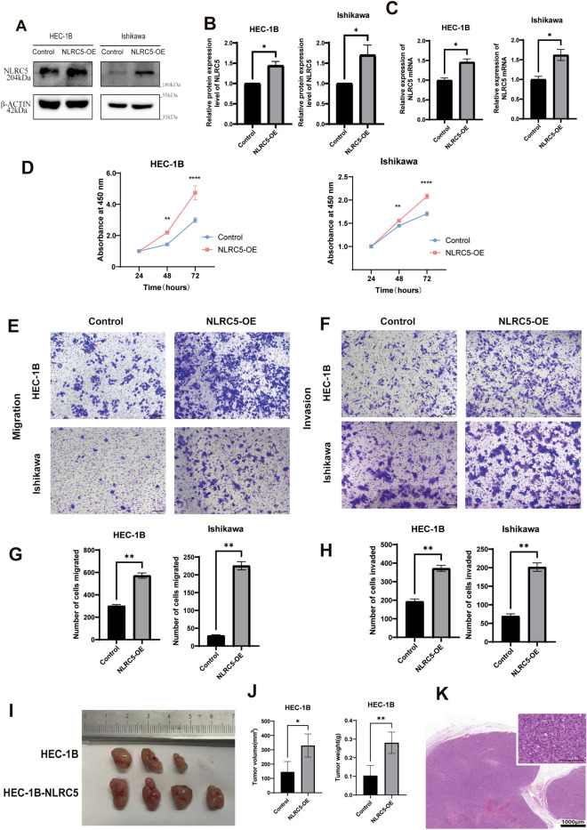
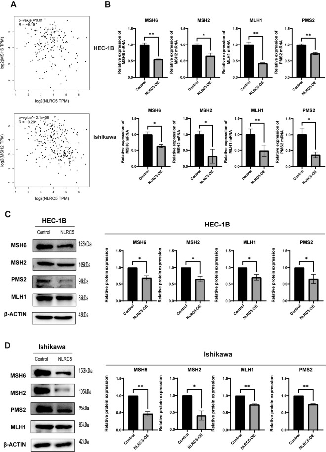
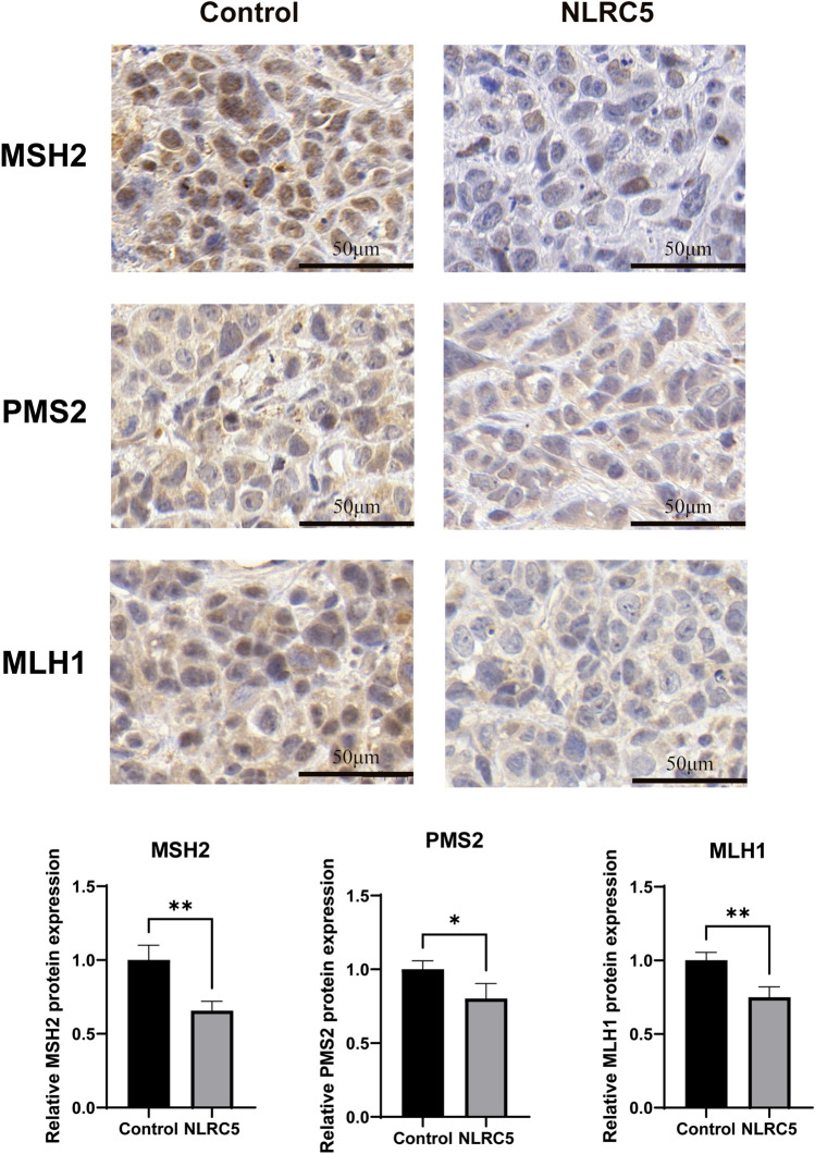
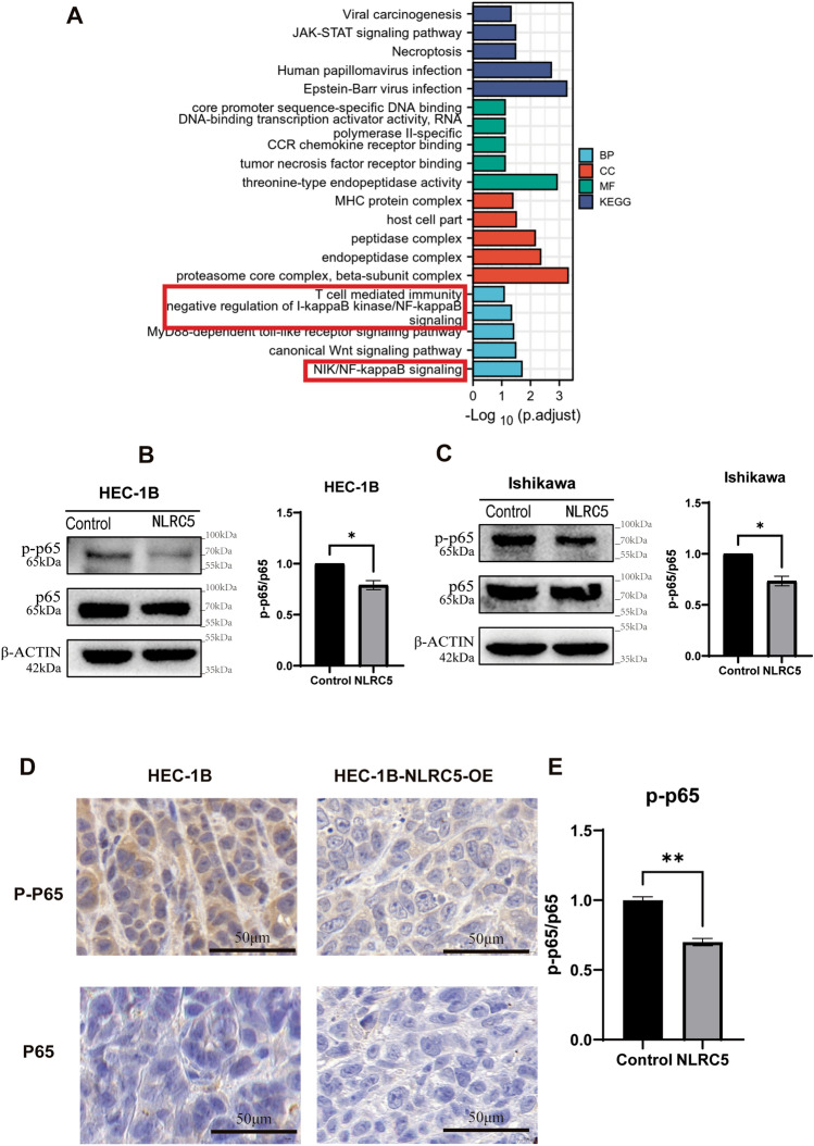
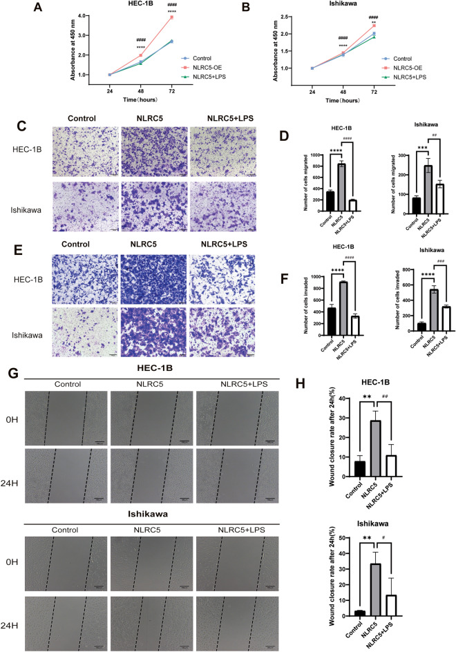
The innate immune molecule NLR family CARD domain-containing 5 (NLRC5) plays a significant role in endometrial carcinoma (EC) immunosurveillance. However, NLRC5 also plays a protumor role in EC cells. Mismatch repair gene deficiency (dMMR) can enable tumors to grow faster and also can exhibit high sensitivity to immune checkpoint inhibitors. In this study, we attempted to determine whether NLRC5-mediated protumor role in EC is via the regulation of dMMR. Our findings revealed that NLRC5 promoted the proliferation, migration, and invasion abilities of EC cells and induced the dMMR status of EC in vivo and in vitro. Furthermore, the mechanism underlying NLRC5 regulated dMMR was also verified. We first found NLRC5 could suppress nuclear factor-kappaB (NF-κB) pathway in EC cells. Then we validated that the positive effect of NLRC5 in dMMR was restricted when NF-κB was activated by lipopolysaccharides in NLRC5-overexpression EC cell lines. In conclusion, our present study confirmed the novel NLRC5/NF-κB/MMR regulatory mechanism of the protumor effect of NLRC5 on EC cells, thereby suggesting that the NLRC5-mediated protumor in EC was depend on the function of MMR.
Keywords: Endometrial carcinoma; Mismatch repair gene deficiency; NLR family CARD domain-containing 5; Nuclear factor-kappaB.
© 2024. The Author(s).
Conflict of interest statement
The authors declare no competing interests.
Figures

Similar articles
-
NLRC5 promotes cell migration and invasion by activating the PI3K/AKT signaling pathway in endometrial cancer.J Int Med Res. 2020 May;48(5):300060520925352. doi: 10.1177/0300060520925352. J Int Med Res. 2020. PMID: 32431202 Free PMC article.
-
PD-L1 Expression in Mismatch Repair-deficient Endometrial Carcinomas, Including Lynch Syndrome-associated and MLH1 Promoter Hypermethylated Tumors.Am J Surg Pathol. 2017 Mar;41(3):326-333. doi: 10.1097/PAS.0000000000000783. Am J Surg Pathol. 2017. PMID: 27984238
-
Molecular Modifiers of Hormone Receptor Action: Decreased Androgen Receptor Expression in Mismatch Repair Deficient Endometrial Endometrioid Adenocarcinoma.Int J Gynecol Pathol. 2019 Jan;38(1):44-51. doi: 10.1097/PGP.0000000000000465. Int J Gynecol Pathol. 2019. PMID: 29210800 Free PMC article.
-
NLRC5: A paradigm for NLRs in immunological and inflammatory reaction.Cancer Lett. 2019 Jun 1;451:92-99. doi: 10.1016/j.canlet.2019.03.005. Epub 2019 Mar 10. Cancer Lett. 2019. PMID: 30867141 Review.
-
Unraveling the Heterogeneity of Deficiency of Mismatch Repair Proteins in Endometrial Cancer: Predictive Biomarkers and Assessment Challenges.Cancers (Basel). 2024 Oct 11;16(20):3452. doi: 10.3390/cancers16203452. Cancers (Basel). 2024. PMID: 39456546 Free PMC article. Review.
Cited by
-
New insights into the structure domain and function of NLR family CARD domain containing 5.Cell Commun Signal. 2025 Jan 23;23(1):42. doi: 10.1186/s12964-024-02012-y. Cell Commun Signal. 2025. PMID: 39849460 Free PMC article. Review.
-
Reassessing the Role of Tissue Factor Pathway Inhibitor 2 in Neoplastic and Non-Neoplastic Lesions.Cancers (Basel). 2025 Apr 25;17(9):1447. doi: 10.3390/cancers17091447. Cancers (Basel). 2025. PMID: 40361374 Free PMC article. Review.
References
-
- Talhouk A, McConechy MK, Leung S, Yang W, Lum A, Senz J, Boyd N, Pike J, Anglesio M, Kwon JS, Karnezis AN, Huntsman DG, Gilks CB, McAlpine JN. Confirmation of ProMisE: A simple, genomics-based clinical classifier for endometrial cancer. Cancer. 2017;123:802–813. doi: 10.1002/cncr.30496. - DOI - PubMed
-
- N. Cancer Genome Atlas Research. Kandoth C, Schultz N, Cherniack AD, Akbani R, Liu Y, Shen H, Robertson AG, Pashtan I, Shen R, Benz CC, Yau C, Laird PW, Ding L, Zhang W, Mills GB, Kucherlapati R, Mardis ER, Levine DA. Integrated genomic characterization of endometrial carcinoma. Nature. 2013;497:67–73. doi: 10.1038/nature12113. - DOI - PMC - PubMed
MeSH terms
Substances
Supplementary concepts
Grants and funding
LinkOut - more resources
Full Text Sources
