. 2024 May 31;14(1):12564.
doi: 10.1038/s41598-024-63014-x.

Leveraging the fundamentals of heat transfer and fluid mechanics in microscale geometries for automated next-generation sequencing library preparation

Affiliations

Leveraging the fundamentals of heat transfer and fluid mechanics in microscale geometries for automated next-generation sequencing library preparation

Olivia Ott et al. Sci Rep. .

Abstract

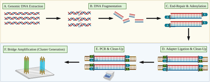
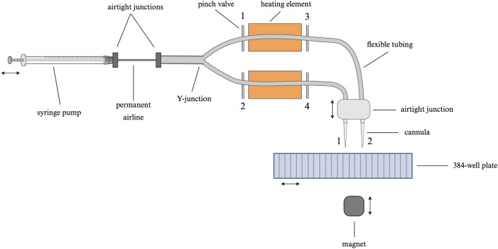
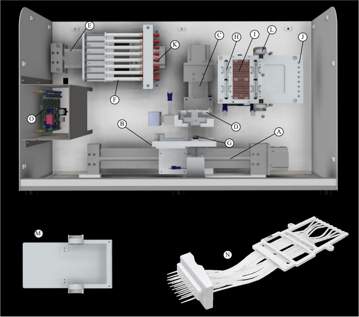
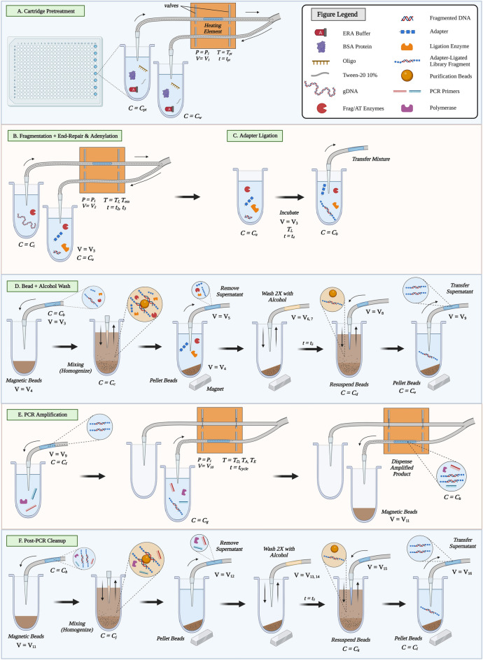
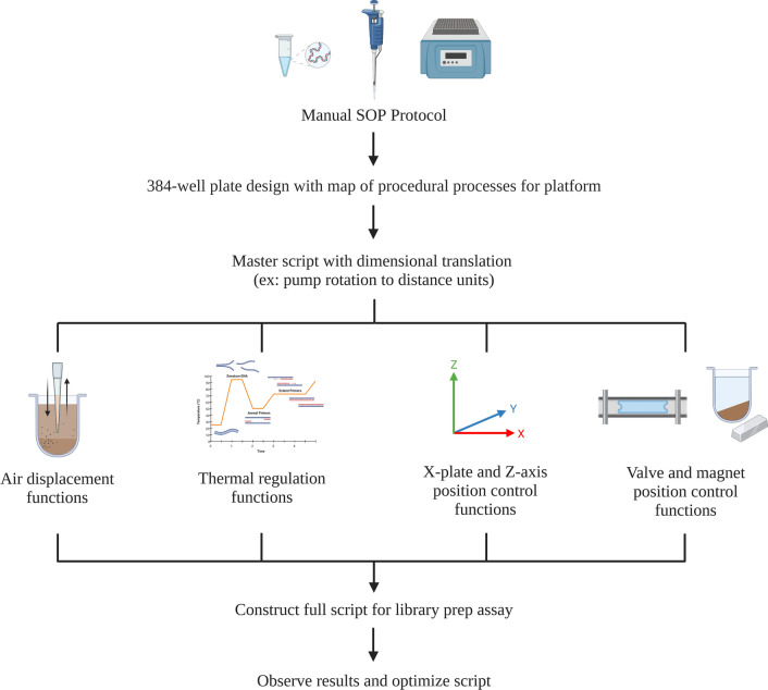
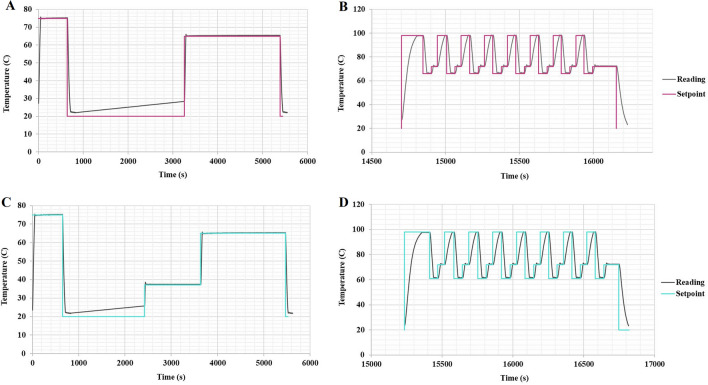
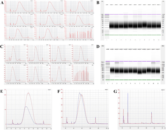
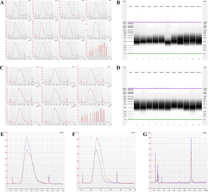
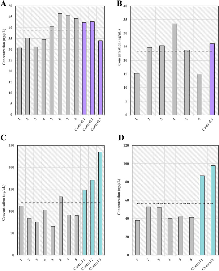
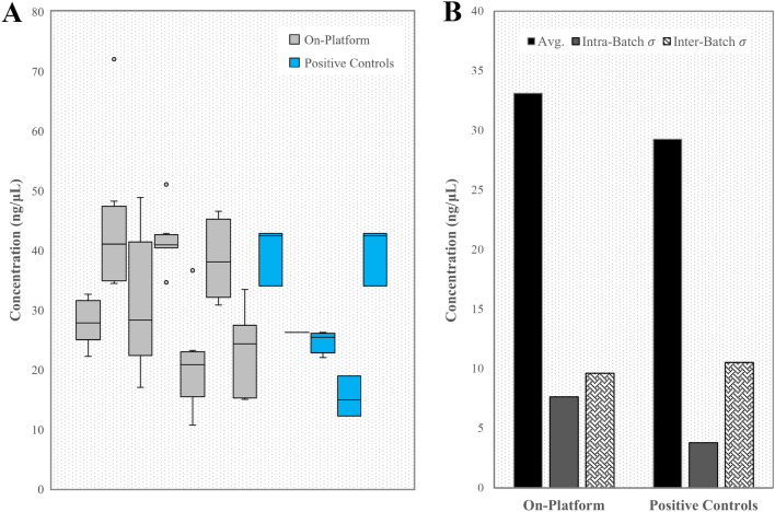
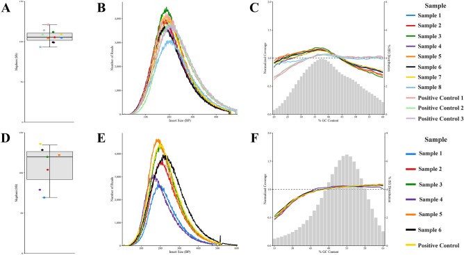
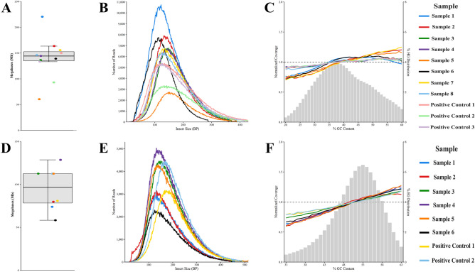
Next-generation sequencing (NGS) is emerging as a powerful tool for molecular diagnostics but remains limited by cumbersome and inefficient sample preparation. We present an innovative automated NGS library preparation system with a simplified mechanical design that exploits both macro- and microfluidic properties for optimizing heat transfer, reaction kinetics, mass transfer, fluid mechanics, adsorption-desorption rates, and molecular thermodynamics. Our approach introduces a unique two-cannula cylindrical capillary system connected to a programmable syringe pump and a Peltier heating element able to execute all steps with high efficiency. Automatic reagent movement, mixing, and magnetic bead-based washing with capillary-based thermal cycling (capillary-PCR) are completely integrated into a single platform. The manual 3-h library preparation process is reduced to less than 15 min of hands-on time via optimally pre-plated reagent plates, followed by less than 6 h of instrument run time during which no user interaction is required. We applied this method to two library preparation assays with different DNA fragmentation requirements (mechanical vs. enzymatic fragmentation), sufficiently limiting consumable use to one cartridge and one 384 well-plate per run. Our platform successfully prepared eight libraries in parallel, generating sequencing data for both human and Escherichia coli DNA libraries with negligible coverage bias compared to positive controls. All sequencing data from our libraries attained Phred (Q) scores > 30, mapping to reference genomes at 99% confidence. The method achieved final library concentrations and size distributions comparable with the conventional manual approach, demonstrating compatibility with downstream sequencing and subsequent data analysis. Our engineering design offers repeatability and consistency in the quality of sequence-able libraries, asserting the importance of mechanical design considerations that employ and optimize fundamental fluid mechanics and heat transfer properties. Furthermore in this work, we provide unique insights into the mechanisms of sample loss within NGS library preparation assays compared with automated adaptations and pinpoint areas in which the principles of thermodynamics, fluid mechanics, and heat transfer can improve future mechanical design iterations.


Conflict of interest statement

The authors declare no conflicts of interest. AT is a paid scientific advisor/consultant for Revvity.

Figures

Figure 1
Figure 1
A typical NGS library preparation workflow. (A) Genomic DNA is extracted from blood or another sample matrix, (B) gDNA is mechanically, enzymatically, or chemically fragmented, (C) DNA fragments are blunted, adenylated, 5' phosphorylated, and (D) ligated to indexed adapters. Primer-dimers are removed in a SPRI bead purification step. (E) Adapter-ligated DNA fragments are PCR amplified and a second SPRI bead purification step removes fragments < 200 bp. (F) Final libraries undergo bridge amplification for cluster generation on the NGS flow cell.
Figure 2
Figure 2
Schematic of our platform hardware, simplified to illustrate components for preparation of one sample. The air pressure syringe pumps are placed on an x-axis motion stage, the cannulas are placed on a z-axis motion stage, the 384-well reagent plate is placed on an x-stage, and the magnet is put on a z-axis motion stage.
Figure 3
Figure 3
Schematic of our platform. (A) X-stage for reagent plate motion, (B) 384 deep-well plate holder, (C) Z-stage for cannula motion, (D) Cartridge cannula spring-loaded clamp, (E) X-axis for pump syringe motion, (F) Syringes, (G) Magnet in raised position for bead-based purification, (H) Pinch valve, (I) Copper heating plate, (J) O-rings for cartridge-airline interface, (K) Airline connection point for plastic tubing (not pictured) to extend to (J), (L) Anchoring point for (M) insulated PCR door, (N) Disposable cartridge with cannula tips forms an airtight seal with (J) and is secured to the device via (M,D). (O) Printed circuit board for platform.
Figure 4
Figure 4
Details of our optimized method. Both the mechanical and enzymatic fragmentation assays follow the protocol depicted below, differing in select incubation times and reaction volumes (See S. Table 1). (A) Surface passivation (Pretreatment) of the cartridge tubing. (B) Fragmentation, end repair, and adenylation reaction at the heating element, (C) adapter ligation reaction, (D) post-ligation SPRI bead purification, (E) Capillary PCR amplification, (F) post-PCR SPRI bead purification. Variables T = temperature, t = time, P = pressure, V = volume, and C = concentration. Note that fragmentation only occurs on-platform for the enzymatic fragmentation assay. Cannula tip positions relative to the well are not exact, as there are multiple z-axis positions per step.
Figure 5
Figure 5
Scripting workflow. Standard operating procedure (SOP) for script and assay development.
Figure 6
Figure 6
Comparison of thermocouple temperature readings and scripted setpoint temperatures vs. time for the duration of the mechanical fragmentation assay (A,B) and the enzymatic fragmentation assay (C,D). (A) Temperature vs. time for PT and ERA, and (B) temperature vs. time for PCR amplification. (C) Temperature vs. time for PT, fragmentation, and ERA, and (D) temperature vs. time for PCR amplification. Note that the temperature drift (A,C) when the setpoint is 20 °C is due to residual heat transferring to the block containing the thermocouple. Reagents are in the 384-well plate during this incubation time, so there is no negative effect on assay performance.
Figure 7
Figure 7
Mechanical fragmentation final library Quality Control. (A) Electropherograms of on-platform (Samples 1–6) and manual positive control (Samples 7–8) human DNA libraries. The bottom right panel shows a DNA ladder for size comparison. (B) Gel image demonstrating fragment size distributions of the human DNA libraries shown in (A). (C) Electropherograms of on-platform (Samples 1–6) and positive control (Sample 7) E. coli DNA libraries. The bottom right panel shows a DNA ladder for size comparison. (D) Gel image demonstrating fragment size distributions of the E. coli DNA libraries shown in C. (E) Human DNA positive control (red) and on-platform (blue) library overlay for yield and size distribution comparison. (F) E. coli DNA positive control (red) and on-platform (blue) library electropherogram overlay for yield and size distribution comparison. (G) Manual (red) and on-platform (blue) negative control electropherogram overlay. All electropherograms consist of lower (first) and upper marker (last) peaks. Adapter-dimer peaks present at ~ 130 bp (or ~ 55 s). Other tiny peaks are either pertaining to primers or noise.
Figure 8
Figure 8
Enzymatic fragmentation final library Quality Control. (A) Electropherograms of on-platform (Samples 1–8) and positive control (Samples 9–11) human DNA libraries. The bottom right panel shows a DNA ladder for size comparison. (B) Gel image demonstrating fragment size distributions of the human DNA libraries shown in (A). (C) Electropherograms of on-platform (Samples 1–6) and positive control (Sample 7–8) E. coli DNA libraries. The bottom right panel shows a DNA ladder for size comparison. (D) Gel image demonstrating fragment size distributions of the E. coli DNA libraries shown in (C). (E) Human DNA positive control (red) and on-platform (blue) library overlay for yield and size distribution comparison. (F) E. coli DNA positive control (red) and on-platform (blue) library electropherogram overlay for yield and size distribution comparison. (G) Manual (red) and on-platform (blue) negative control electropherogram overlay.
Figure 9
Figure 9
Positive control and on-platform final library concentrations in ng/µL for (A) 20 ng input human DNA libraries produced by the mechanical fragmentation assay, (B) 20 ng input E. coli DNA libraries produced by the mechanical fragmentation assay, (C) 50 ng input human DNA libraries produced by the enzymatic fragmentation assay, and (D) 50 ng input E. coli DNA libraries produced by the enzymatic fragmentation assay. Dashed lines represent the average library concentration across samples and positive controls for each group. Note that all Qubit concentration measurements have a standard error of 12%.
Figure 10
Figure 10
Final library concentrations across multiple sample preparation sessions for mechanical fragmentation assay with assorted DNA input. (A) Automated plates compared to several batches of manually prepared positive controls. Each box represents a separate plate (for on-platform samples) or separate batch of manual samples (for positive controls). (B) Average library concentration across 54 on-platform samples and 14 manual positive controls. Sample-to-sample yield standard deviation, as well as standard deviation between average batch performances were determined to examine consistency between samples and between platform runs.
Figure 11
Figure 11
Sequencing data analysis for the mechanical fragmentation assay. Data yield per sample in Mb for (A) human DNA input and (D) E. coli DNA input. Number of reads vs. insert size for (B) human DNA input and (E) E. coli DNA input. Sequencing bias analysis in terms of normalized coverage vs. % GC content for (C) human DNA input and (F) E. coli DNA input.
Figure 12
Figure 12
Sequencing data analysis for the enzymatic fragmentation assay. Data yield per sample in Mb for (A) human DNA input and (D) E. coli DNA input. Number of reads vs. insert size for (B) human DNA input and (E) E. coli DNA input. Sequencing bias analysis in terms of normalized coverage vs. % GC content for (C) human DNA input and (F) E. coli DNA input.

Similar articles

References

    1. Christofyllakis, K. et al. Cost-effectiveness of precision cancer medicine-current challenges in the use of next generation sequencing for comprehensive tumour genomic profiling and the role of clinical utility frameworks (Review). Mol. Clin. Oncol.16(1), 21 (2022). - PMC - PubMed
    1. Onoyama, T., Ishikawa, S. & Isomoto, H. Gastric cancer and genomics: Review of literature. J. Gastroenterol.57(8), 505–516 (2022). - PMC - PubMed
    1. Schweiger, M. R., Kerick, M., Timmermann, B. & Isau, M. The power of NGS technologies to delineate the genome organization in cancer: From mutations to structural variations and epigenetic alterations. Cancer Metastasis Rev.30(2), 199–210 (2011). - PubMed
    1. Servetto, A. et al. A review of the use of next generation sequencing methodologies to identify biomarkers of resistance to CDK4/6 inhibitors in ER+/HER2- breast cancer. Crit. Rev. Oncol. Hematol.157, 103191 (2021). - PubMed
    1. Cinque, L. et al. Novel pathogenic variants of the AIRE gene in two autoimmune polyendocrine syndrome type i cases with atypical presentation: Role of the ngs in diagnostic pathway and review of the literature. Biomedicines8(12), 631 (2020). - PMC - PubMed

LinkOut - more resources