. 2024 Dec;96(7):1707-1717.
doi: 10.1038/s41390-024-03224-1. Epub 2024 May 31.

Novel biomarkers of preterm brain injury from blood transcriptome in sheep model of intrauterine asphyxia

Affiliations

Novel biomarkers of preterm brain injury from blood transcriptome in sheep model of intrauterine asphyxia

C Joakim Ek et al. Pediatr Res. 2024 Dec.

Abstract

Background: Infants born preterm have a higher incidence of neurological deficits. A key step in finding effective treatments is to identify biomarkers that reliably predict outcome.

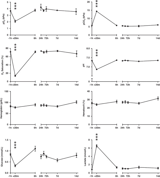
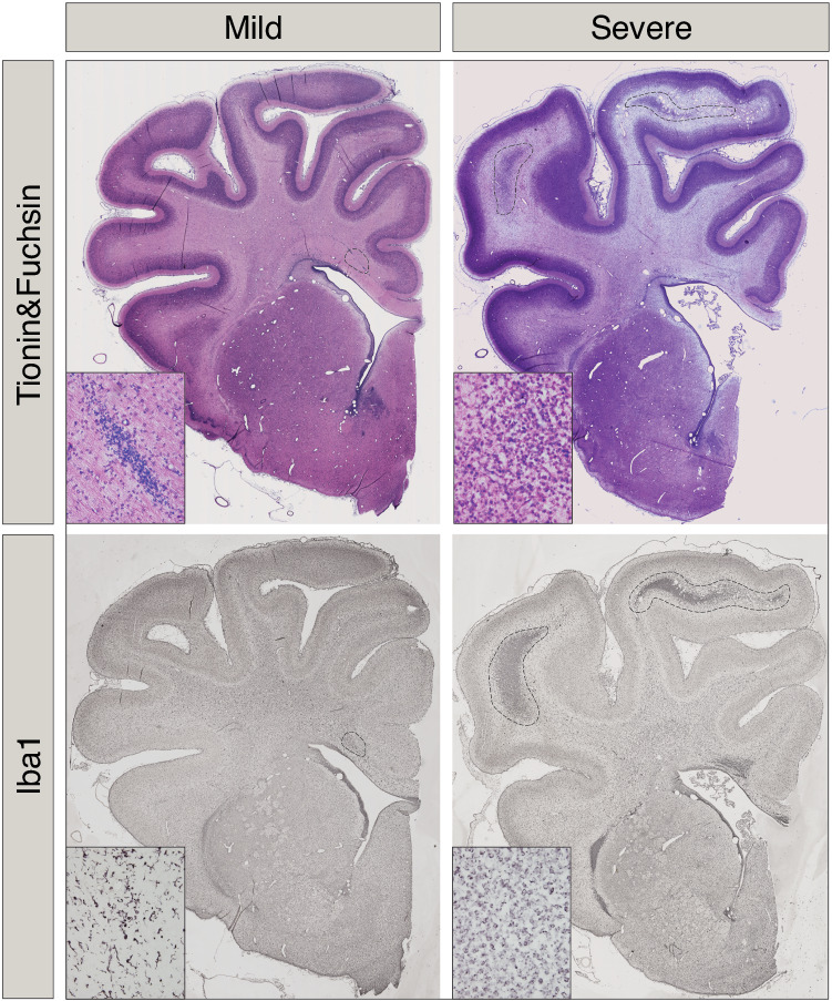
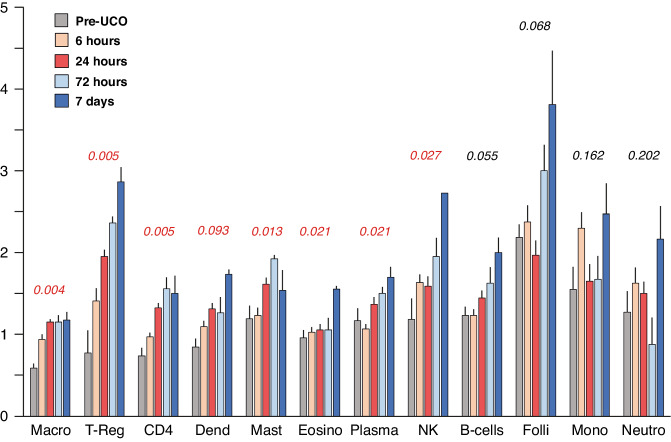
Methods: Following umbilical cord occlusion (UCO) in pregnant sheep, whole fetal blood RNA was sequenced pre- and post-UCO, brain injury outcome was determined by battery of neuropathology scoring and the transcriptome signature correlated to the degree of brain injury. Additionally, we developed a novel analytical procedure to deduce cell blood composition over time.

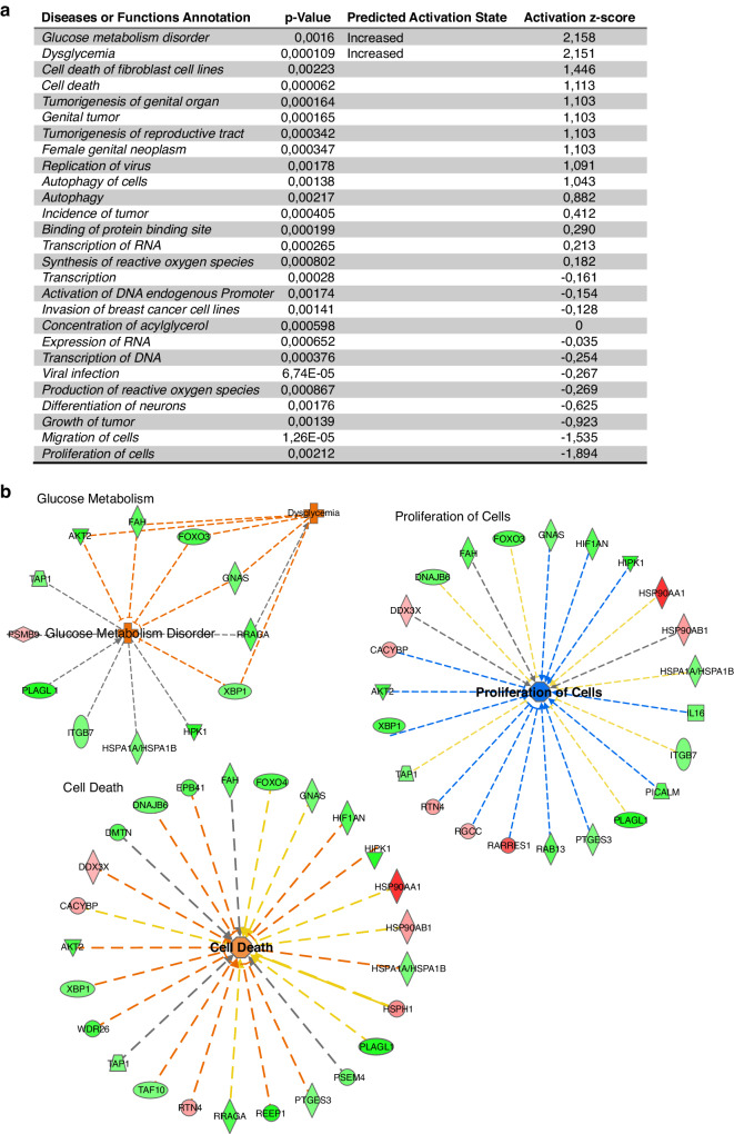
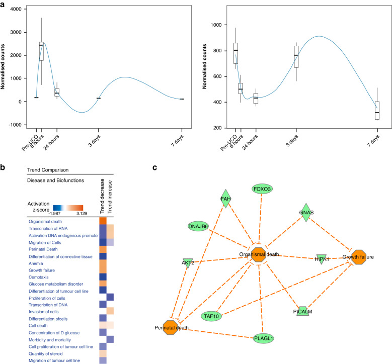
Results: Sixty-one genes were identified with significant altered expression after UCO. In pre-UCO blood, the level of three mRNAs (Trex2, Znf280b, novel miRNA) and in post-UCO, four mRNAs (Fam184a, Angptl2, novel lincRNA and an unknown protein-coding gene) were associated to brain injury (FDR < 0.01). Several of these mRNAs are related to inflammation and angiogenesis. Pathway analysis highlighted genes playing a role in perinatal death and growth failure. Results also indicate that several leukocyte populations undergo significant changes after UCO.

Conclusion: We have used a whole transcriptomic approach to uncover novel biomarkers in fetal blood that correlate to neuropathology in the preterm sheep brain. The current data forms a basis for future studies to investigate mechanisms of these mRNAs in the injury progression.

Impact: Trend analysis of genes following asphyxia reveal a group of genes associated with perinatal death and growth failure. Several pre-asphyxia transcripts were associated to brain injury severity suggesting genomic susceptibility to injury. Several post-asphyxia transcripts were correlated to brain injury severity, thus, serve as potential novel biomarkers of injury outcome. Successfully adaptation of cell profiling algorithms suggests significant changes in blood cell composition following asphyxia.


Conflict of interest statement

Competing interests: The authors declare no competing interests.

Figures

Fig. 1
Fig. 1. Blood analysis before and after umbilical cord occlusion (UCO).
Blood gases, hemoglobin, hematocrit, glucose and lactate levels in pre- (−1 h), during (+20 min) and post-UCO blood samples (+6 h, +24 h, +72 h, +7 days and +14 days). n = 7–10 per time-point. Data are mean ± SD, repeated measures ANOVA (compared to pre-UCO levels), ***p < 0.001.
Fig. 2
Fig. 2. Pathway analysis of gene changes after UCO.
a Downstream effect analysis using IPA of post-UCO regulated genes listing all biofunctions with p < 0.05. Predicted increase of glucose metabolism disorder and dysglycemia (z-score = +2.16 and +2.15, respectively). Cell death shows second highest z-score (+1.11) while proliferation of cells shows highest negative z-score (−1.86). b Networks of molecules related to dysglycemia, cell death and proliferation of cells.
Fig. 3
Fig. 3. Trend analysis of pre/post-UCO genes.
a Polynomial regression models for pre-/post-UCO regulated genes. b Trends comparison (using IPA) of down-stream effect analysis generated from genes up- or down-regulated at 6 h post UCO. c Molecular network for perinatal death, organismal death and growth failure.
Fig. 4
Fig. 4. Brain sections, at the level of the lateral ventricle, were stained with acid fuchsin/thionine or Iba-1 (microglia marker).
In the severe injury group note regions in the subcortical white matter with pyknotic cells and microgliosis (dashed areas), see also Table 1.
Fig. 5
Fig. 5. Genes associated with mild or severe injury.
a Pre-UCO gene levels in blood significantly associated with mild or severe brain injury. b Six hour post-UCO gene levels in blood associated to mild or severe brain injury. FDR < 0.01 was used to identify these genes.
Fig. 6
Fig. 6. Changes in blood cell composition inferred by RNAseq expression profile post-UCO using validated leukocyte markers (CIBERSORT).
Numbers indicate q-values.

References

    1. Braun, P. E., Sandillon, F., Edwards, A., Matthieu, J. M. & Privat, A. Immunocytochemical localization by electron microscopy of 2'3’-cyclic nucleotide 3’-phosphodiesterase in developing oligodendrocytes of normal and mutant brain. J. Neurosci.8, 3057–3066 (1988). - PMC - PubMed
    1. Morgan, A. S. et al. Birth outcomes between 22 and 26 weeks’ gestation in national population-based cohorts from Sweden, England and France. Acta Paediatr.111, 59–75 (2022). - PMC - PubMed
    1. Morsing, E. et al. Neurodevelopmental disorders and somatic diagnoses in a national cohort of children born before 24 weeks of gestation. Acta Paediatr.111, 1167–1175 (2022). - PMC - PubMed
    1. Serenius, F. et al. Neurodevelopmental outcomes among extremely preterm infants 6.5 years after active perinatal care in Sweden. JAMA Pediatr.170, 954–963 (2016). - PubMed
    1. Volpe, J. J. Brain injury in premature infants: a complex amalgam of destructive and developmental disturbances. Lancet Neurol.8, 110–124 (2009). - PMC - PubMed

LinkOut - more resources