. 2024 May 31;21(1):144.
doi: 10.1186/s12974-024-03126-3.

Therapeutic role of interferon-γ in experimental autoimmune encephalomyelitis is mediated through a tolerogenic subset of splenic CD11b+ myeloid cells

Affiliations

Therapeutic role of interferon-γ in experimental autoimmune encephalomyelitis is mediated through a tolerogenic subset of splenic CD11b+ myeloid cells

Gabriel Arellano et al. J Neuroinflammation. .

Abstract

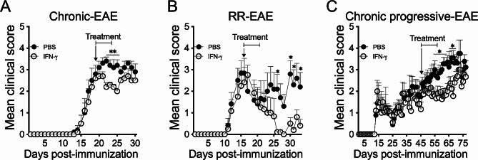
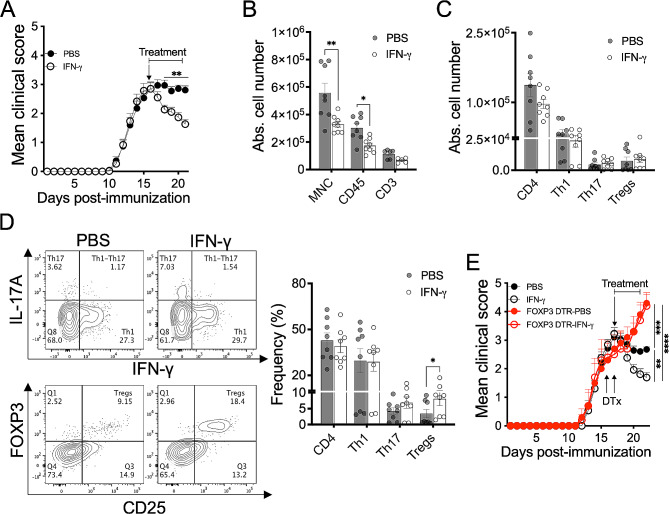
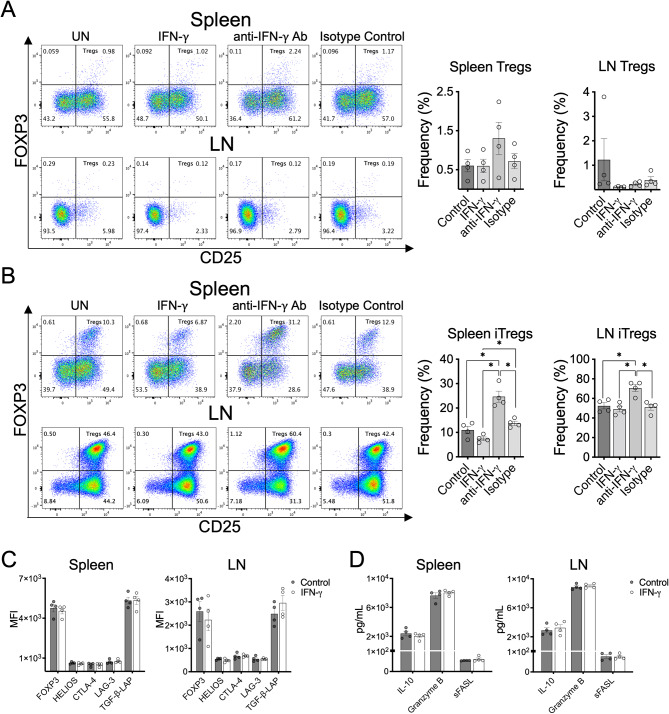
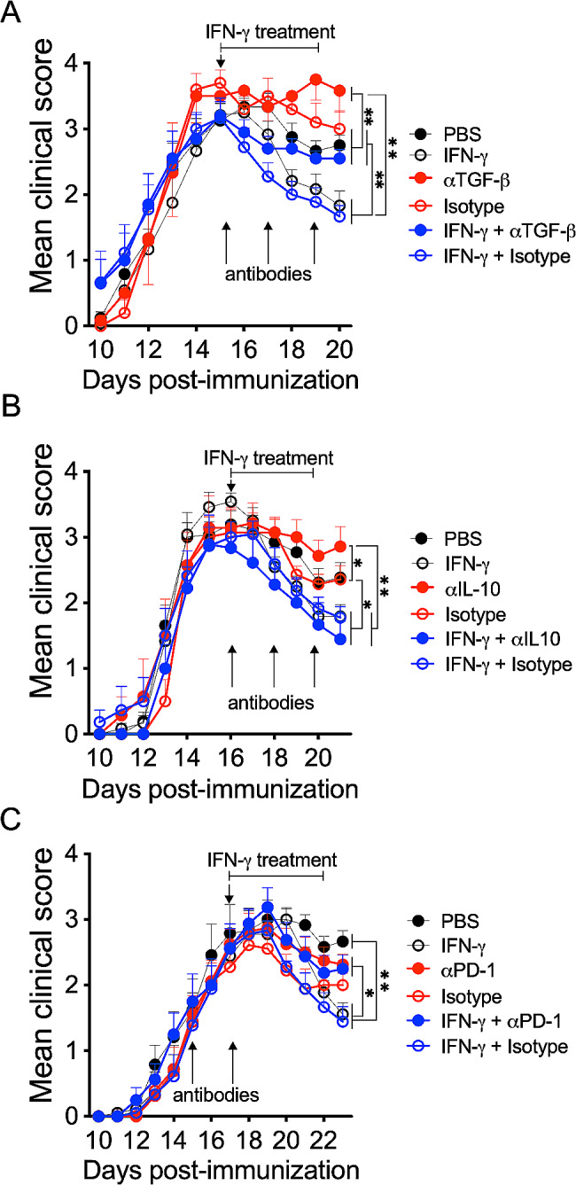
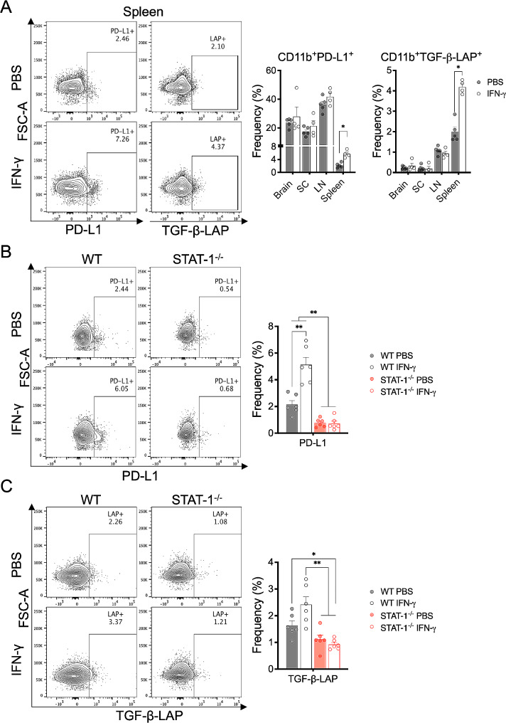
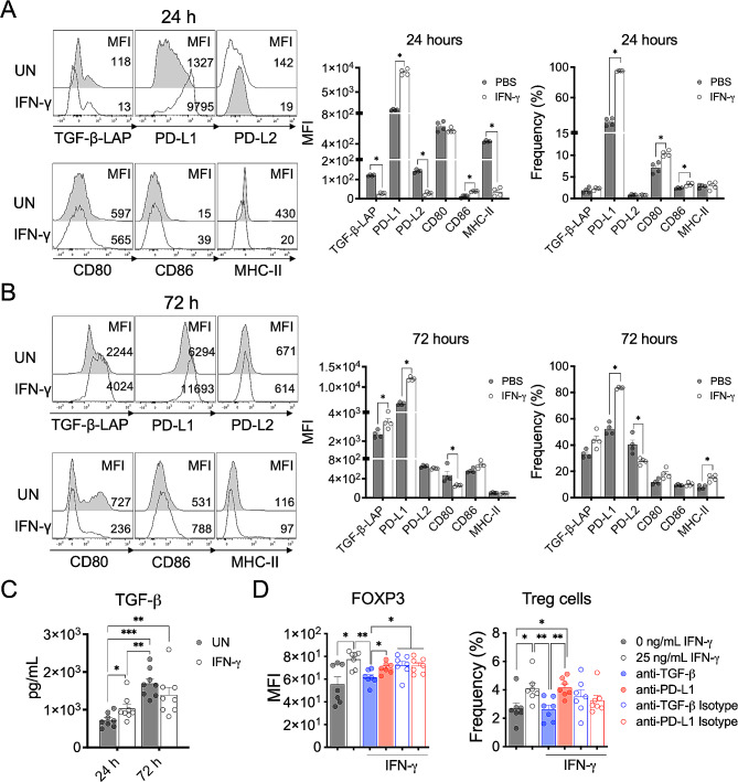
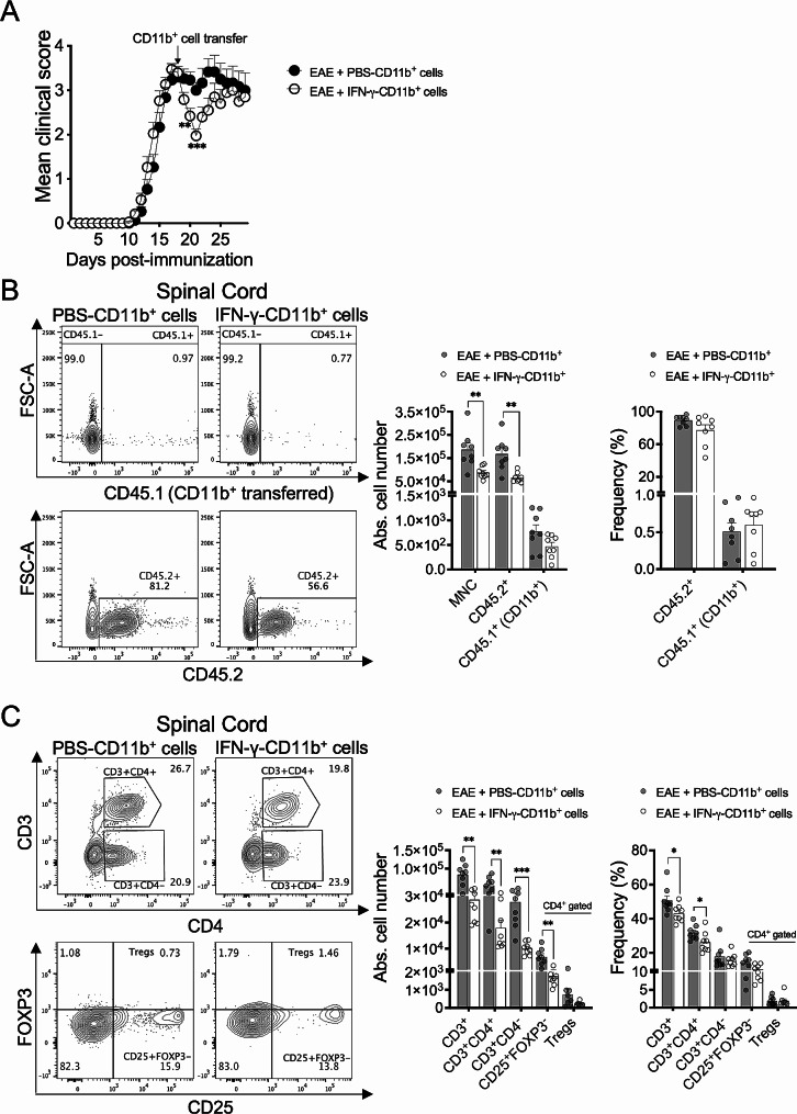
Cumulative evidence has established that Interferon (IFN)-γ has both pathogenic and protective roles in Multiple Sclerosis and the animal model, Experimental Autoimmune Encephalomyelitis (EAE). However, the underlying mechanisms to the beneficial effects of IFN-γ are not well understood. In this study, we found that IFN-γ exerts therapeutic effects on chronic, relapsing-remitting, and chronic progressive EAE models. The frequency of regulatory T (Treg) cells in spinal cords from chronic EAE mice treated with IFN-γ was significantly increased with no effect on Th1 and Th17 cells. Consistently, depletion of FOXP3-expressing cells blocked the protective effects of IFN-γ, indicating that the therapeutic effect of IFN-γ depends on the presence of Treg cells. However, IFN-γ did not trigger direct in vitro differentiation of Treg cells. In vivo administration of blocking antibodies against either interleukin (IL)-10, transforming growth factor (TGF)-β or program death (PD)-1, revealed that the protective effects of IFN-γ in EAE were also dependent on TGF-β and PD-1, but not on IL-10, suggesting that IFN-γ might have an indirect role on Treg cells acting through antigen-presenting cells. Indeed, IFN-γ treatment increased the frequency of a subset of splenic CD11b+ myeloid cells expressing TGF-β-Latency Associated Peptide (LAP) and program death ligand 1 (PD-L1) in a signal transducer and activator of transcription (STAT)-1-dependent manner. Furthermore, splenic CD11b+ cells from EAE mice preconditioned in vitro with IFN-γ and myelin oligodendrocyte glycoprotein (MOG) peptide exhibited a tolerogenic phenotype with the capability to induce conversion of naïve CD4+ T cells mediated by secretion of TGF-β. Remarkably, adoptive transfer of splenic CD11b+ cells from IFN-γ-treated EAE mice into untreated recipient mice ameliorated clinical symptoms of EAE and limited central nervous system infiltration of mononuclear cells and effector helper T cells. These results reveal a novel cellular and molecular mechanism whereby IFN-γ promotes beneficial effects in EAE by endowing splenic CD11b+ myeloid cells with tolerogenic and therapeutic activities.

Keywords: CD11b+ cells; Experimental autoimmune encephalomyelitis; Interferon-γ; Multiple sclerosis; Regulatory T cells; TGF-β.


Conflict of interest statement

SDM is a co-founder, paid consultant, scientific advisory board member, and grantee of COUR Pharmaceuticals. SDM is also a consultant for NextCure and Takeda Pharmaceuticals. The other authors have declared that no conflicts of interest exist.

Figures

Fig. 1
Fig. 1
Therapeutic administration of IFN-γ ameliorates clinical severity in chronic, relapsing-remitting, and chronic progressive-EAE. (A) Two groups of C57BL/6 mice were induced with chronic EAE (n = 9/group) and daily treated with 1 µg IFN-γ (empty black circles) or PBS (solid black circles) at peak of disease for 5 days. (B) SJL mice were induced with relapsing-remitting (RR)-EAE (n = 6/group) and daily treated with 1 µg IFN-γ (empty black circles) or PBS (solid black circles) at peak of the first flare for 5 days. (C) Chronic progressive-EAE was induced in NOD mice (n = 13/group) and treated with 1 µg IFN-γ (empty black circles) or PBS (solid black circles) during the chronic phase of the disease (starting at 45 days post-immunization) for 10 days. Clinical symptoms were monitored daily using the standard scoring scale. Results are shown as the mean ± SEM. Data were analyzed using two-way ANOVA followed by Bonferroni post-hoc test. *p < 0.05, **p < 0.01
Fig. 2
Fig. 2
Therapeutic effect of IFN-γ depends on the presence of FOXP3+ Treg cells in EAE. (A) Mice induced with chronic EAE were treated daily with 1 µg IFN-γ (empty black circles) or PBS (solid black circles) at the peak of disease for five days (n = 10/group, mean of three independent experiment). (B-D) At the end of treatment, spinal cords were isolated and the absolute number of (B) mononuclear cells (MNC), CD45+ cells, and CD3+ cells, and (C) the absolute number and (D) frequency of CD4+ T cells, Th1, Th17 and Treg cells were determined by flow cytometry. (E) Wild type (n = 13/group) and FOXP3-DTR mice (n = 5/group) were induced with EAE and treated with 1 µg IFN-γ (empty black and red circles) or PBS (solid black and red circles) at the peak of disease for five days. To deplete FOXP3+ Treg cells, FOXP3-DTR mice were i.p. treated with one dose 2.5 µg of diphtheria toxin (DTx) two days before starting IFN-γ treatment and with another similar dose two days later. Three independent experiments were pooled. Results are shown as the mean ± SEM. Data were analyzed using two-way ANOVA followed by Bonferroni post-hoc test (A), Mann–Whitney U test (B-D), or one-way ANOVA followed by Bonferroni post-hoc test (E). *p < 0.05, **p < 0.01, ***p < 0.001, ****p < 0.0001
Fig. 3
Fig. 3
IFN-γ does not exert a direct effect on Treg cell differentiation. (A) CD4+CD25 T cells isolated (purity > 90%) from spleen or draining lymph nodes (LN) of EAE mice at the peak of disease were stimulated for 3 days with 2 µg/mL plate-bound anti-CD3 and 1 µg/mL soluble anti-CD28 in the absence (control) or presence of either 25 ng/mL IFN-γ, 5 µg/mL anti-IFN-γ antibody or 5 µg/mL isotype control antibody. (B) Similar assay as described in (A) in the presence of 2 ng/mL TGF-β and 10 ng/mL IL-2 to promote differentiation of inducible Treg cells (iTregs). The frequency of Treg cells (CD3+CD4+CD25+FOXP3+) was determined by flow cytometry. (C) Purified fraction of CD4+CD25+ cells (containing > 82% of Treg cells) obtained during the purification of CD4+CD25 T cells were stimulated with 2 µg/mL plate-bound anti-CD3 and 1 µg/mL soluble anti-CD28 for 3 days in the absence (control, solid gray bars) or presence of 25 ng/mL IFN-γ (empty gray bars). The expression of regulatory markers FOXP3, HELIOS, CTLA-4, LAG-3 and TGF-β-LAP was determined as median fluorescence intensity (MFI) by flow cytometry. (D) Secretion of IL-10, Granzyme B and sFASL in cell culture supernatants was assessed by multiplex assay. Results are shown as the mean ± SEM (n = 4). Data were analyzed using Mann–Whitney U test. *p < 0.05
Fig. 4
Fig. 4
Therapeutic effect of IFN-γ is dependent on TGF-β and PD-1. EAE mice were treated at the peak of disease for 5 days with 1 µg/day IFN-γ alone (empty black circles) or along with (A) 100 µg of neutralizing antibody against TGF-β (solid blue circles, n = 6) or (B) IL-10 (solid blue circles, n = 7) administered at days 0, 2 and 4 after IFN-γ treatment, or with (C) 500 µg neutralizing anti-PD-1 antibody (solid blue circles, n = 8) administered two days before starting IFN-γ treatment and at the same time of IFN-γ treatment. Control EAE mice were treated with PBS (vehicle, solid black circles) or corresponding isotype antibody alone (empty red circles) or along with IFN-y (empty blue circles). Two independent experiments were pooled. Results are shown as the mean ± SEM. Data were analyzed using one-way ANOVA followed by the Bonferroni post-hoc test. *p < 0.05, **p < 0.01
Fig. 5
Fig. 5
IFN-γ/STAT-1 axis induces splenic CD11b+ myeloid cells expressing PD-L1 and TGF-β-LAP. (A) Frequency of PD-L1+ and TGF-β-LAP+ CD11b+ cells in brain, spinal cord (SC), draining lymph nodes (LN) and spleen from EAE mice treated at the peak of disease with IFN-γ (empty gray bars, n = 4) or PBS (solid gray bars, n = 5) for 5 days was determined by flow cytometry. (B) Frequency of CD11b+PD-L1+ cells and (C) CD11b+TGF-β-LAP+ cells were determined by flow cytometry in spleen from wild-type mice (solid and empty gray bars, n = 6) and STAT-1 deficient mice (solid and empty red bars, n = 6) induced with EAE and treated at the peak of disease with IFN-γ (empty bars) or PBS (vehicle, solid bars) for 5 days. Three independent experiments were pooled. Results are shown as the mean ± SEM. Data were analyzed using Mann–Whitney U or Kruskal-Wallis test. *p < 0.05, **p < 0.01
Fig. 6
Fig. 6
IFN-γ directly induces a tolerogenic phenotype and function in splenic CD11b+ cells from EAE mice. Splenic CD11b+ cells isolated from EAE mice were in vitro stimulated with 25 ng/mL IFN-γ (empty gray bars) or left untreated (UN, solid gray bars) for (A) 24 and (B) 72 h in the presence of 10 µg/mL MOG33 − 55 peptide. Frequency and median fluorescence intensity (MFI) of TGF-β-LAP, PD-L1, PD-L2, CD80, CD86 and MHC-II were analyzed by flow cytometry (n = 4). (C) Cell culture supernatants were collected at 24 and 72 h and concentration of TGF-β1 was measured by multiplex assay (n = 8). (D) Splenic CD11b+ cells from EAE mice were preconditioned with 10 µg/mL MOG33 − 55 peptide and 25 ng/mL IFN-γ for 24 h, washed, and co-cultured with naïve CD4+ T cells (ratio 1:1) from healthy mice in the presence of 1 µg/mL soluble anti-CD3 antibody and with either anti-TGF-β antibody (solid blue bar), anti-PD-L1 antibody (solid red bar), or corresponding isotype control antibodies (empty blue and red bars). After 72 h, the frequency of Treg cells (CD4+CD25+FOXP3+) was determined by flow cytometry. Results are shown as the mean ± SEM. Data were analyzed using Mann–Whitney U test. *p < 0.05, **p < 0.01
Fig. 7
Fig. 7
Adoptive transfer of splenic CD11b+ cells from IFN-γ-treated EAE mice into recipient EAE mice suppresses disease progression. (A) Splenic CD11b+ cells (106) from CD45.1 EAE mice treated for 5 days with IFN-γ (empty black circles) or PBS (solid black circles) were i.v. transferred into CD45.2 EAE mice at the peak of disease (n = 15). Clinical symptoms were monitored daily. Four independent experiments were pooled. (B-C) After three days of transfer, mononuclear cells from spinal cords of recipient EAE mice receiving CD45.1+CD11b+ cells from EAE mice treated with IFN-γ (empty gray bars, n = 8) or PBS (solid gray bars, n = 8) were isolated and absolute number and frequencies of (B) mononuclear cells (MNC), CD45.2+ cells, and CD45.1+CD11b+ cells, and (C) CD3+ T cells, CD3+CD4+ T cells, CD3+CD4 T cells, CD3+CD4+CD25+FOXP3 T cells, and Treg cells (CD3+CD4+CD25+FOXP3+), were determined by flow cytometry (n = 8, two different experiments were pooled). Results are shown as the mean ± SEM. Data were analyzed using two-way ANOVA followed by the Bonferroni post-hoc test (A) or Mann–Whitney U test (B-C). *p < 0.05, **p < 0.01, ***p < 0.001

Similar articles

Cited by

References

    1. Dendrou CA, Fugger L, Friese MA. Immunopathology of multiple sclerosis. Nat Rev Immunol. 2015;15:545–58. doi: 10.1038/nri3871. - DOI - PubMed
    1. Yadav SK, Mindur JE, Ito K, Dhib-Jalbut S. Advances in the immunopathogenesis of multiple sclerosis. Curr Opin Neurol. 2015;28:206–19. doi: 10.1097/WCO.0000000000000205. - DOI - PubMed
    1. Compston A, Coles A. Multiple sclerosis. Lancet. 2008;372:1502–17. doi: 10.1016/S0140-6736(08)61620-7. - DOI - PubMed
    1. Mahad DH, Trapp BD, Lassmann H. Pathological mechanisms in progressive multiple sclerosis. Lancet Neurol. 2015;14:183–93. doi: 10.1016/S1474-4422(14)70256-X. - DOI - PubMed
    1. Confavreux C, Vukusic S. The clinical course of multiple sclerosis. Handb Clin Neurol. 2014;122:343–69. doi: 10.1016/B978-0-444-52001-2.00014-5. - DOI - PubMed

MeSH terms

Substances