. 2024 May 31;57(1):36.
doi: 10.1186/s40659-024-00518-w.

The crucial role of HFM1 in regulating FUS ubiquitination and localization for oocyte meiosis prophase I progression in mice

Affiliations

The crucial role of HFM1 in regulating FUS ubiquitination and localization for oocyte meiosis prophase I progression in mice

Chenyi Zhong et al. Biol Res. .

Abstract

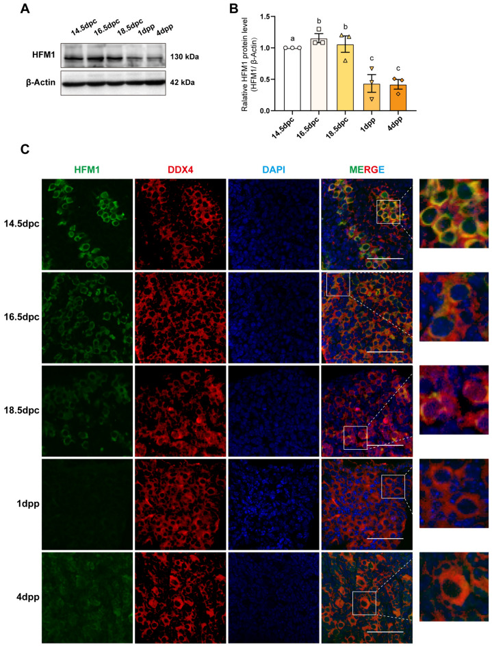
Background: Helicase for meiosis 1 (HFM1), a putative DNA helicase expressed in germ-line cells, has been reported to be closely associated with premature ovarian insufficiency (POI). However, the underlying molecular mechanism has not been clearly elucidated. The aim of this study was to investigate the function of HFM1 in the first meiotic prophase of mouse oocytes.

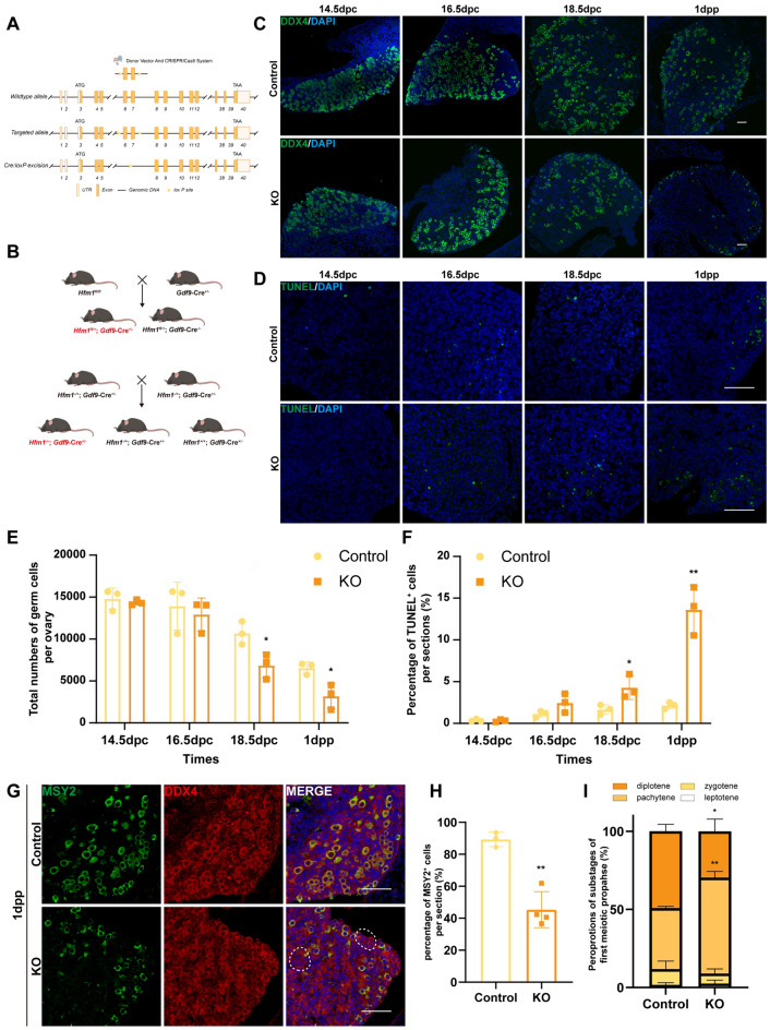
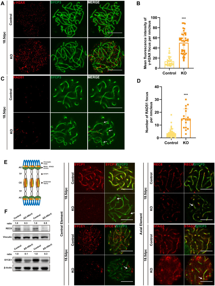
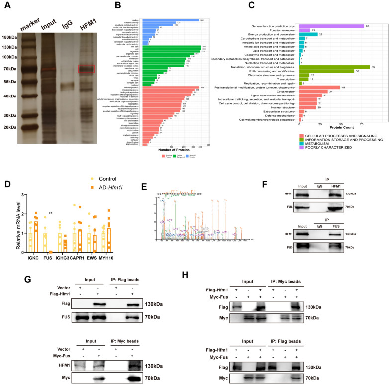
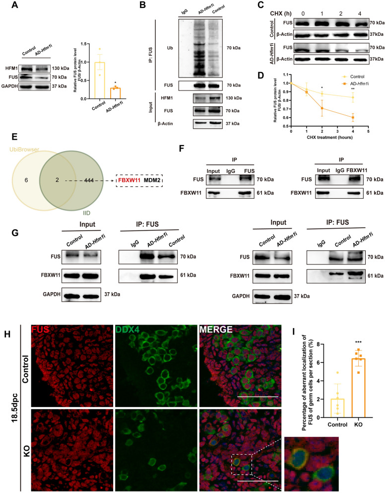
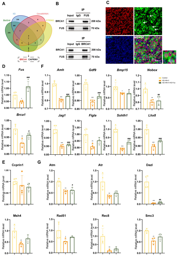
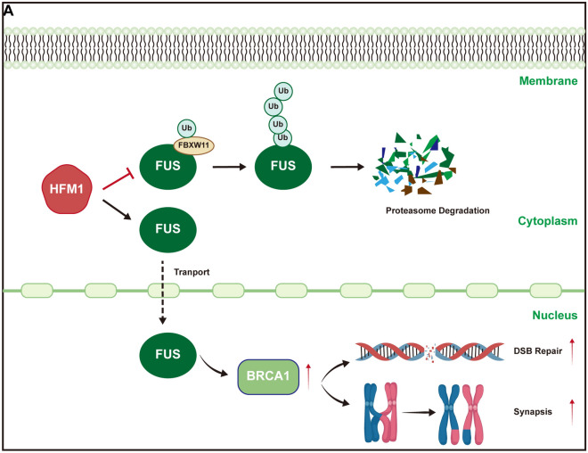
Results: The results suggested that the deficiency of HFM1 resulting in increased apoptosis and depletion of oocytes in mice, while the oocytes were arrested in the pachytene stage of the first meiotic prophase. In addition, impaired DNA double-strand break repair and disrupted synapsis were observed in the absence of HFM1. Further investigation revealed that knockout of HFM1 promoted ubiquitination and degradation of FUS protein mediated by FBXW11. Additionally, the depletion of HFM1 altered the intranuclear localization of FUS and regulated meiotic- and oocyte development-related genes in oocytes by modulating the expression of BRCA1.

Conclusions: These findings elaborated that the critical role of HFM1 in orchestrating the regulation of DNA double-strand break repair and synapsis to ensure meiosis procession and primordial follicle formation. This study provided insights into the pathogenesis of POI and highlighted the importance of HFM1 in maintaining proper meiotic function in mouse oocytes.

Keywords: FUS; HFM1; Meiosis prophase I; Oocyte; Premature ovarian failure/insufficiency.


Conflict of interest statement

The authors declare that they have no competing interests.

Figures

Fig. 1
Fig. 1
HFM1 predominantly expressed in the prenatal embryonic mouse ovary. A Immunoblotting staining showed that HFM1 was relatively highly expressed in the ovaries of embryonic mice at 14.5 days post coitum (dpc), 16.5 dpc, and 18.5 dpc, while the expression was significantly lower at 1 day post-partum (dpp) and 4 dpp after birth. β-actin was used as a loading control. B Quantification of HFM1 gray value. n = 3 biologically independent experiments. Data represented as mean ± standard error of the mean and the different letters (a-c) indicate the difference between the groups was statistically significant (two-sided ANOVA test), P (a, b) = ns, P (a, c) < 0.05, P (b, c) < 0.01. C HFM1 was mainly expressed in the cytoplasm of oocytes in mouse ovaries. Embryonic and neonatal mouse ovaries were stained for HFM1 (green) and germ cell-specific marker DDX4 (red). The nucleus was dyed with DAPI (blue). Scale bars: 50 μm
Fig. 2
Fig. 2
Deficiency of Hfm1 expression impeded oocyte meiotic process and cell survival in mouse ovaries. A Engineered a conditional floxed allele for Hfm1 and a Cre-mediated recombination to delete exons 6 and 7 of Hfm1 in mice. B Schematic of the specific mating and breeding method to obtain systemic Hfm1-KO mice. C Deletion of Hfm1 disrupted oocyte survival and early folliculogenesis in mice. Immunofluorescence staining showed ovaries of the Control and KO mice at the indicated developmental stages (14.5, 16.5, and 18.5 dpc and 1 dpp). Oocytes were stained with DDX4 (green). The nucleus was stained using DAPI (blue). Scale bars: 50 μm. D Apoptotic cells increased in KO ovaries compared with the Control ovaries with the development of oocytes. TUNEL signals (green) marked apoptotic cells, while the nucleus was stained using DAPI (blue). Scale bars: 50 μm. E, F Statistical analysis of total numbers of germ cells per ovary. (E) and the percentages of TUNEL+ cells per section (F) between Control and KO mice in the indicated developmental stages. *P < 0.05, **P < 0.01 (t test), n = 3. G First meiotic prophase in the Hfm1-KO mice at 1 dpp was arrested before the diplotene phase. The sections were stained with MSY2 (green) which specifically presented in oocytes of the diplotene and afterward stages and DDX4 (red). The nucleus was stained using DAPI (blue). The oocytes circled in the dashed line highlighted oocytes with no expression of MSY2. Scale bars: 50 μm. H Statistical analysis showed that the percentage of MSY2+ oocytes (number of cells both MSY2+ and DDX4+/number of cells DDX4+) per section decreased significantly following HFM1 deprivation. **P < 0.01 (t test), n = 3. I Chromatin spread of 18.5 dpc ovaries showed that the KO group had more oocytes in the pachytene stage and fewer in the diplotene stage than the Control group. *P < 0.05, **P < 0.01 (t test), n = 3
Fig. 3
Fig. 3
Depletion of Hfm1 expression damaged the repair of DNA double-strand breaks and synaptonemal complex formation. A, B Deletion of Hfm1 caused DSB repair deficiency in embryonic mouse ovaries. (A) Immunoblotting staining of the meiotic spread showed repaired or unrepaired DSBs in 18.5-dpc Control or KO ovaries. γ-H2AX (red) indicates unrepaired DSB sites. SYCP3 (green) demonstrates axial elements. Scale bars: 10 μm. (B) Statistical analysis showed that the mean fluorescence intensity of γ-H2AX on chromosomes per nucleus increased significantly following HFM1 deprivation. ***P < 0.001 (t test), WT: n = 31; KO: n = 41. C, D Deletion of Hfm1 resulted in the ectopic expression of RAD51. (C) Immunoblotting staining showed normal or ectopic RAD51 (DNA break repair protein) focus in 18.5-dpc Control or KO ovaries. Oocyte chromosomes were co-stained with RAD51 (red) and SYCP3 (green). Scale bars: 10 μm. (D) Statistical analysis showed that the number of RAD51 foci on chromosomes per slide increased significantly following HFM1 deprivation. ***P < 0.001, WT: n = 42; KO: n = 19. E Schematic demonstrating the synaptonemal complex during the first meiotic prophase (left). Deletion of Hfm1 impacted the formation of the synaptonemal complex. Immunoblotting staining of the meiotic spread showed abnormal expression of synaptonemal complex proteins SYCP1, SYCE1, REC8, and STAG3. Arrows demonstrated loose bivalent chromosome (right). Scale bars: 10 μm. F Immunoblotting showed significantly reduced expression in REC8 and SYCE1 of KO group (n = 3). β-Actin or Vinculin was used as a loading control
Fig. 4
Fig. 4
HFM1 and FUS interacted with each other. A Co-IP and silver staining showed the proteins bound to HFM1. B, C Functional analysis of enriched genes by Co-IP. Gene Ontology (GO) analysis (B) and eukaryotic orthologous groups (KOGs) analysis (C) of enriched genes described the Molecular Function, Cellular Component and Biological Process of the enriched genes. D Real-time PCR showed that the interference of HFM1 expression decreased the expression of FUS, but not IGKC, IGHG3, CAPR1, EWS, and MYH10. **P < 0.01, n = 6. E Mass spectrometry (MS) analysis to determine which proteins bound to HFM1 showed FUS to be an interacting protein. F Endogenous protein interactions of HFM1 and FUS were assessed in embryonic mouse ovary lysates by immunoprecipitation with anti-HFM1 or anti-FUS and evaluated using immunoblotting with indicated antibodies. IgG was used as a negative control. G, H Exogenous protein interactions demonstrated in HEK 293 T cells. HEK 293 T cells transfected with indicated plasmid (Flag-tagged HFM1 plasmid, Myc-tagged FUS plasmid and plasmid vector, separately) and treated with proteasome inhibitor MG132 (10 μM). Cells were lysed with NP-40 and analyzed using Co-IP with Flag or Myc beads followed by immunoblotting
Fig. 5
Fig. 5
HFM1 acted on the ubiquitination and degradation of FUS and the cytoplasmic–cytosolic localization of FUS. A HFM1 silencing led to a decrease in FUS protein expression (*P < 0.05, n = 3). GAPDH was used as a loading control. B Lysates from embryonic ovaries transfected with control or AD-Hfm1i, followed by treatment with MG132 before harvest, were immunoprecipitated and examined with indicated antibodies. Quantification of relative ubiquitin-FUS levels showed that the ubiquitination level of FUS increased after HFM1 knockdown. C, D Embryonic ovaries were cultured with control or AD-Hfm1i, treated with cycloheximide (CHX, 100 μg/mL), and collected for immunoblotting analysis at the indicated time points (C). Quantification of FUS band intensity was presented (D). *P < 0.05, **P < 0.01 (t test), n = 3. E Venn diagram showed that FBXW11 and MDM2 may be the potential ligating (E3) enzymes during the ubiquitination of FUS using UbiBrowser database (http://ubibrowser.bio-it.cn/ubibrowser/) and the Integrated Interactions Database (http://iid.ophid.utoronto.ca/search_by_proteins/). F Endogenous protein interactions of FBXW11 and FUS were assessed in embryonic mouse ovary lysates by immunoprecipitation with anti-FBXW11 or anti-FUS and evaluated using immunoblotting with indicated antibodies. IgG was used as a negative control. G Lysates from ovaries transfected with control or AD-Hfm1i were collected for Co-IP. The binding of FUS and FBXW11 increased with the knockdown of Hfm1. H, I HFM1 maintained the nuclear localization of FUS in embryonic mouse oocytes. 18.5-dpc embryonic mouse ovaries were stained for FUS (red) and germ cell-specific marker DDX4 (green), while the nucleus was stained using DAPI (blue). The area boxed by dotted line was the oocytes with aberrant localization of FUS (H). Scale bars: 50 μm. Statistical analysis showed the number of germ cells with aberrant-localized FUS per section (I). ***P < 0.001 (t test), n = 6
Fig. 6
Fig. 6
BRCA1 might be a possible target of the HFM1-FUS axis. A Venn diagram showed that BRCA1 and CAPRIN1 were the binding proteins of FUS using BioGrid, IID, GeneMANIA, and STRING. B Lysates from embryonic ovaries were immunoprecipitated and examined with indicated antibodies to assess the endogenous protein interactions of BRCA1 and FUS. C BRCA1 co-localized with FUS in some oocytes of 18.5-dpc embryonic mouse ovaries. BRCA1 was stained with green, and FUS was stained with red. The nucleus was stained using DAPI (blue). The oocytes in which BRCA1 co-localized with FUS are highlighted in dashed boxes or pointed by arrows. Scale bars: 10 μm. D Real-time PCR showed that the interference of HFM1 expression decreased the expression of Brca1, and the overexpression of FUS restored the mRNA level of Brca1. E Real-time PCR showed interference of HFM1 expression would not change the expression of ccprin1. F, G HFM1 regulated the expression of oocyte development-related factors (F) and meiosis-related factors (G) by affecting FUS-BRCA1. Real-time PCR showed that the interference of HFM1 expression decreased the expression of those genes, while the overexpression of FUS restored the expression of these genes appropriately. Compared with the control group, *P < 0.05, **P < 0.01, ***P < 0.001; compared with the AD-Hfm1-RNAi group, #P < 0.05, ##P < 0.01,.###P < 0.001, n = 4
Fig. 7
Fig. 7
Schematic model of HFM1 functioning in oocytes during the first meiotic prophase. HFM1 regulated E3 ubiquitin ligase FBXW11-mediated ubiquitination degradation of FUS and maintained the nuclear localization of FUS in oocytes, thereby regulating the expression of BRCA1 and affecting oocyte DSB repair and synapsis

Similar articles

Cited by

References

    1. Webber L, Davies M, Anderson R, Bartlett J, Braat D, et al. ESHRE Guideline: management of women with premature ovarian insufficiency. Hum Reprod. 2016;31(5):926–937. doi: 10.1093/humrep/dew027. - DOI - PubMed
    1. Rahman R, Panay N. Diagnosis and management of premature ovarian insufficiency. Best Pract Res Clin Endocrinol Metab. 2021;35(6):101600. doi: 10.1016/j.beem.2021.101600. - DOI - PubMed
    1. Coulam CB, Adamson SC, Annegers JF. Incidence of premature ovarian failure. Obstet Gynecol. 1986;67(4):604–606. - PubMed
    1. Chon SJ, Umair Z, Yoon M-S. Premature ovarian insufficiency: past, present, and future. Front Cell Dev Biol. 2021;9:672890. doi: 10.3389/fcell.2021.672890. - DOI - PMC - PubMed
    1. Molina JR, Barton DL, Loprinzi CL. Chemotherapy-induced ovarian failure: manifestations and management. Drug Saf. 2005;28(5):401–416. doi: 10.2165/00002018-200528050-00004. - DOI - PubMed

LinkOut - more resources