. 2024 May 31;16(1):75.
doi: 10.1186/s13073-024-01347-y.

Knockout mice with pituitary malformations help identify human cases of hypopituitarism

Affiliations

Knockout mice with pituitary malformations help identify human cases of hypopituitarism

Julian Martinez-Mayer et al. Genome Med. .

Abstract

Background: Congenital hypopituitarism (CH) and its associated syndromes, septo-optic dysplasia (SOD) and holoprosencephaly (HPE), are midline defects that cause significant morbidity for affected people. Variants in 67 genes are associated with CH, but a vast majority of CH cases lack a genetic diagnosis. Whole exome and whole genome sequencing of CH patients identifies sequence variants in genes known to cause CH, and in new candidate genes, but many of these are variants of uncertain significance (VUS).

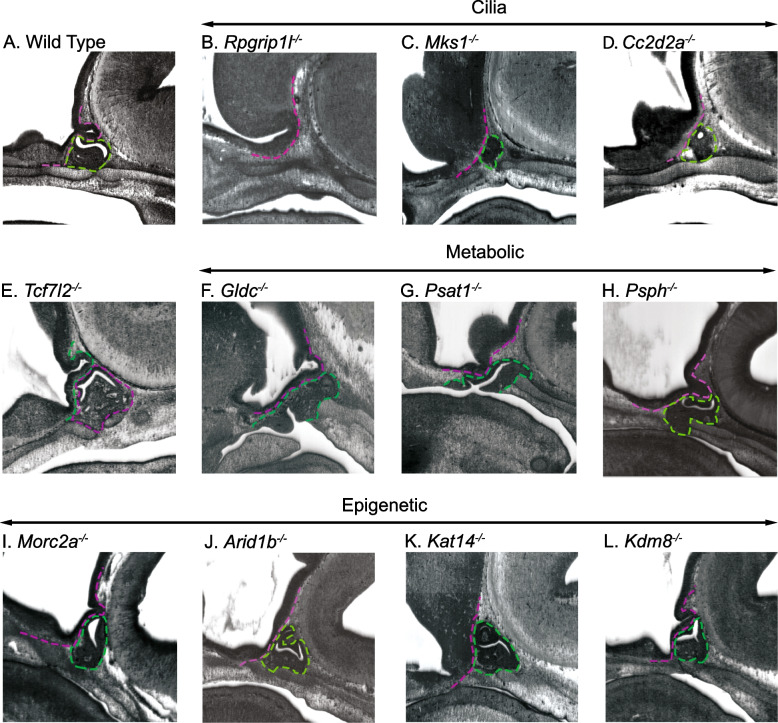
Methods: The International Mouse Phenotyping Consortium (IMPC) is an effort to establish gene function by knocking-out all genes in the mouse genome and generating corresponding phenotype data. We used mouse embryonic imaging data generated by the Deciphering Mechanisms of Developmental Disorders (DMDD) project to screen 209 embryonic lethal and sub-viable knockout mouse lines for pituitary malformations.

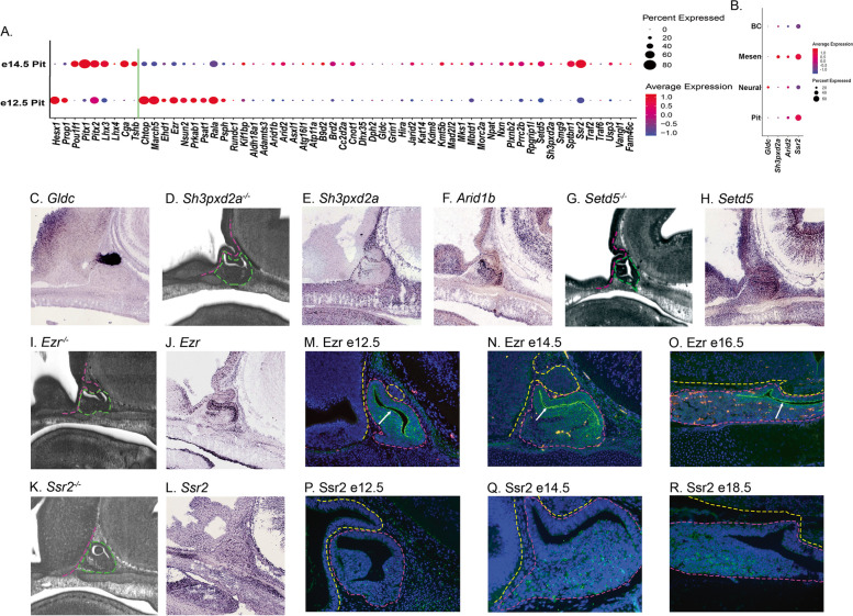
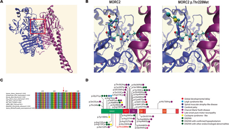
Results: Of the 209 knockout mouse lines, we identified 51 that have embryonic pituitary malformations. These genes not only represent new candidates for CH, but also reveal new molecular pathways not previously associated with pituitary organogenesis. We used this list of candidate genes to mine whole exome sequencing data of a cohort of patients with CH, and we identified variants in two unrelated cases for two genes, MORC2 and SETD5, with CH and other syndromic features.

Conclusions: The screening and analysis of IMPC phenotyping data provide proof-of-principle that recessive lethal mouse mutants generated by the knockout mouse project are an excellent source of candidate genes for congenital hypopituitarism in children.

Keywords: MORC2; SETD5; Cleft palate; Development; Growth hormone; Holoprosencephaly; Hypothalamus; International Mouse Phenotyping Consortium (IMPC); Pituitary; Septo-optic dysplasia.


Conflict of interest statement

The authors declare that they have no competing interests.

Figures

Fig. 1
Fig. 1
Pituitary gland malformations observed in candidate CH genes. Sagittal images captured from the DMDD HREM stackviewer for a e14.5 A wild type embryo and homozygous null embryos for Rpgrip1l, CMks1, Cc2d2a, Tcf7l2, Gldc, Psat1, Psph, Morc2a, Arid1b, Kat14, and Kdm8. In all images, the pituitary anterior and intermediate lobes are outlined in green and the basal side of the ventral diencephalon near the pituitary anterior lobe is outlined in magenta
Fig. 2
Fig. 2
Expression analysis of the CH candidate genes. A scRNA-seq dot plots for all candidate genes (to the right of the green line) from dissected e12.5 and e14.5 pituitary glands. Note that Tent5c uses the alternate gene symbol Fam46c in this analysis. Dot size represents the percent of cells in each condition that express the candidate gene, while the color represents the relative expression level in those cells. Nine genes (Hesx1 to Tshb to the left of the green line) with known expression in the developing pituitary gland are included for qualitative comparison. B Dot plots for expression in cells sorted into mesenchyme (Mesen), endothelial cells (BC), ventral diencephalon (Neural), and pituitary anterior lobe (Pit) at e14.5 for select candidate CH genes. Dot size and color are the same as in A. C GenePaint RNA in situ hybridization image for Gldc.D DMDD HREM stackviewer image for Sh3pxd2a−/−. E and F GenePaint RNA in situ hybridization images for Sh3pxd2a and Arid1b. G DMDD HREM stackviewer image for Setd5−/−−. H GenePaint RNA in situ hybridization image for Setd5−/−. I DMDD HREM stackviewer image for Ezr−/−. J GenePaint RNA in situ hybridization image for Ezr. K DMDD HREM stackviewer image for Ssr2.−/−. L GenePaint RNA in situ hybridization image for Ssr2.MO Immunofluorescence images for Ezr at e12.5 (M), e14.5 (N), and e18.5 (O). PR Immunofluorescence images for Ssr2 at e12.5 (P), e14.5 (Q), and e18.5 (R)
Fig. 3
Fig. 3
Heterozygous DNA sequence variant in MORC2 identified in a patient with CPHD. A Predicted protein structure of a MORC2 dimer, with the monomers shown in blue and purple. The boxed region indicates the interaction domain that contains Thr 228. B Close-up view of the boxed region in A showing the interactions between Thr 228 and Gly 227 and between Thr 228 and Arg 437 (left) and how those bonds are disrupted in MORC2 p.Thr228Met (right). C Protein alignment of MORC2 with select mammalian species. The red box indicates Thr 228, which is conserved across mammals. D Schematic of MORC2, showing the domain structure and location of known heterozygous variants and the associated phenotypes
Fig. 4
Fig. 4
Gene structure of SETD5 and splicing analysis of the mutant transcript. Exons are shown as black boxes, except for exons 8 to 10 that form the SET domain, depicted in blue, and the affected exon 16, depicted in red. Wildtype splicing events between exons 15 and 16 are shown as black lines. The affected intronic base is highlighted in bold and underlined in the wildtype splicing event. The G to A variant generates two possible splicing events. Cryptic splicing between exon 15 and the cryptic splice site in exon 16 is diagrammed in 1 with the red box indicating the deleted bases. Exon skipping from exon 15 to 17 is diagrammed in 2. The spliceAI score for the wildtype and mutated acceptor site (including the cryptic acceptor site) are shown with an illustrative light blue bar. The resulting premature stop codons and the lost acceptor site in the alternative splicing events are highlighted in red
Fig. 5
Fig. 5
A Schematic of a primary cilium. Primary cilia consist of an axoneme extending from the apical surface of cells and a basal body. Transition fibers connect the basal body to the plasma membrane. Primary cilia form from centrosomes when the mother centriole becomes the basal body while retaining its attachment to the daughter centriole. The transition zone is located between the axoneme and the basal body and contains Y-shaped linkages that connect the microtubules to the plasma membrane. The transition zone is important for regulating transport of molecules into and out of the cilium. IFT-A and IFT-B are polymeric complexes that cooperate with the molecular motors, kinesin, and dynein, to transport cilia molecules. The role of the BBSome is not well understood, but studies suggest it has some role in cilial trafficking. *Analysis of HREM images from DMDD identifies pituitary hypoplasia in mice deficient in these genes. #Patients have been identified who have mutations in these genes and exhibit ciliopathy phenotypes as well as hypopituitarism. This panel created using Biorender.com. (B) Schematic of glycine and serine metabolism. In the cytoplasm, the enzymes PHGDH, PSAT, and PSPH convert 3-phosphoglycerate into serine. SHMT1 can convert serine into glycine, while generating CH2-THF in the 1-carbon cycle. Products from the 1-carbon cycle are used for purine and methionine synthesis. In the mitochondria, SHMT2 can convert serine into glycine, while generating CH2-THF in the 1-carbon cycle. The glycine cleavage system, consisting of the enzymes GLDC, AMT, and DLD, also generates CH2-THF for the 1-carbon cycle. Abbreviations GLY (glycine), SER (serine), 3PG (3-phosphoglycerate), 3PHP (3-phosphohydroxypyruvate), pSER (phosphoserine), H-am (H-protein aminomethylated), H-ox (H-protein oxidized), H-red (H-protein reduced), THF (tetrahydrofolate), CH2-THF (5,10-methylene tetrafhydrofolate)

References

    1. Vishnopolska SA, Mercogliano MF, Camilletti MA, Mortensen AH, Braslavsky D, Keselman A, et al. Comprehensive identification of pathogenic gene variants in patients with neuroendocrine disorders. J Clin Endocrinol Metab. 2021;106(7):1956–1976. doi: 10.1210/clinem/dgab177. - DOI - PMC - PubMed
    1. Fang Q, George AS, Brinkmeier ML, Mortensen AH, Gergics P, Cheung LY, et al. Genetics of combined pituitary hormone deficiency: roadmap into the genome era. Endocr Rev. 2016;37(6):636–675. doi: 10.1210/er.2016-1101. - DOI - PMC - PubMed
    1. Dusatkova P, Pfäffle R, Brown MR, Akulevich N, Arnhold IJ, Kalina MA, et al. Genesis of two most prevalent PROP1 gene variants causing combined pituitary hormone deficiency in 21 populations. Eur J Human Genet: EJHG. 2016;24(3):415–420. doi: 10.1038/ejhg.2015.126. - DOI - PMC - PubMed
    1. Gregory LC, Cionna C, Cerbone M, Dattani MT. Identification of genetic variants and phenotypic characterization of a large cohort of patients with congenital hypopituitarism and related disorders. Genet Med. 2023;25(9):100881. doi: 10.1016/j.gim.2023.100881. - DOI - PubMed
    1. Blum WF, Klammt J, Amselem S, Pfäffle HM, Legendre M, Sobrier ML, et al. Screening a large pediatric cohort with GH deficiency for mutations in genes regulating pituitary development and GH secretion: Frequencies, phenotypes and growth outcomes. EBioMedicine. 2018;36:390–400. doi: 10.1016/j.ebiom.2018.09.026. - DOI - PMC - PubMed

Publication types

LinkOut - more resources