

The dynamics and regulation of PARP1 and PARP2 in response to DNA damage and during replication
- PMID: 38823186
- PMCID: PMC12217778
- DOI: 10.1016/j.dnarep.2024.103690
The dynamics and regulation of PARP1 and PARP2 in response to DNA damage and during replication
Abstract
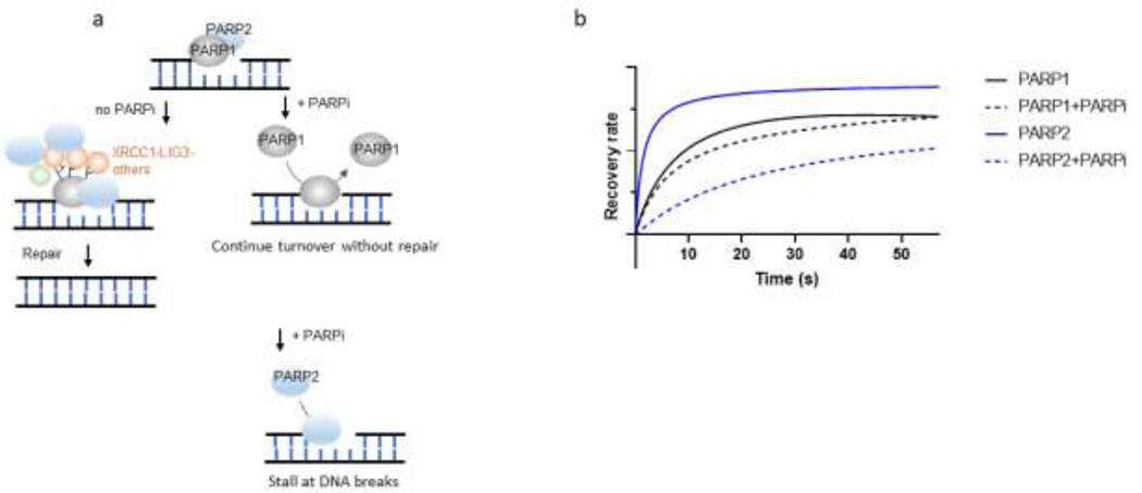
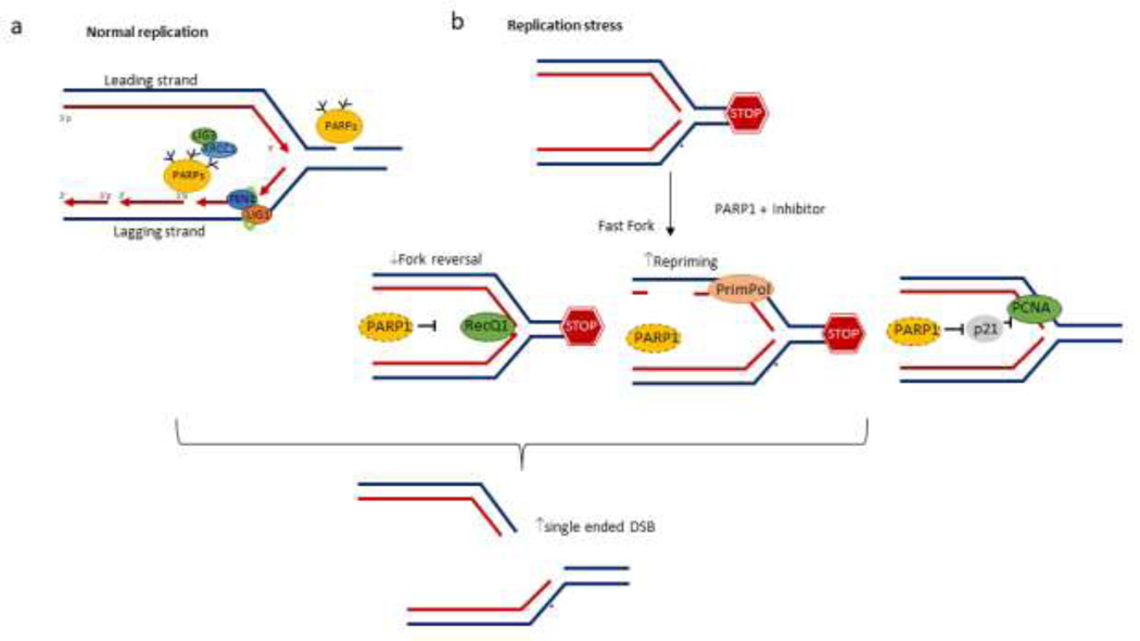
DNA strand breaks activate Poly(ADP-ribose) polymerase (PARP) 1 and 2, which use NAD+ as the substrate to covalently conjugate ADP-ribose on themselves and other proteins (e.g., Histone) to promote chromatin relaxation and recruit additional DNA repair factors. Enzymatic inhibitors of PARP1 and PARP2 (PARPi) are promising cancer therapy agents that selectively target BRCA1- or BRCA2- deficient cancers. As immediate early responders to DNA strand breaks with robust activities, PARP1 and PARP2 normally form transient foci (<10 minutes) at the micro-irradiation-induced DNA lesions. In addition to enzymatic inhibition, PARPi also extend the presence of PARP1 and PARP2 at DNA lesions, including at replication forks, where they may post a physical block for subsequent repair and DNA replication. The dynamic nature of PARP1 and PARP2 foci made live cell imaging a unique platform to detect subtle changes and the functional interaction among PARP1, PARP2, and their regulators. Recent imaging studies have provided new understandings of the biological consequence of PARP inhibition and uncovered functional interactions between PARP1 and PARP2 and new regulators (e.g., histone poly(ADP-ribosylation) factor). Here, we review recent advances in dissecting the temporal and spatial Regulation of PARP1 and PARP2 at DNA lesions and discuss their physiological implications on both cancer and normal cells.
Keywords: Live cell quantitative imaging; PAPR1; PARP2; PARylation; Replication.
Copyright © 2024 Elsevier B.V. All rights reserved.
Conflict of interest statement
Declaration of Competing Interest The authors declare that they have no known competing financial interests or personal relationships that could have appeared to influence the work reported in this paper.
Figures
Similar articles
-
XRCC1 mediates PARP1- and PAR-dependent recruitment of PARP2 to DNA damage sites.Nucleic Acids Res. 2025 Feb 8;53(4):gkaf086. doi: 10.1093/nar/gkaf086. Nucleic Acids Res. 2025. PMID: 39970298 Free PMC article.
-
Inactive Parp2 causes Tp53-dependent lethal anemia by blocking replication-associated nick ligation in erythroblasts.Mol Cell. 2024 Oct 17;84(20):3916-3931.e7. doi: 10.1016/j.molcel.2024.09.020. Epub 2024 Oct 8. Mol Cell. 2024. PMID: 39383878
-
Oncometabolite 2-hydroxyglutarate suppresses basal protein levels of DNA polymerase beta that enhances alkylating agent and PARG inhibition induced cytotoxicity.DNA Repair (Amst). 2024 Aug;140:103700. doi: 10.1016/j.dnarep.2024.103700. Epub 2024 Jun 4. DNA Repair (Amst). 2024. PMID: 38897003 Free PMC article.
-
The Molecular Mechanisms of Actions, Effects, and Clinical Implications of PARP Inhibitors in Epithelial Ovarian Cancers: A Systematic Review.Int J Mol Sci. 2022 Jul 23;23(15):8125. doi: 10.3390/ijms23158125. Int J Mol Sci. 2022. PMID: 35897700 Free PMC article.
-
Joining of DNA breaks- interplay between DNA ligases and poly (ADP-ribose) polymerases.DNA Repair (Amst). 2025 May;149:103843. doi: 10.1016/j.dnarep.2025.103843. Epub 2025 May 2. DNA Repair (Amst). 2025. PMID: 40347914 Review.
Cited by
-
Transient Poly(ADP-Ribose) Triggers FUS Condensation Hysteresis via a Prion-Like Mechanism.bioRxiv [Preprint]. 2025 Jul 5:2025.07.03.659157. doi: 10.1101/2025.07.03.659157. bioRxiv. 2025. PMID: 40631075 Free PMC article. Preprint.
-
XRCC1 mediates PARP1- and PAR-dependent recruitment of PARP2 to DNA damage sites.Nucleic Acids Res. 2025 Feb 8;53(4):gkaf086. doi: 10.1093/nar/gkaf086. Nucleic Acids Res. 2025. PMID: 39970298 Free PMC article.
-
Protein-Protein Interactions in Base Excision Repair.Biomolecules. 2025 Jun 18;15(6):890. doi: 10.3390/biom15060890. Biomolecules. 2025. PMID: 40563529 Free PMC article. Review.
-
Targeting PCNA/PARP1 axis inhibits the malignant progression of hepatocellular carcinoma.Front Pharmacol. 2025 Apr 17;16:1571786. doi: 10.3389/fphar.2025.1571786. eCollection 2025. Front Pharmacol. 2025. PMID: 40313621 Free PMC article.
-
Nicotinamide: A Multifaceted Molecule in Skin Health and Beyond.Medicina (Kaunas). 2025 Feb 1;61(2):254. doi: 10.3390/medicina61020254. Medicina (Kaunas). 2025. PMID: 40005371 Free PMC article. Review.
References
Publication types
MeSH terms
Substances
Grants and funding
LinkOut - more resources
Full Text Sources
Research Materials
Miscellaneous
