. 2024 Jun 11;57(6):1413-1427.e9.
doi: 10.1016/j.immuni.2024.05.002. Epub 2024 May 31.

Isolation of human antibodies against influenza B neuraminidase and mechanisms of protection at the airway interface

Affiliations

Isolation of human antibodies against influenza B neuraminidase and mechanisms of protection at the airway interface

Rachael M Wolters et al. Immunity. .

Abstract

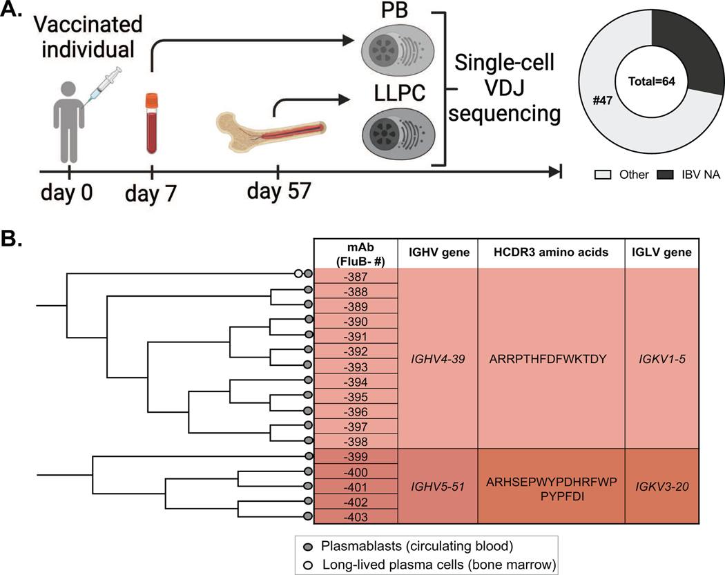
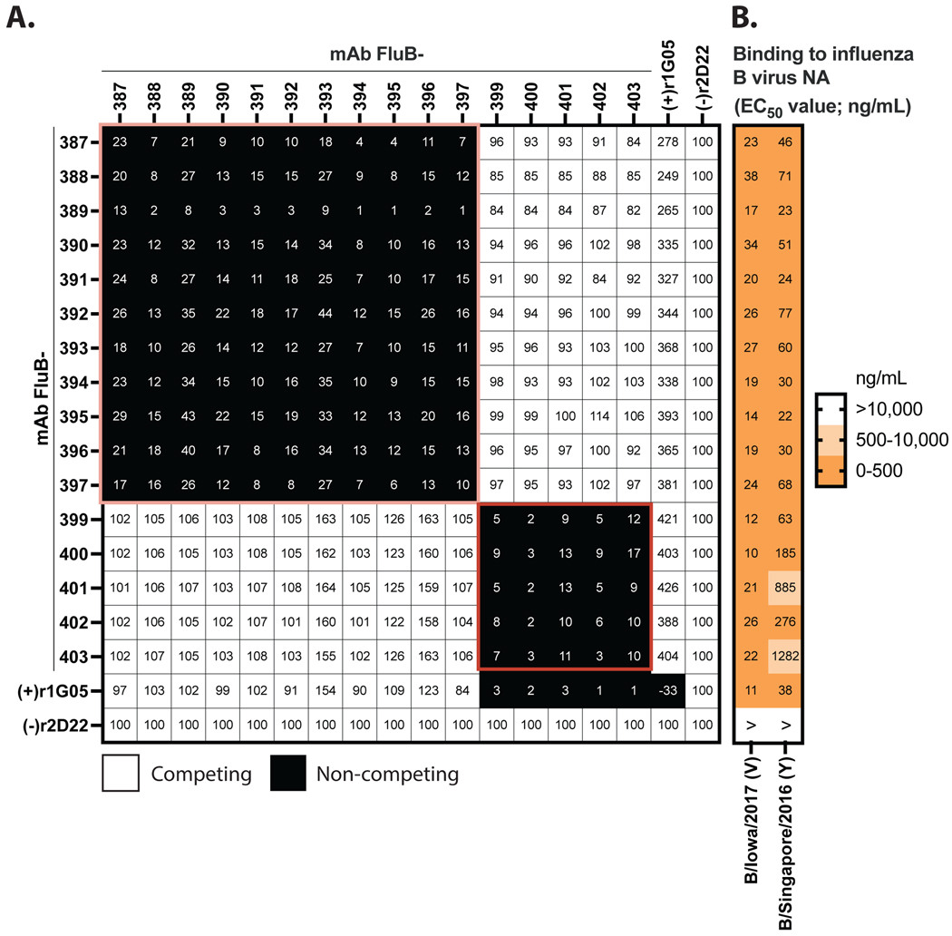
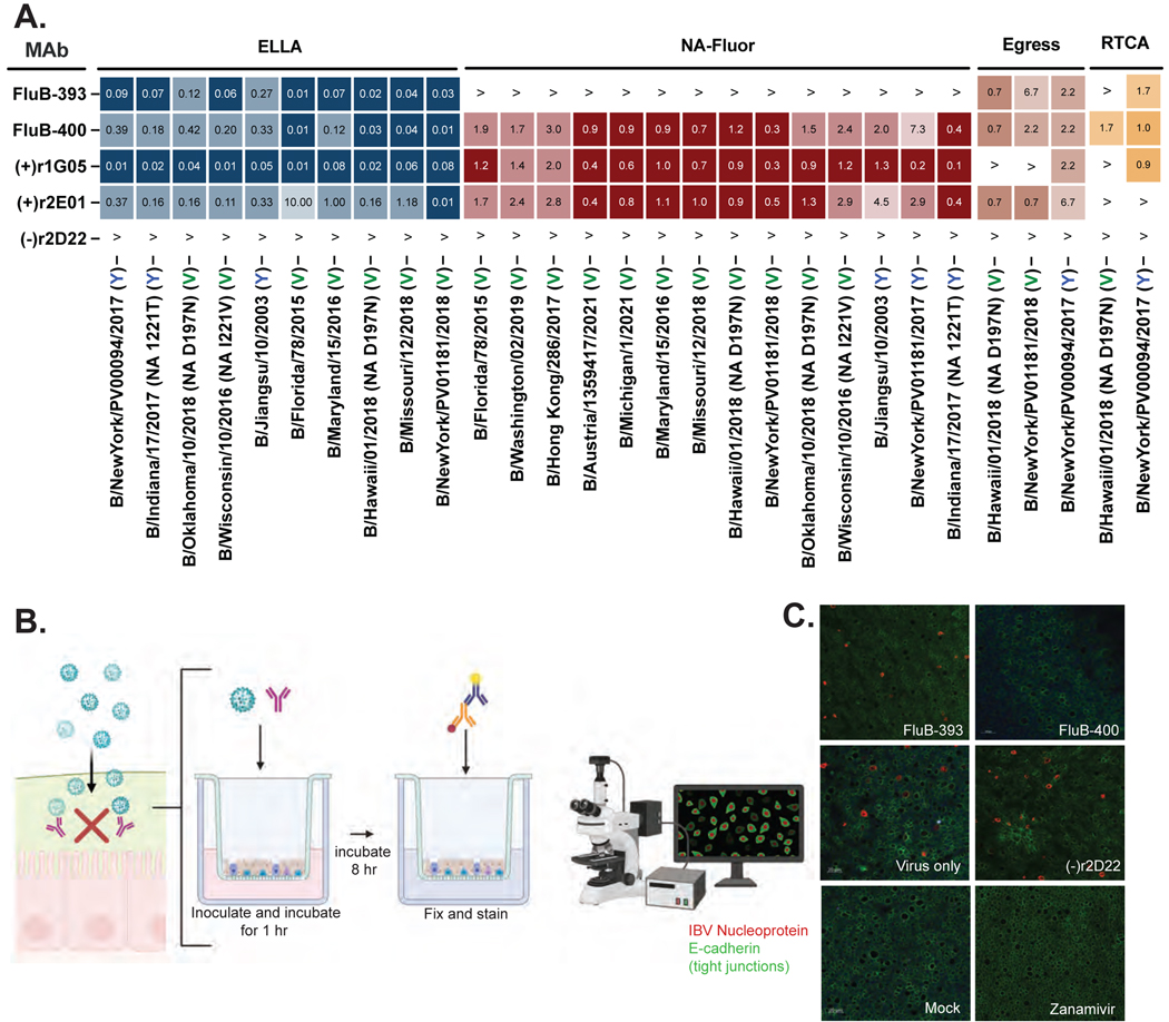
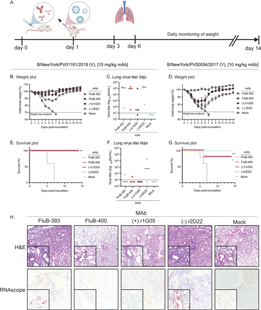
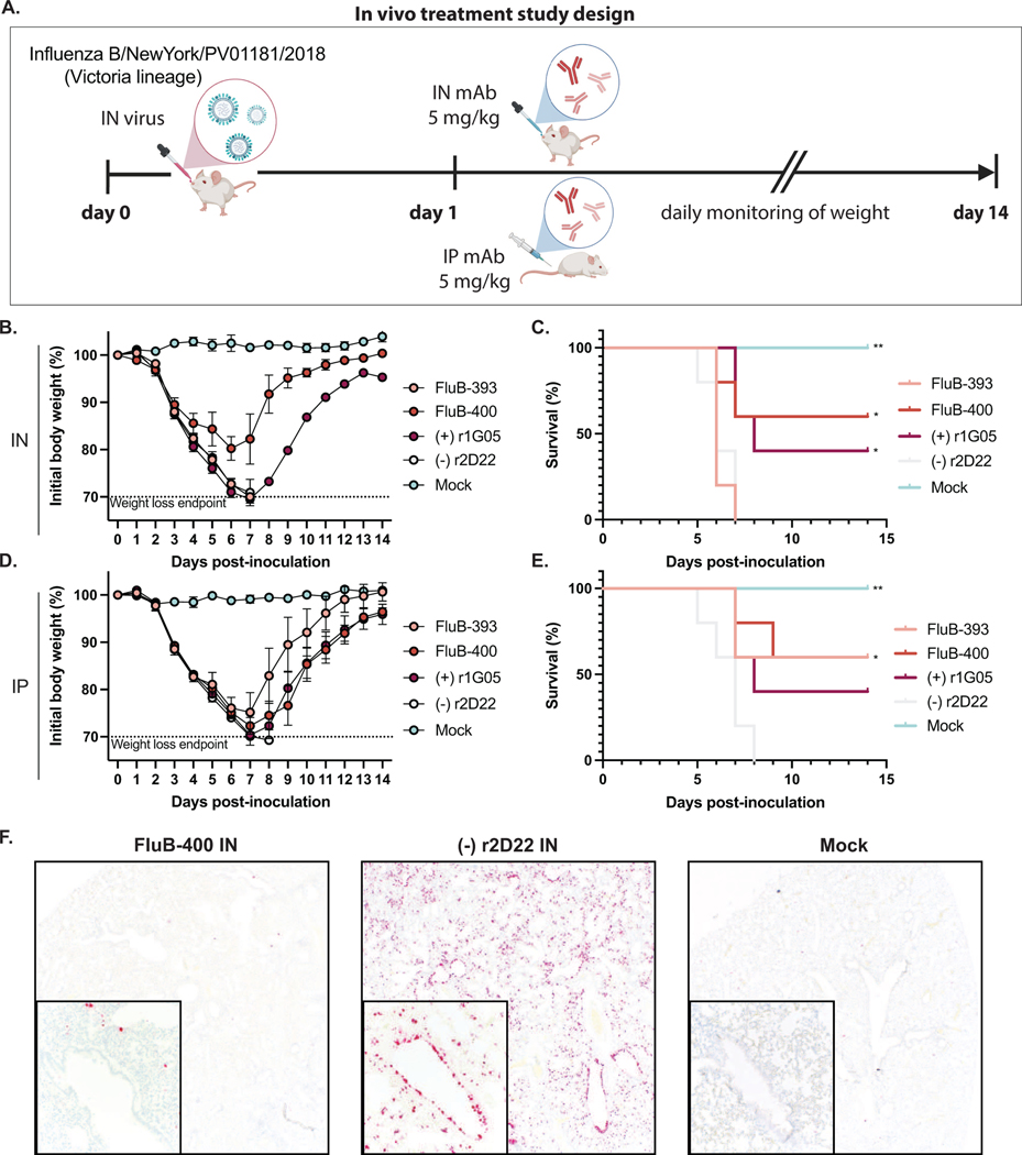
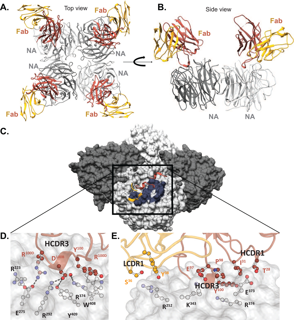
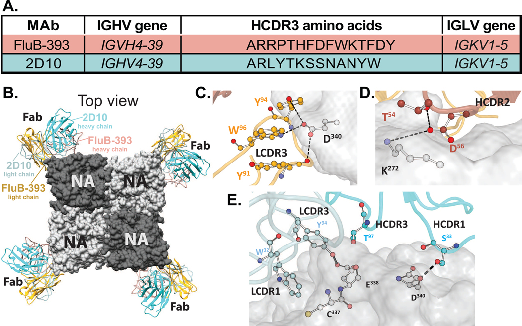
Influenza B viruses (IBVs) comprise a substantial portion of the circulating seasonal human influenza viruses. Here, we describe the isolation of human monoclonal antibodies (mAbs) that recognized the IBV neuraminidase (NA) glycoprotein from an individual following seasonal vaccination. Competition-binding experiments suggested the antibodies recognized two major antigenic sites. One group, which included mAb FluB-393, broadly inhibited IBV NA sialidase activity, protected prophylactically in vivo, and bound to the lateral corner of NA. The second group contained an active site mAb, FluB-400, that broadly inhibited IBV NA sialidase activity and virus replication in vitro in primary human respiratory epithelial cell cultures and protected against IBV in vivo when administered systemically or intranasally. Overall, the findings described here shape our mechanistic understanding of the human immune response to the IBV NA glycoprotein through the demonstration of two mAb delivery routes for protection against IBV and the identification of potential IBV therapeutic candidates.

Keywords: adaptive immunity; antibodies; human; influenza B; monoclonal; neuraminidase.


Conflict of interest statement

Declaration of interests L.E.W. serves as a consultant for BigHat Biosciences. The content of this article is solely the responsibility of the authors and does not represent the official views of BigHat Biosciences. C.B.C. serves as a consultant to GlaxoSmithKline, Sanofi, TDCowen Investments, Guidepoint Global, Debiopharm, and CommenseBio and receives royalties from UpToDate. The laboratory of C.B.C. receives funding for unrelated work from Moderna. J.E.C. has served as a consultant for Luna Labs USA, Merck Sharp & Dohme Corporation, Emergent Biosolutions, GlaxoSmithKline, and BTG International Inc; is a member of the Scientific Advisory Board of Meissa Vaccines; a former member of the Scientific Advisory Board of Gigagen (Grifols); and is founder of IDBiologics. The laboratory of J.E.C. received unrelated sponsored research agreements from AstraZeneca, Takeda, and IDBiologics during the conduct of the study. The opinions, interpretations, conclusions, and recommendations contained herein are those of the authors and are not necessarily endorsed by the US Department of Defense. E.D., T.E.E., and B.J.D. are employees of Integral Molecular, B.J.D. is a shareholder of Integral Molecular. Vanderbilt University has applied for a patent pertinent to some of the materials in this paper.

Figures

Figure 1.
Figure 1.. Vaccinated donor mounts influenza B (IBV) anti-NA response.
(A) Study design. A healthy adult individual was studied following intramuscular administration of the quadrivalent 2018–19 seasonal influenza vaccine Flucelvax. Blood (day 7) and bone marrow aspirates (day 57) were collected after vaccination for plasmablasts (PBs) and long-lived plasma cells (LLPCs) secreting antibodies to IBV and the resulting mAb from single cells were expressed in microscale format. The circle plot shows n = 17 of the total 64 clones secreted antibodies that reacted with the IBV NA protein. (B) Phylogenetic tree of the antibody variable genes for the 17 anti-NA mAbs with corresponding variable gene usage indication. Phylogenetic tree clonal families were constructed on the heavy chain V-D-J-REGION by a neighbor-joining method using the VH germline gene as an outgroup. Alignments were performed in MUSCLE. Trees were inferred using PHYLIP and visualized using FigTree. Circles correspond with the tissue sample from which the sequence was isolated: plasmablasts (PB) or long-lived plasma cells (LLPC) in the bone marrow, or both.
Figure 2.
Figure 2.. The two clonally expanded families bind NA proteins from both IBV lineages and recognize distinct binding sites.
(A) Competition binding analysis of the panel NA-reactive FluB mAbs to B/Iowa/06/2017 (V). Competition binding was measured in the presence of saturating concentrations of competitor mAbs via a competition binding ELISA and relative maximal signal for binding was normalized to the binding observed in the presence of the negative control Dengue virus-specific mAb, (−) r2D22. The mAb, (+) r1G05, was used as a reference mAb for NA active site binding mAbs. Black denotes full competition (<70% binding of reference mAb), and white denotes no competition (>71% binding of reference mAb). The orange box represents the clonally expanded family of mAbs that bind to the NA active site as indicated by competition with (+) r1G05. The pale pink box represents a family of clonally expanded mAbs that recognize a different antigenic site. (B) Binding of 16 NA-reactive mAbs, a positive control mAb (+) r1G05, or an isotype-matched negative control mAb, (−) r2D22, to recombinant NA proteins of the indicated IBV strains in ELISA. Representative EC50 values (ng/mL) are plotted as a heatmap. Data are representative of two independent experiments.
Figure 3.
Figure 3.. Human anti-NA mAbs broadly inhibit viral activity.
(A) Heatmap indicating potency of mAbs: enzyme-linked lectin assays (ELLA-NI; IC50 values); NA-Fluor assay (IC50 values); egress assay (IC100 values); real-time cell analysis (RTCA) neutralization (IC50 values). Lower IC50 or IC100 values shown in darker shades, each lineage specified as Victoria (V) or Yamagata (Y). Data are representative of two independent experiments. (B) Experimental design of the air-liquid interface (ALI) culture system of primary human tracheal respiratory epithelial cells. IBV was inoculated at an MOI of 0.1 and treated with 10 μg/mL of anti-NA mAb, an isotype-matched negative control mAb, or a virus-only condition. Incubation time was restricted to 8 hours post-inoculation to allow for viral attachment but not egress. Cells were fixed and stained with anti-IBV nucleoprotein (NP; red) or anti-E-cadherin mAb (green). (C) Confocal photomicrographs of ALI culture membranes. Images were captured on a Zeiss 710 confocal microscope. Data are representative of 9 fields of view, and two independent experiments were performed.
Figure 4.
Figure 4.. Anti-NA mAbs mediate protection in vivo against lethal IBV challenge in mice.
(A) Study design. Groups of BALB/c mice were inoculated intraperitoneally (i.p.) 12 hours before virus challenge with 10 mg/kg of anti-NA mAb, an irrelevant negative-control mAb (r2D22), or a positive control mAb (r1G05) recognizing the IBV NA protein. Mice were challenged intranasally (i.n.) with a lethal dose of indicated IBV virus and monitored daily for survival. (B and D) The body weights of the mice are represented as the group mean ± SEM. The lower dotted line indicates the no-recovery threshold (>30% weight loss) and endpoint for euthanasia. (E and G) Survival curves were estimated using the Kaplan-Meier method. Survival of each group treated with an anti-NA or r1G05 (+) mAb were compared to the r2D22 treatment group using the log-rank (Mantel-Cox) test, * p < 0.05, ** p < 0.005. n = 5 mice per group. (C and F) Lung titers of mice challenged 3 days and 6 days after inoculation. n = 3 mice per group. (G) Photomicrographs of left lung lobes 6 dpi. The lobes were insufflated and prepared for hematoxylin and eosin (H&E) staining and RNAscope in situ hybridization assays.
Figure 5.
Figure 5.. Airway delivery of IBV anti-NA active site mAbs is effective.
(A) Study design. Groups of BALB/c mice were inoculated by the intranasal (i.n.) route with a lethal dose of influenza B/New York/PV01181/2018 (V) virus. (C and E) One day later (1 dpi) mice were administered by the i.n. or intraperitoneal (i.p.) route with 5 mg/kg of the indicated anti-NA mAb, the r2D22 (−) control mAb, or the r1G05 (+) control mAb, and monitored for protection. Survival curves were estimated using the Kaplan-Meier method and compared as indicated using log-rank (Mantel-Cox) test, * p < 0.05, ** p < 0.005 (B and D) The body weights of mice represent the group mean ± SEM. The lower dotted line indicates the no-recovery threshold (> 30% weight loss) and endpoint for euthanasia. Data represent one experiment, n = 5 mice per group. (F) Photomicrographs of the left lung lobes 6-dpi. The lobes were insufflated and prepared for RNAscope, n = 3 mice per group.
Figure 6.
Figure 6.. Cryo-EM reconstruction of FluB-400 in complex with NA.
Top (A) and side (B) view of the cryo-EM reconstruction of FluB-400 Fab molecules in complex with the NA protein B/Iowa/06/2017 (V). One NA tetramer (gray) bound to four Fab molecules (Fv: red and Fc: yellow). (C) Epitope footprints profile of NA-FluB-400 (D, E) Specific interactions of FluB-400 with NA.
Figure 7.
Figure 7.. Comparative cryo-EM reconstructions of FluB-393 and 2D10 in complex with NA.
(A) IGHV and IGLV gene usage, and amino acid sequence of the HCDR3 loop for mAbs FluB-393 and 2D10. (B) Cryo-EM reconstructions of FluB-393 and 2D10 Fab molecules in complex with the NA protein B/Iowa/06/2017 (V). One NA tetramer (gray) is shown in a top view. Four individual Fabs (for each of two mAbs [FluB-393 and 2D10]) are shown in overlay as colored ribbon structures bound to a single NA protomer (light or dark gray space-filling structures). Mab FluB-393 Fab heavy chain is shown in salmon color ribbon, and the light chain in gold ribbon. MAb 2D10 Fab heavy chain is shown in dark cyan ribbon, and the light chain in light cyan ribbon. The specific interactions of FluB-393 with NA are shown in (C) [light chain] and (D) [heavy chain]. The specific interactions of 2D10 with NA are shown in (E).

Similar articles

Cited by

References

    1. Schneider C, and Coughlan L. (2023). Always a bridesmaid, never a bride: Committing to flu neuraminidase as a vaccine target. Mol Ther. 10.1016/j.ymthe.2023.04.014. - DOI - PMC - PubMed
    1. Matrosovich MN, Matrosovich TY, Gray T, Roberts NA, and Klenk H-D (2004). Neuraminidase is important for the initiation of influenza virus infection in human airway epithelium. J. Virol 78, 12665–12667. 10.1128/jvi.78.22.12665-12667.2004. - DOI - PMC - PubMed
    1. Cohen M, Zhang X-Q, Senaati HP, Chen H-W, Varki NM, Schooley RT, and Gagneux P. (2013). Influenza A penetrates host mucus by cleaving sialic acids with neuraminidase. Virol J 10, 321. 10.1186/1743-422x-10-321. - DOI - PMC - PubMed
    1. DiLillo DJ, Palese P, Wilson PC, and Ravetch JV (2016). Broadly neutralizing anti-influenza antibodies require Fc receptor engagement for in vivo protection. J Clin Invest 126, 605–610. 10.1172/jci84428. - DOI - PMC - PubMed
    1. Momont C, Dang HV, Zatta F, Hauser K, Wang C, Iulio J. di, Minola A, Czudnochowski N, Marco AD, Branch K, et al. (2023). A pan-influenza antibody inhibiting neuraminidase via receptor mimicry. Nature, 1–8. 10.1038/s41586-023-06136-y. - DOI - PMC - PubMed

Publication types

MeSH terms