Review
. 2024 Jul;300(7):107428.
doi: 10.1016/j.jbc.2024.107428. Epub 2024 May 30.

Lipid switches in the immunological synapse

Affiliations
Review

Lipid switches in the immunological synapse

Gillian Griffiths et al. J Biol Chem. 2024 Jul.

Abstract

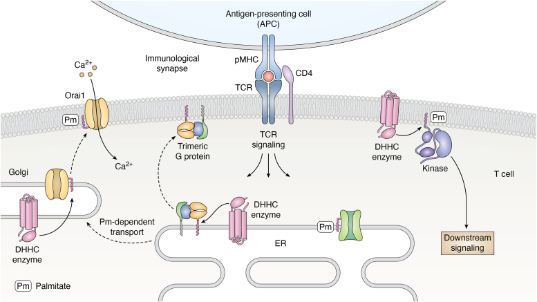
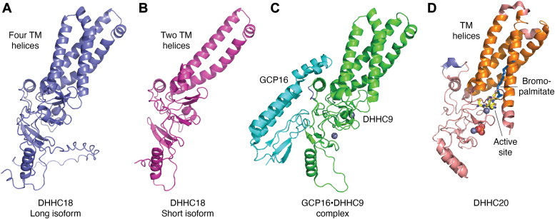
Adaptive immune responses comprise the activation of T cells by peptide antigens that are presented by proteins of the Major Histocompatibility Complex (MHC) on the surface of an antigen-presenting cell. As a consequence of the T cell receptor interacting productively with a certain peptide-MHC complex, a specialized cell-cell junction known as the immunological synapse forms and is accompanied by changes in the spatiotemporal patterning and function of intracellular signaling molecules. Key modifications occurring at the cytoplasmic leaflet of the plasma and internal membranes in activated T cells comprise lipid switches that affect the binding and distribution of proteins within or near the lipid bilayer. Here, we describe two major classes of lipid switches that act at this critical water/membrane interface. Phosphoinositides are derived from phosphatidylinositol, an amphiphilic molecule that contains two fatty acid chains and a phosphate group that bridges the glycerol backbone to the carbohydrate inositol. The inositol ring can be variably (de-)phosphorylated by dedicated kinases and phosphatases, thereby creating phosphoinositide signatures that define the composition and properties of signaling molecules, molecular complexes, or whole organelles. Palmitoylation refers to the reversible attachment of the fatty acid palmitate to a substrate protein's cysteine residue. DHHC enzymes, named after the four conserved amino acids in their active site, catalyze this post-translational modification and thereby change the distribution of proteins at, between, and within membranes. T cells utilize these two types of molecular switches to adjust their properties to an activation process that requires changes in motility, transport, secretion, and gene expression.

Keywords: DHHC enzymes; S-acylation; T cell; immunological synapse; palmitoylation; phosphoinositides.


Conflict of interest statement

Conflict of interest The authors declare that they have no known competing financial interests or personal relationships that could have appeared to influence the work reported in this paper.

Figures

Figure 1
Figure 1
Depiction of the T cell synapse highlighting important phosphoinositide switches. In particular, PI(4,5)P2 conversion at the synapse leads to rapid changes in the second messengers, inositol-1,4,5-trisphosphate (IP3) and diacylglycerol (DAG), most prominently affecting Ca2+ signaling and protein kinase signaling. Note that the IP3 receptor IP3R and the store-operated calcium entry channel Orai1 are required to be palmitoylated (Pm) for functionality.
Figure 2
Figure 2
Depiction of the T cell synapse highlighting some of the most prominent palmitoylation targets in the synapse. The DHHC enzymes catalyzing palmitoylation (Pm) are located at the ER, the Golgi, or the plasma membrane. Palmitoylation may alter the transport, activity, or clustering of target proteins within membranes, as shown here for the Gα subunit of a trimeric G protein, the Ca2+ channel Orai1, or a kinase (Lck).
Figure 3
Figure 3
A TCR-induced PIP-switch triggers membrane specialization and actin depletion across the immunological synapse. Lipid and phosphoinositide-specific bio-probes (green) revealed the “membrane signature” across the immunological synapse (∗) formed with the APC (blue) when actin (labeled with Lifeact, red) is depleted and granule secretion can occur. Images are single slices of 3D images as described in (30). Scale bars: 5 μm. The scheme on the right side shows the changes in lipid species that occur upon T cell stimulation, leading to TCR signaling and actin flow away from the center of the synapse.
Figure 4
Figure 4
Structures and structural models of DHHC enzymes.A and B, comparison of the structures of the long and short isoforms of DHHC18 as predicted by Alphafold (124, 125). The long isoform (A) contains four transmembrane helices while the short isoform comprises two (B). C, electron microscopy structure of DHH9 (in green) in complex with the accessory protein GCP16 (in cyan) (126). The geometry of the GCP16 interaction site is conserved in the DHHC18 isoforms, while it is partially occupied by an internal short helix (shown in blue) in the crystal structure of DHHC20 (62) (D). In the DHHC20 structure the transmembrane helices are depicted in orange and the active site (DHHC) in yellow with the cysteine-attached bromo-palmitate in marine blue. Zinc ions are shown as blue spheres.
Figure 5
Figure 5
Workflows for MS-based phosphoinositide analytics. For the relative quantification of phosphoinositides, specific PIPx-class standards are spiked in. The lipid extract can be analyzed either directly by direct injection (DI)-MS/MS or by liquid chromatography (LC)-coupled MS/MS analysis. Phosphate methylation of the lipid extracts significantly increases the sensitivity of the PIPx analysis. In addition to LC-MS/MS analysis of methylated lipids, recent work has shown that PIPx regioisomers can be separated using chiral chromatography. A preceding chromatography step can further improve the sensitivity of the analysis. MS spectra evaluation is done with dedicated software tools. PIPx, mono-, di- or triphosphorylated phosphatidylinositol.

References

    1. Grakoui A., Bromley S.K., Sumen C., Davis M.M., Shaw A.S., Allen P.M., et al. The immunological synapse: a molecular machine controlling T cell activation. Science. 1999;285:221–227. - PubMed
    1. Dustin M.L., Choudhuri K. Signaling and polarized communication across the T cell immunological synapse. Annu. Rev. Cell Dev. Biol. 2016;32:303–325. - PubMed
    1. Wu W., Shi X., Xu C. Regulation of T cell signalling by membrane lipids. Nat. Rev. Immunol. 2016;16:690–701. - PubMed
    1. Muntjewerff E.M., Meesters L.D., van den Bogaart G., Revelo N.H. Reverse signaling by MHC-I molecules in immune and non-immune cell types. Front. Immunol. 2020;11 - PMC - PubMed
    1. Sun Y., Dandekar R.D., Mao Y.S., Yin H.L., Wulfing C. Phosphatidylinositol (4,5) bisphosphate controls T cell activation by regulating T cell rigidity and organization. PLoS One. 2011;6 - PMC - PubMed

Substances

LinkOut - more resources