. 2024 Jun 1;15(1):4697.
doi: 10.1038/s41467-024-49002-9.

An axonemal intron splicing program sustains Plasmodium male development

Affiliations

An axonemal intron splicing program sustains Plasmodium male development

Jiepeng Guan et al. Nat Commun. .

Abstract

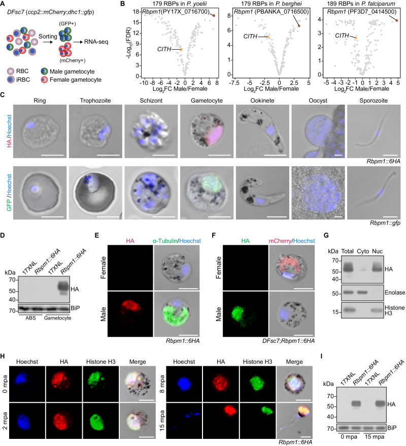
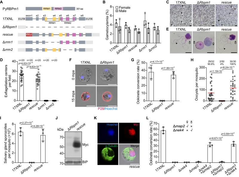
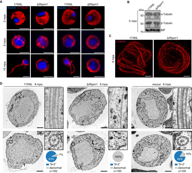
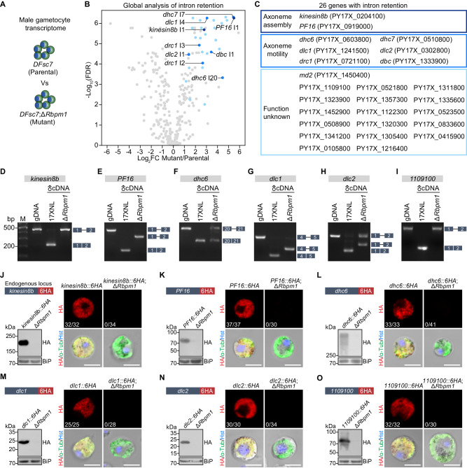
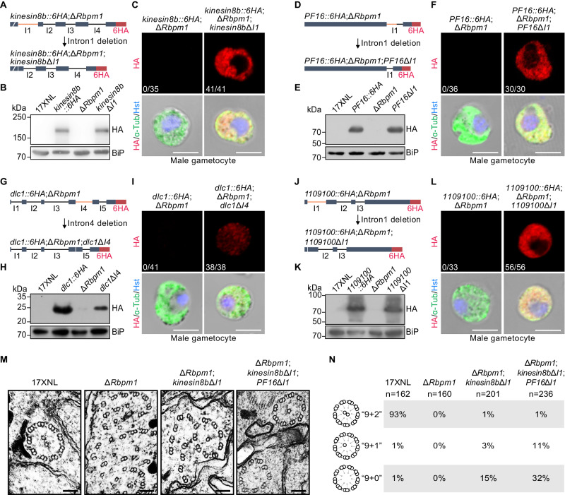
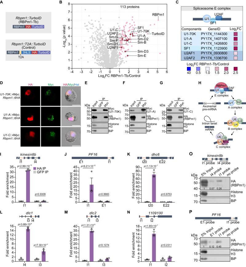
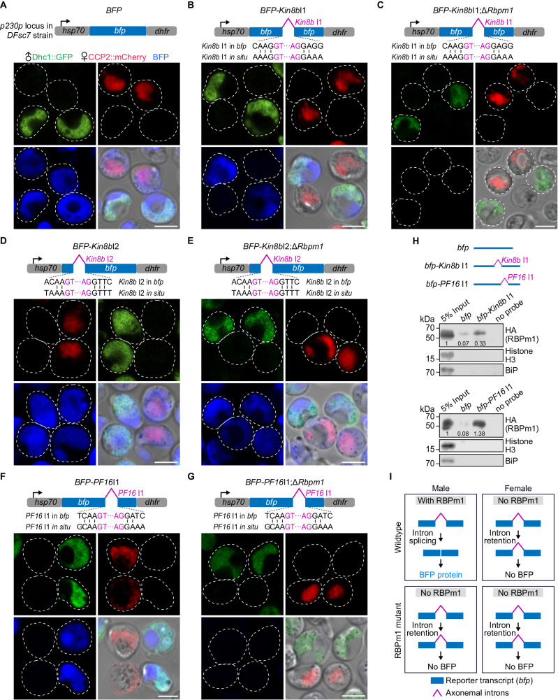
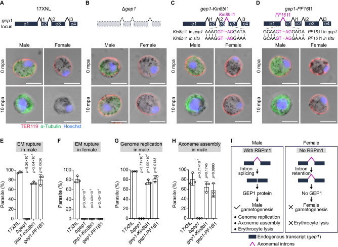
Differentiation of male gametocytes into flagellated fertile male gametes relies on the assembly of axoneme, a major component of male development for mosquito transmission of the malaria parasite. RNA-binding protein (RBP)-mediated post-transcriptional regulation of mRNA plays important roles in eukaryotic sexual development, including the development of female Plasmodium. However, the role of RBP in defining the Plasmodium male transcriptome and its function in male gametogenesis remains incompletely understood. Here, we performed genome-wide screening for gender-specific RBPs and identified an undescribed male-specific RBP gene Rbpm1 in the Plasmodium. RBPm1 is localized in the nucleus of male gametocytes. RBPm1-deficient parasites fail to assemble the axoneme for male gametogenesis and thus mosquito transmission. RBPm1 interacts with the spliceosome E complex and regulates the splicing initiation of certain introns in a group of 26 axonemal genes. RBPm1 deficiency results in intron retention and protein loss of these axonemal genes. Intron deletion restores axonemal protein expression and partially rectifies axonemal defects in RBPm1-null gametocytes. Further splicing assays in both reporter and endogenous genes exhibit stringent recognition of the axonemal introns by RBPm1. The splicing activator RBPm1 and its target introns constitute an axonemal intron splicing program in the post-transcriptional regulation essential for Plasmodium male development.


Conflict of interest statement

The authors declare no competing interests.

Figures

Fig. 1
Fig. 1. RNA-binding protein RBPm1 is expressed in the nucleus of male gametocytes.
A Flowchart showing the purification and transcriptome analysis of male (green, GFP+) and female (red, mCherry+) gametocytes from a P. yoelii parasite reporter line DFsc7. B Gender analysis of gene transcription for the Plasmodium genome-wide putative RBPs between male and female gametocytes. The top male gene PY17X_0716700, RBPm1, is marked in red. CITH (orange dot) is a known female RBP. The results of P. berghei (middle panel) and P. falciparum (right panel) were based on the published gametocyte transcriptomes contributed by Yeoh, L.M. 2017 and Lasonder, E. 2016. The p-values were calculated by quasi-likelihood F-test and adjusted by false discovery rate (FDR). C Stage expression of RBPm1 during the P. yoelii life cycle. Immunofluorescence assay (IFA) of RBPm1 expression in the Rbpm1::6HA parasites stained with anti-HA antibody (top panel). Live cell imaging of the RBPm1::GFP protein in the Rbpm1::gfp parasites (bottom panel). Nuclei were stained with Hoechst 33342. Three independent experiments with similar results. Scale bars: 5 µm. D Immunoblot of RBPm1 in the asexual blood stage (ABS) and gametocyte of the Rbpm1::6HA parasites. BiP as a loading control. Three independent experiments with similar results. E IFA of HA-tagged RBPm1 and α-Tubulin (male gametocyte marker protein) in Rbpm1::6HA gametocytes. Three independent experiments with similar results. Scale bars: 5 µm. F IFA of HA-tagged RBPm1 and mCherry (expressed in female gametocytes) in the DFsc7;Rbpm1::6HA gametocytes. Three independent experiments with similar results. Scale bars: 5 µm. G Immunoblot of RBPm1 in cytosolic and nuclear fractions of Rbpm1::6HA gametocytes. Enolase (cytoplasmic/Cyto) and histone H3 (nuclear/Nuc) proteins used as controls respectively. Two independent experiments with similar results. H IFA of HA-tagged RBPm1 and histone H3 during male gametogenesis of the Rbpm1::6HA parasites. mpa: minute post activation. Three independent experiments with similar results. Scale bars: 5 µm. I Immunoblot of RBPm1 expression in the Rbpm1::6HA parasites during male gametogenesis. Two independent experiments with similar results.
Fig. 2
Fig. 2. RBPm1 is essential for male gametogenesis and mosquito transmission of parasite.
A A schematic showing genetic modification at the Rbpm1 locus in the P. yoelii parasite. The top panel depicts the protein structure of RBPm1 with two RNA recognition motifs RRM1 (residues 119-190, yellow) and RRM2 (residues 203-274, purple). ΔRbpm1, deletion of the whole coding sequence from the 17XNL (wild type) strain; rescue, the ΔRbpm1 line complemented with Rbpm1 fused with a 4Myc tag; Δrrm1 and Δrrm2, deletion each of RRM1 and RRM2 from the 17XNL. B Female and male gametocyte formation in mice for the modified parasite. Data are means ± SEM of three independent experiments. C Exflagellation center (EC) formation of activated male gametocytes at 10 mpa. Cell clusters representing the EC are marked with white arrows. Four independent experiments with similar results. Scale bars: 20 μm. D Quantification of EC formation. The ECs were counted within a 1 × 1-mm square area in the hemocytometer under a light microscope. n represents the number of fields counted. Means ± SEM, one-way ANOVA with Tukey multiple pairwise-comparisons. Three independent experiments. E Light microscope images of the exflagellated male gametes (black arrow) after Giemsa staining. Four independent experiments with similar results. Scale bars: 5 μm. F Female gamete formation assayed by P28 staining. P28 is a female gamete plasma membrane protein. n = 32 and 35 female gametocytes in 17XNL and ΔRbpm1 respectively. Scale bars: 5 μm. G Ookinete formation in vitro. Data are means ± SEM from three independent experiments, one-way ANOVA with Tukey multiple pairwise-comparisons. H Midgut oocyst formation in mosquitoes at 7 days after blood feeding. x/y at the top represents the number of mosquitoes containing oocysts/the number of dissected mosquitoes, and the percentage represents the infection prevalence of mosquitoes. Red lines show the mean value of oocyst numbers, one-way ANOVA with Tukey multiple pairwise-comparisons. Three independent experiments with similar results. I Salivary gland sporozoite formation in mosquitoes at 14 days after blood feeding. At least 20 infected mosquitoes were counted in each group. Data are means ± SEM of three independent experiments, one-way ANOVA with Tukey multiple pairwise-comparisons. J Immunoblot analysis of RBPm1 expression in gametocytes of the complemented line rescue. BiP as a loading control. Two independent experiments with similar results. K IFA of Myc-tagged RBPm1 and α-Tubulin in gametocytes of the rescue parasite. Three independent experiments with similar results. Scale bars: 5 μm. L Gender gamete fertility assay of the ΔRbpm1 by parasite genetic cross. Fertility was determined by ookinete development of ΔRbpm1 gametes after cross-fertilization with mutant lines that are defective in either female (Δnek4) or male (Δmap2) gametes. Data are means ± SEM of three independent experiments, one-way ANOVA with Tukey multiple pairwise-comparisons.
Fig. 3
Fig. 3. Defective axonemal assembly in RBPm1-null male gametogenesis.
A Detection of formation and exflagellation of axonemes during male gametogenesis (0, 8, and 15 mpa) by staining α-Tubulin (left panels) and β-Tubulin (right panels). Nuclei were stained with Hoechst 33342. Four independent experiments with similar results. Scale bars: 5 µm. B Immunoblot of α- and β-Tubulins in gametocytes. The numbers indicate the relative intensities of the bands in the immunoblots. BiP as a loading control. Two independent experiments with similar results. C Ultrastructure expansion microscopy (U-ExM) of the axonemes in male gametocytes stained with α-Tubulin antibody at 8 mpa. Three independent experiments with similar results. Scale bars: 5 μm. D Transmission electron microscopy of axoneme architecture in male gametocytes at 8 mpa. Inset panels show longitudinal sections (top panels) and cross sections (bottom panels) of axonemes. The enclosed area (black box) was zoomed in. Pie charts show the quantification of axoneme (“9 + 2” microtubules) in the mutant parasites. n is the total number of intact and defective axoneme structures observed in each group. Three independent experiments with similar results. Scale bars: 1 μm.
Fig. 4
Fig. 4. RBPm1 deficiency causes intron retention and protein loss of axonemal genes.
A A schematic showing the transcriptome analysis of RBPm1-null male gametocytes. DFsc7Rbpm1 is a DFsc7-derived RBPm1 mutant line. DFsc7 (parental) and DFsc7Rbpm1 (mutant) male gametocytes were sorted by FACS for RNA-seq. B Global analysis of differential intron retention identified 30 retained introns (blue dots) in the mutant versus the parental line. Retained introns with log2FC ≥ 1 and p ≤ 0.05 were further verified manually by visualization in IGV. The p-values were calculated by quasi-likelihood F-test and adjusted by FDR. These introns were originated from 26 genes. Detailed information of these genes and introns is provided in Supplementary Fig. 4A. C List of the 26 genes with intron retention from (B), categorized by protein function. DI. RT-PCR confirmation of intron retention in 6 selected genes kinesin8b, PF16, dhc6, dlc1, dlc2, and PY17X_1109100. Genomic DNA (gDNA) from 17XNL parasite, complementary DNA (cDNA) from male gametocytes of parental and mutant parasites were analyzed. Exons are indicated by boxes and introns by lines. Three independent experiments with similar results. RT-PCR analysis for all 26 genes is presented in Supplementary Fig. 5A. JO. Protein expression analysis of the 6 genes shown in (DI) in male gametocytes after loss of RBPm1. Each endogenous gene was tagged with a 6HA at the C-terminus in both 17XNL and ΔRbpm1 parasites (the schematic in the top left panel), generating two tagged lines. Immunoblot of the 6HA-tagged protein in gametocytes with and without RBPm1 (bottom left panel). IFA of the 6HA-tagged protein in male gametocytes with and without RBPm1 (right panel). x/y at bottom-left represents the number of HA-positive male gametocytes/the total number of male gametocytes tested. Three independent experiments with similar results. Scale bars: 5 µm.
Fig. 5
Fig. 5. Intron deletion restores axonemal protein expression and partially rectifies axoneme assembly defects in RBPm1-null male gametocytes.
A A schematic showing the genomic deletion of retained intron (kinesin8b intron1, orange) in the kinesin8b::6HARbpm1 parasite (abbreviated as ΔRbpm1), generating the mutant kinesin8b::6HARbpm1;kinesin8bΔintron1 (abbreviated as kinesin8bΔI1). B Immunoblot of 6HA-tagged Kinesin8B protein in gametocytes. Three independent experiments with similar results. C IFA of 6HA-tagged Kinesin8B in male gametocytes. x/y represents the number of HA-positive male gametocytes/the total number of male gametocytes tested. Three independent experiments with similar results. Scale bars: 5 µm. D, E, F Effect of intron deletion (PF16 intron1) on the restoration of PF16 protein in RBPm1-null male gametocytes. Similar analysis as in (A, B, C). G, H, I Effect of intron deletion (dlc1 intron4) on the restoration of Dlc1 protein in RBPm1-null male gametocytes. J, K, L Effect of intron deletion (PY17X_1109100 intron1) on the restoration of PY17X_1109100 protein in RBPm1-null male gametocytes. M Transmission electron microscopy of axoneme architecture in male gametocytes at 8 mpa. ΔRbpm1;kinesin8bΔI1 is a ΔRbpm1-derived modified line with deletion of kinesin8b intron1. ΔRbpm1;kinesin8bΔI1;PF16ΔI1 is a ΔRbpm1 derived modified line with deletion of both kinesin8b intron1 and PF16 intron1. Scale bars: 100 nm. N Quantification of axoneme formation from parasites in (M). n is the total number of the intact and defective axoneme structures observed in each group. Three independent experiments with similar results.
Fig. 6
Fig. 6. RBPm1 interacts with spliceosome E complex and introns of axonemal genes.
A A schematic showing two modified parasite lines generated for searching RBPm1-interacting proteins by TurboID-based proximity labeling and mass spectrometry. The motif of HA::TurboID and T2A::3NLS::HA::TurboID, respectively, was inserted at the C-terminus of the endogenous RBPm1, generating the line Rbpm1::TurboID and the control line Rbpm1::T2A::TurboID. B Volcano plot displaying 113 significantly enriched proteins (pink dot, cutoffs log2FC ≥ 1 and p ≤ 0.05) in the Rbpm1::TurboID versus Rbpm1::T2A::TurboID. Among them, 13 subunits (red dot) of the early spliceosome E complex were included. The p-values were calculated by two-sided t-test and adjusted by FDR. C Protein interaction analysis between RBPm1 and six spliceosome E complex subunit proteins (U1-70K, U1-A, U1-C, SF1, U2AF1, and U2AF2) from (B). D IFA of 6HA-tagged RBPm1 and 4Myc-tagged U1 snRNP proteins (U1-70K, U1-A, and U1-C) in male gametocytes of three double-tagged parasites. Three independent experiments with similar results. Scale bars: 5 µm. E Co-immunoprecipitation of RBPm1 and U1-70K in gametocytes of the double-tagged parasite U1-70K::4Myc;Rbpm1::6HA. Anti-Myc nanobody was used. Bip as a loading control. Three independent experiments with similar results. F Co-immunoprecipitation of RBPm1 and U1-A in gametocytes of the double-tagged parasite U1-A::4Myc;Rbpm1::6HA. Three independent experiments with similar results. G Co-immunoprecipitation of RBPm1 and U1-C in gametocytes of the double-tagged parasite U1-C::4Myc;Rbpm1::6HA. Three independent experiments with similar results. H Proposed model showing the interaction between RBPm1 and early spliceosome E complex for intron splicing of axonemal genes. IN. UV-RIP detection of RBPm1 interaction with the retained introns of 6 axonemal genes (kinesin8b, PF16, dhc6, dlc1, dlc2, and PY17X_1109100). A top schematic shows the exon-intron structure of the RBPm1 target axonemal genes. The retained introns are indicated with orange lines, and the genomic regions for qPCR amplicon are shown. UV-RIP was performed in Rbpm1::6HA lines using anti-HA nanobody. Anti-GFP nanobody was used as a control. Bound RNA was analyzed by RT-qPCR. Means ± SEM from three independent experiments, two-sided t-test. O RNA pull-down assay detecting RBPm1 interaction with kinesin8b intron1. A top schematic shows the exon-intron structure of the kinesin8b gene. The retained introns are indicated in orange lines. A biotinylated 500 nt RNA probe I1 (comprising intron1 and its flanking sequences) and a control probe I4 (comprising intron4 and its flanking sequences) were used. Proteins via RNA pull-down were immunoblot with anti-HA antibody. The numbers are the relative intensities of bands in the blot. Histone H3 and Bip were used as negative controls. Two independent experiments with similar results. P RNA pull-down assay detecting RBPm1 interaction with PF16 intron1. A top schematic shows the exon-intron structure of the PF16 gene. The retained introns are indicated in orange lines. A biotinylated 500 nt RNA probe I1 (comprising intron1 and its flanking sequences) and a control probe E1 in exon1 were used. Two independent experiments with similar results.
Fig. 7
Fig. 7. RBPm1 directs splicing of axonemal introns inserted in a reporter gene.
A A top schematic shows a transgenic line BFP with a bfp reporter expression cassette integrated at the p230p locus of the DFsc7 reporter line. The intact bfp is driven by the 5′UTR of hsp70 and the 3′UTR of dhfr, allowing expression of BFP in both male (GFP+) and female (mCherry+) gametocytes. Live cell imaging was shown. Three independent experiments with similar results. Scale bars: 5 µm. B. A transgenic line BFP-Kin8bI1 with a kinesin8b intron 1 (Kin8b I1, purple line)-inserted bfp cassette integrated at the p230p locus of the DFsc7 line. Kin8b I1 (purple) was inserted into the bfp gene at the nucleotides 396-397 to mimic the splice site (vertical lines) of in situ Kin8b I1. BFP expression was detected specifically in male gametocytes of the BFP-Kin8bI1 parasites. Three independent experiments with similar results. Scale bars: 5 µm. C A BFP-Kin8bI1 derived RBPm1 mutant line BFP-Kin8bI1;ΔRbpm1. No BFP expression was detected in male gametocytes of the BFP-Kin8bI1;ΔRbpm1 parasites. Three independent experiments. Scale bars: 5 µm. D Effect of the kinesin8b intron 2 (Kin8b I2) insertion on the gametocyte expression of BFP. Similar analysis as in (B). BFP expression was detected in both male and female gametocytes of the BFP-Kin8bI2 parasites. E A BFP-Kin8bI2 derived RBPm1 mutant line BFP-Kin8bI2;ΔRbpm1. Similar analysis as in (C). BFP expression was detected in both male and female gametocytes of the BFP-Kin8bI2;ΔRbpm1 parasites. F Effect of the PF16 intron1 (PF16 I1) insertion on the gametocyte expression of BFP. Similar analysis as in (B). BFP expression was detected specifically in male gametocytes of the BFP-PF16I1 parasites. G A BFP-PF16I1 derived RBPm1 mutant line BFP-PF16I1;ΔRbpm1. Similar analysis as in (C). No BFP expression was detected in male gametocytes of the BFP-PF16I1;ΔRbpm1 parasites. H RNA pull-down assay detecting RBPm1 interaction with the Kin8b I1 and PF16 I1 -inserted bfp transcripts from BFP-Kin8bI1 and BFP-PF16I1 gametocytes, respectively. Three biotinylated RNA probes bfp, bfp-Kin8bI1 (corresponding to the Kin8b I1-inserted bfp transcript), bfp-PF16I1 (corresponding to the PF16 I1-inserted bfp transcript) were used. Proteins via RNA pull-down were immunoblotted with anti-HA antibody. The numbers are the relative intensities of bands in the blot. Histone H3 and Bip were used as negative controls. Two independent experiments with similar results. I A schematic of RBPm1-dependent splicing of axonemal introns inserted in the reporter transcript.
Fig. 8
Fig. 8. RBPm1 directs splicing of axonemal introns inserted in the endogenous gene.
A A top schematic shows the genomic locus of a 4-exon gene gep1, which is expressed in both gender gametocytes and essential for both genders’ gametogenesis. Erythrocyte plasma membrane (EM) rupture, genome replication, and cytoplasmic assembly of the axoneme were analyzed in gametocytes of the 17XNL parasites at 10 mpa. The parasites were co-stained with anti-TER-119 and anti-α-Tubulin antibodies and Hoechst 33342. TER-119 (red) negative gametocytes were recognized as EM rupture. Enlarged nuclei represent the genome replication in male gametocytes. Enhanced α-Tubulin (green) signal represents cytoplasmic assembly of the axoneme in male gametocytes. Three independent experiments with similar results. Scale bars: 5 µm. B. A top schematic shows a modified line Δgep1, in which the endogenous gep1 gene was deleted in the 17XNL. Similar analysis for the Δgep1 parasites as in (A). C A top schematic shows a modified line gep1-Kin8bI1, in which the kinesin8b intron1 (Kin8bI1) was inserted into the exon3 of gep1 locus at the nucleotides 273-274 in the 17XNL. Similar analysis for the gep1-Kin8bI1 parasites as in (A). D A top schematic shows a modified line gep1-PF16I1, in which the PF16 intron1 (PF16I1) was inserted into the exon1 of gep1 locus at the nucleotides 885-886 in the 17XNL. Similar analysis for the gep1-PF16I1 parasites as in (A). E Quantification of EM rupture in male gametocytes of the four parasites tested. Data are means ± SEM from three independent experiments, two-sided t-test. F Quantification of EM rupture in female gametocytes of the four parasites tested. Data are means ± SEM from three independent experiments, two-sided t-test. G Quantification of genome replication in male gametocytes of the four parasites tested. Data are means ± SEM from three independent experiments, two-sided t-test. H Quantification of axoneme assembly in male gametocytes of the four parasites tested. Data are means ± SEM from three independent experiments, two-sided t-test. I Schematic of RBPm1-dependent splicing of axonemal introns inserted in the endogenous gene gep1. Male-specific RBPm1 could recognize and splice the axonemal introns (kinesin8b intron1 or PF16 intron1) inserted in the gep1 transcript, allowing male-specific GEP expression and thus male gametogenesis.

Similar articles

Cited by

References

    1. WHO. World Malaria Report 2019. (WHO, 2019).
    1. Baker DA. Malaria gametocytogenesis. Mol. Biochem. Parasitol. 2010;172:57–65. doi: 10.1016/j.molbiopara.2010.03.019. - DOI - PMC - PubMed
    1. Guttery DS, et al. Commit and transmit: molecular players in plasmodium sexual development and zygote differentiation. Trends Parasitol. 2015;31:676–685. doi: 10.1016/j.pt.2015.08.002. - DOI - PubMed
    1. Bennink, S., Kiesow, M. & Pradel, G. Malaria parasite development in the mosquito midgut. Cell. Microbiol. 18, 905–918 (2016). - PMC - PubMed
    1. Guttery DS, et al. Division and transmission: malaria parasite development in the mosquito. Annu Rev. Microbiol. 2022;76:113–134. doi: 10.1146/annurev-micro-041320-010046. - DOI - PubMed