. 2024 Jun 1;15(1):4683.
doi: 10.1038/s41467-024-49132-0.

Structural basis for human mitochondrial tRNA maturation

Affiliations

Structural basis for human mitochondrial tRNA maturation

Vincent Meynier et al. Nat Commun. .

Abstract

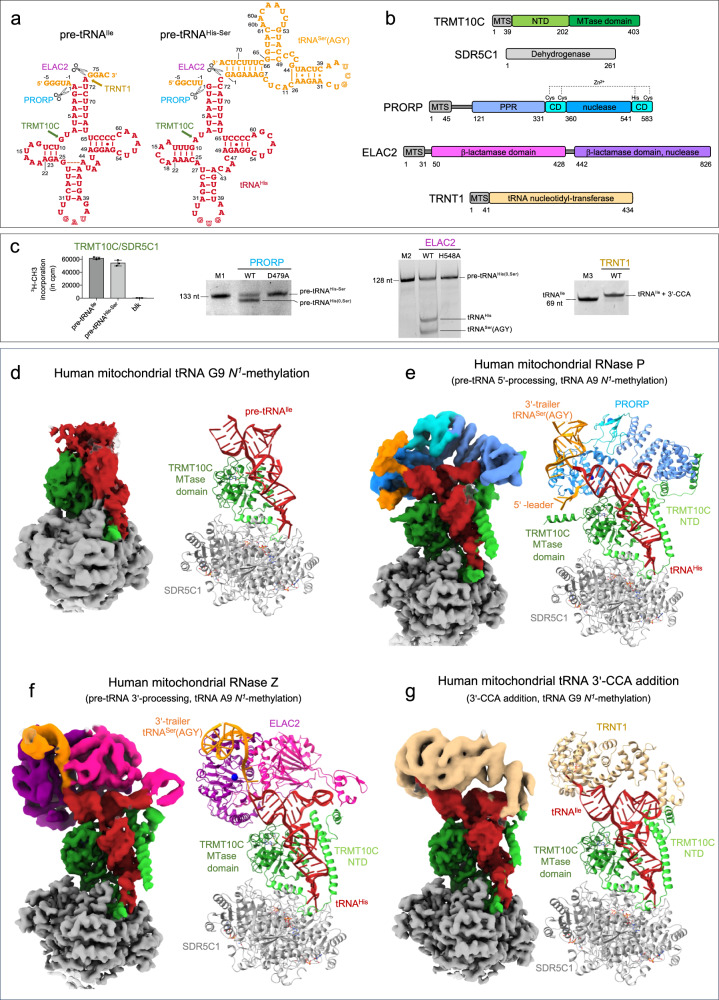
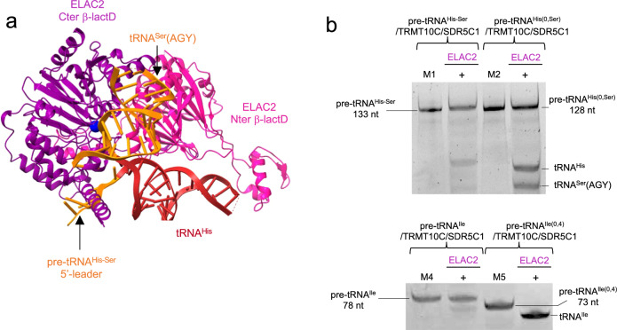
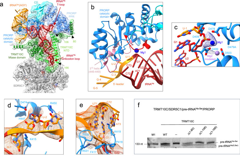
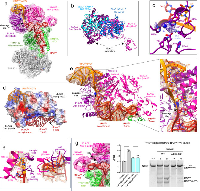
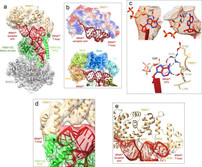
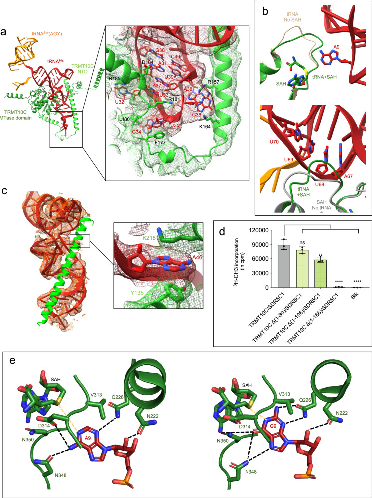
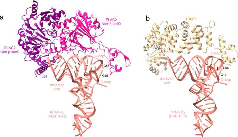
The human mitochondrial genome is transcribed into two RNAs, containing mRNAs, rRNAs and tRNAs, all dedicated to produce essential proteins of the respiratory chain. The precise excision of tRNAs by the mitochondrial endoribonucleases (mt-RNase), P and Z, releases all RNA species from the two RNA transcripts. The tRNAs then undergo 3'-CCA addition. In metazoan mitochondria, RNase P is a multi-enzyme assembly that comprises the endoribonuclease PRORP and a tRNA methyltransferase subcomplex. The requirement for this tRNA methyltransferase subcomplex for mt-RNase P cleavage activity, as well as the mechanisms of pre-tRNA 3'-cleavage and 3'-CCA addition, are still poorly understood. Here, we report cryo-EM structures that visualise four steps of mitochondrial tRNA maturation: 5' and 3' tRNA-end processing, methylation and 3'-CCA addition, and explain the defined sequential order of the tRNA processing steps. The methyltransferase subcomplex recognises the pre-tRNA in a distinct mode that can support tRNA-end processing and 3'-CCA addition, likely resulting from an evolutionary adaptation of mitochondrial tRNA maturation complexes to the structurally-fragile mitochondrial tRNAs. This subcomplex can also ensure a tRNA-folding quality-control checkpoint before the sequential docking of the maturation enzymes. Altogether, our study provides detailed molecular insight into RNA-transcript processing and tRNA maturation in human mitochondria.


Conflict of interest statement

The authors declare no competing interests.

Figures

Fig. 1
Fig. 1. Structures of the human mitochondrial tRNA maturation complexes.
a Secondary structure of pre-tRNAIle and pre-tRNAHis-Ser, the 5′-leader and 3′-trailer are indicated in orange, tRNASer(AGY) is the 3′-trailer of tRNAHis. The sequence of the other pre-tRNAs used in this study and the nucleotide numbering used in the atomic coordinates deposited in the PDB are shown in Supplementary Fig. 1a. The maturation sites of the enzymes studied here are indicated. b Domain organisations of TRMT10C, SDR5C1, PRORP, ELAC2 and TRNT1; MTS: mitochondrial transport signal, NTD: N-terminal domain, MTase: methyltransferase, PPR: pentatrico-peptide repeat, CD: central domain. c Activity assays of reconstituted complexes showing methylation of pre-tRNAIle and pre-tRNAHis-Ser by the TRMT10C/SDR5C1 complex, processing of pre-tRNAHis-Ser by PRORP, processing of pre-tRNAHis(0,Ser) by ELAC2 and 3′-CCA addition to tRNAIle by TRNT1 when pre-tRNAs are bound to the TRMT10C/SDR5C1 complex, PRORPD479A and ELAC2H548A are not active. pre-tRNAHis(0,Ser) has no 5′-leader sequence, Blk is an assay without TRMT10C/SDR5C1. Marker M1 is pre-tRNAHis-Ser (133 nucleotides, nt), M2 is pre-tRNAHis(0,Ser) (128 nt) and M3 is tRNAIle (69 nt). Source data are provided as a Source Data file. For methylation assay, error bars represent standard deviation, n = 3 technical replicates, gels are representative of three experiments. d Cryo-EM density map and refined model of the complex responsible for human mitochondrial tRNA G9 N1-methylation, composed of TRMT10C(SAH)/SDR5C1(NADH)/pre-tRNAIle (Complex 1). e Composite map and refined model of the human mitochondrial RNase P with pre-tRNAHis-Ser, composed of TRMT10C(SAH)/SDR5C1(NADH)/pre-tRNAHis-Ser/PRORPD479A (Complex 2). f Composite map and refined model of the human mitochondrial RNase Z with pre-tRNAHis(0,Ser), composed of TRMT10C(SAH)/SDR5C1(NADH)/pre-tRNAHis(0,Ser)/ELAC2H548A (Complex 3). g Composite map and refined model of the complex responsible for the human mitochondrial tRNA 3’-CCA addition, composed of TRMT10C(SAH)/SDR5C1(NADH)/RNAIle/TRNT1/CDP (Complex 4). Domains of enzymes are colored as in b.
Fig. 2
Fig. 2. ELAC2 cleavage is inhibited by the pre-tRNA 5′-leader.
a The pre-tRNAHis-Ser from the mt-RNase P complex makes steric clashes with ELAC2 at the level of the pre-tRNA 5′-leader, indicated by the black arrow. b Cleavage assays by ELAC2 of pre-tRNAHis-Ser, pre-tRNAHis(0,Ser), pre-tRNAIle and pre-tRNAIle(0,4) showing that ELAC2 cleavage is efficient on pre-tRNA without a 5′-leader. Markers M1, M2 are the same as in Fig. 1c, M4 is pre-tRNAIle (78 nt) and M5 is pre-tRNAIle(0,4) (73 nt). Gels are representative of three experiments, uncropped gels are in Source Data file.
Fig. 3
Fig. 3. Structure of the human mitochondrial RNase P complex processing pre-tRNAHis-Ser.
a Cryo-EM density map and cartoon representation of the structure of PRORP poised to process the TRMT10C/SDR5C1-bound pre-tRNAHis-Ser, with the color code indicated in Figure 1a, b. b Substrate-bound PRORP active site, in red loop interacting with the 5′-cleavage site and in pink, loops interacting with the 5′-leader of tRNAHis. c PRORP metal-ion binding in the active site. Residues in the active site and the pre-tRNAHis-Ser are shown in stick representation. Mg1 is shown as a marine sphere (with the density map in marine), and the Mg2+ ion labelled Mg2 as a light-purple sphere, was modeled on the basis of the PRORP1 crystal structure (PDB 4G24). d Interactions between the 5′-leader of pre-tRNAHis-Ser and PRORP. e Interactions between the 3′-cleavage site of pre-tRNAHis-Ser and PRORP. f Cleavage assays showing that PRORP activity is dependent on the interaction between PRORP and TRMT10C residues (1-106). Marker M1 is the same as in Fig. 1c, the gel is representative of three experiments, uncropped gel is in Source Data file. WT: TRMT10C/SDR5C1/PRORP/pre-tRNAHis-Ser, (-): PRORP/pre-tRNAHis-Ser, Δ(xx-yy): TRMT10C Δ(xx-yy)/SDR5C1/PRORP/ pre-tRNAHis-Ser, the location on the TRMT10C structure of the NTD residues 80, 106 and 166 are shown in a.
Fig. 4
Fig. 4. Structure of the human mitochondrial RNase Z in pre-cleavage complex with pre-tRNAHis(0,Ser).
a Cryo-EM density map and cartoon representation of the structure of ELAC2 poised to process the TRMT10C/SDR5C1-bound pre-tRNAHis(0,Ser), with the color code indicated in Fig. 1a, b. b Superimposition of ELAC2 with ELAC1 homodimer (PDB 3ZWF). c Substrate-bound ELAC2 active site and metal-ion binding. Residues in the active site and the pre-tRNAHis(0,Ser) are shown in stick representation. Zn1 is shown as a solid marine sphere (with the density map in purple), and Zn2 was modeled on the basis of the ELAC1 crystal structure (PDB 3ZFW), and is shown as a light-purple sphere. d Vacuum electrostatics surface representation of ELAC2 in the complex with TRMT10C/SDR5C1-bound pre-tRNAHis(0,Ser). Positive charges are in blue, whereas negative ones are in red with the maximum color saturation corresponding to −5 kT/e (red) and +5 kT/e (blue). pre-tRNAHis(0,Ser) is represented as ribbon and sticks with the color code indicated in Fig. 1a. e Substrate-bound ELAC2 active site and pre-tRNAHis(0,Ser) binding, the insets shows the binding of C73 from tRNAHis and, G1 and A2 from tRNASer(AGY) at the interface between the ELAC2 β-lactamase domains with the density map for the tRNAs in orange. f Interactions between ELAC2 and, C73 from tRNAHis and G1 from tRNASer(AGY); Interactions between ELAC2 and G1 from tRNAHis; the density map is represented in orange for the tRNAs. g Density map showing the interactions between ELAC2 and TRMT10C at the level of the T-loop of tRNAHis; DSF assays testing the ability of ELAC2 and deletion mutant Δ(249-302) to bind pre-tRNAHis(0,Ser). Data were analysed by one-way ANOVA using a F test statistic. P-values are indicated as follows: ns for P = 0.61 and **** for P < 0.0001, n = 3 technical replicates; the error bars are SD. Cleavage assays of ELAC2 and deletion mutant Δ(249-302) on pre-tRNAHis(0,Ser) for 5 and 30 minutes, marker M2 is as in Fig. 1c. The gel is representative of three experiments. Source data are provided as a Source Data file.
Fig. 5
Fig. 5. Structure of the human mitochondrial CCA-adding enzyme maturing tRNAIle.
a Cryo-EM density map and cartoon representation of the structure of TRNT1 poised to add the first cytosine to tRNAIle, with the color code of Fig. 1a, b. b Vacuum electrostatics surface representation of TRNT1 in the TRMT10C(SAH)/SDR5C1(NADH)/tRNAIle/TRNT1 structure. Positive charges are in blue, whereas negative ones are in red with the maximum color saturation corresponding to −5 kT/e (red) and +5 kT/e (blue). tRNAIle is represented as red ribbon and sticks. The different domains of TRNT1 are indicated. c Cryo-EM density map showing the stacking of A73 of tRNAIle with the CDP in the TRNT1 active site and base-specific interactions between TRNT1 and CDP. Potential hydrogen bonds are shown as dashed black lines. The residues of the catalytic site are represented as beige sticks. d, Interaction between TRNT1 (in beige), the T-loop of tRNAIle (in red) and TRMT10C (in green). e Interactions between the body and tail domains of TRNT1 (in beige) and tRNAIle (in red).
Fig. 6
Fig. 6. TRMT10C interactions with pre-tRNA for purine-9 N1-methylation.
a Overview of the TRMT10C/pre-tRNAHis-Ser structure (Complex 2), SDR5C1 and PRORP have been removed for clarity. TRMT10C is represented as cartoon with the color code indicated in Fig. 1 and the pre-tRNAHis-Ser as red ribbon and sticks. The inset shows a close-up of the anticodon loop and TRMT10C structure in the cryo-EM density map. b Conformation of the SAM/SAH-binding loop in the TRMT10C MTase domain upon binding of SAH and tRNA (complex 2, in green) compared to the conformation in the structure with tRNA and without SAM/SAH (PDB 7ONU, in beige); Conformation of the RNA-binding loop in the TRMT10C MTase domain upon binding of tRNA (complex 2, in green) compared to the conformation in the structure of TRMT10C with no tRNA, but with SAM (PDB 5NFJ, in grey). c Interactions between tRNAHis (in red) and the helix (126-166, in green) of TRMT10C, the inset shows the interaction of nucleotide A46 with Y135 from TRMT10C NTD and K218 from TRMT10C MTase domain. d Methyltransferase assays with TRMT10C with different N-terminal deletions, Blk: assay without pre-tRNA. Data were analysed by one-way ANOVA using a Dunnett’s multiple comparison test. P-values are indicated as follows: ns for P = 0.15, *** for P = 0.0004 and **** for P < 0.0001, n = 3 technical replicates. The error bars are SD. Source data are provided as a Source Data file. e Interactions between A9 of pre-tRNAHis-Ser and G9 of pre-tRNAIle with conserved residues in the active site of TRMT10C. Potential hydrogen bonds are shown as dashed black lines.
Fig. 7
Fig. 7. ELAC2 and TRNT1 interactions with human cytosolic tRNAs.
a Model of the ELAC2/human cytosolic tRNALys3 complex. b Model of the TRNT1/human cytosolic tRNALys3 complex.

Similar articles

Cited by

References

    1. Anderson S, et al. Sequence and organization of the human mitochondrial genome. Nature. 1981;290:457–465. doi: 10.1038/290457a0. - DOI - PubMed
    1. Ojala D, Merkel C, Gelfand R, Attardi G. The tRNA genes punctuate the reading of genetic information in human mitochondrial DNA. Cell. 1980;22:393–403. doi: 10.1016/0092-8674(80)90350-5. - DOI - PubMed
    1. Ojala D, Montoya J, Attardi G. tRNA punctuation model of RNA processing in human mitochondria. Nature. 1981;290:470–474. doi: 10.1038/290470a0. - DOI - PubMed
    1. Montoya J, Christianson T, Levens D, Rabinowitz M, Attardi G. Identification of initiation sites for heavy-strand and light-strand transcription in human mitochondrial DNA. Proc. Natl Acad. Sci. USA. 1982;79:7195–7199. doi: 10.1073/pnas.79.23.7195. - DOI - PMC - PubMed
    1. Montoya J, Ojala D, Attardi G. Distinctive features of the 5’-terminal sequences of the human mitochondrial mRNAs. Nature. 1981;290:465–470. doi: 10.1038/290465a0. - DOI - PubMed

MeSH terms