

VCA supercooling in a swine partial hindlimb model
- PMID: 38824189
- PMCID: PMC11144209
- DOI: 10.1038/s41598-024-63041-8
VCA supercooling in a swine partial hindlimb model
Abstract
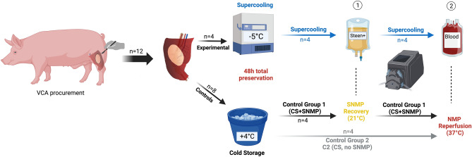
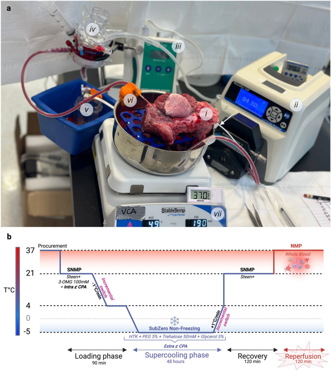
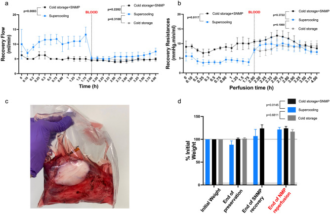
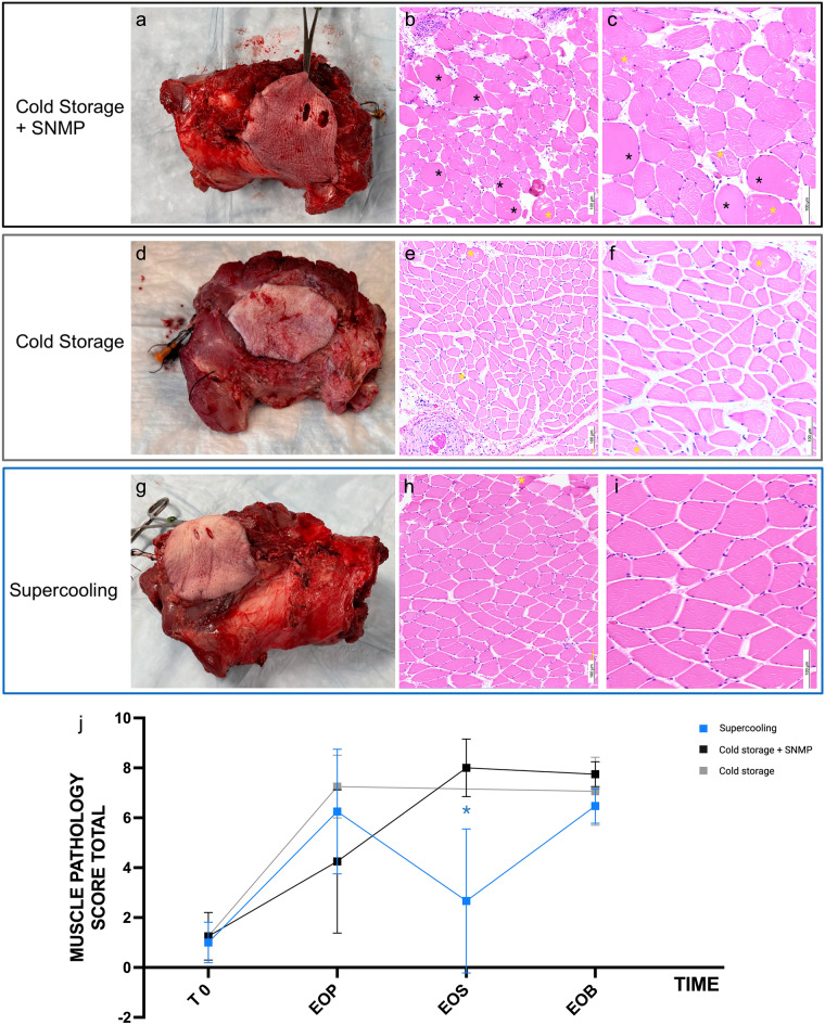
Vascularized composite allotransplantations are complex procedures with substantial functional impact on patients. Extended preservation of VCAs is of major importance in advancing this field. It would result in improved donor-recipient matching as well as the potential for ex vivo manipulation with gene and cell therapies. Moreover, it would make logistically feasible immune tolerance induction protocols through mixed chimerism. Supercooling techniques have shown promising results in multi-day liver preservation. It consists of reaching sub-zero temperatures while preventing ice formation within the graft by using various cryoprotective agents. By drastically decreasing the cell metabolism and need for oxygen and nutrients, supercooling allows extended preservation and recovery with lower ischemia-reperfusion injuries. This study is the first to demonstrate the supercooling of a large animal model of VCA. Porcine hindlimbs underwent 48 h of preservation at - 5 °C followed by recovery and normothermic machine perfusion assessment, with no issues in ice formation and favorable levels of injury markers. Our findings provide valuable preliminary results, suggesting a promising future for extended VCA preservation.
© 2024. The Author(s).
Conflict of interest statement
K.U., C.L.C, A.G.L., M.T and Y.B. have patent applications relevant to this field. K.U. and M.T. have financial interests in and serve on the Scientific Advisory Board for Sylvatica Biotech Inc., a private company developing high subzero organ preservation technologies. Competing interests for Massachusetts General Hospital investigators are managed by the MGH and MGB in accordance with their conflict-of-interest policies.
Figures

Similar articles
-
Enhanced VCA Storage: A Pilot Study Demonstrating Supercooling in Orthotopic Rodent Hindlimb Transplantation.Transplant Proc. 2024 Nov;56(9):2039-2045. doi: 10.1016/j.transproceed.2024.10.006. Epub 2024 Oct 28. Transplant Proc. 2024. PMID: 39490377
-
Sub-zero non-freezing of vascularized composite allografts in a rodent partial hindlimb model.Cryobiology. 2024 Sep;116:104950. doi: 10.1016/j.cryobiol.2024.104950. Epub 2024 Aug 24. Cryobiology. 2024. PMID: 39134131 Free PMC article.
-
Supercooling: A Promising Technique for Prolonged Organ Preservation in Solid Organ Transplantation, and Early Perspectives in Vascularized Composite Allografts.Front Transplant. 2023;2:1269706. doi: 10.3389/frtra.2023.1269706. Epub 2023 Oct 23. Front Transplant. 2023. PMID: 38682043 Free PMC article.
-
Advances in machine perfusion, organ preservation, and cryobiology: potential impact on vascularized composite allotransplantation.Curr Opin Organ Transplant. 2018 Oct;23(5):561-567. doi: 10.1097/MOT.0000000000000567. Curr Opin Organ Transplant. 2018. PMID: 30080697 Free PMC article. Review.
-
Subzero organ preservation: the dawn of a new ice age?Curr Opin Organ Transplant. 2017 Jun;22(3):281-286. doi: 10.1097/MOT.0000000000000403. Curr Opin Organ Transplant. 2017. PMID: 28266941 Free PMC article. Review.
Cited by
-
Enhanced VCA Storage: A Pilot Study Demonstrating Supercooling in Orthotopic Rodent Hindlimb Transplantation.Transplant Proc. 2024 Nov;56(9):2039-2045. doi: 10.1016/j.transproceed.2024.10.006. Epub 2024 Oct 28. Transplant Proc. 2024. PMID: 39490377
-
Vascular Microphysiological System for Investigating Endothelial Barrier Function During Organ Preservation and Reperfusion.Small. 2025 Mar;21(11):e2410168. doi: 10.1002/smll.202410168. Epub 2025 Feb 19. Small. 2025. PMID: 39972937 Free PMC article.
-
Experimental Swine Models for Vascularized Composite Allotransplantation and Immunosuppression: A Systematic Review and Case Report of a Novel Heterotopic Hemifacial Swine Model.Transpl Int. 2025 Jul 29;38:14520. doi: 10.3389/ti.2025.14520. eCollection 2025. Transpl Int. 2025. PMID: 40799314 Free PMC article. Review.
-
Dielectric properties of individual cryoprotective agents and cocktails VS55, M22, DP6 at subzero temperatures for cryopreservation.Sci Rep. 2025 Jul 1;15(1):20734. doi: 10.1038/s41598-025-07207-y. Sci Rep. 2025. PMID: 40593022 Free PMC article.
-
Beyond the icebox: modern strategies in organ preservation for transplantation.Clin Transplant Res. 2024 Dec 31;38(4):377-403. doi: 10.4285/ctr.24.0039. Clin Transplant Res. 2024. PMID: 39743232 Free PMC article. Review.
References
MeSH terms
Substances
Grants and funding
- W81XWH-17-1-0440/U.S. Army Medical Research Acquisition Activity
- R56 AI171958/AI/NIAID NIH HHS/United States
- Prix mobilité 2021/Centre Hospitalier Universitaire de Rennes
- R01 EB028782/EB/NIBIB NIH HHS/United States
- W81XWH-17-1-0437/U.S. Army Medical Research Acquisition Activity
- 09-2022/Fondation des Gueules Cassées
- EEC 1941543/National Science Foundation
- 84308-BOS-22/Shriners Hospitals for Children
- R01EB028782/National Institute for Health Care Management Foundation
- R56AI171958/National Institute for Health Care Management Foundation
- 85105-BOS-23/Shriners Hospitals for Children
LinkOut - more resources
Full Text Sources
