. 2024 Jun 1;15(1):4681.
doi: 10.1038/s41467-024-49072-9.

A persistent variant telomere sequence in a human pedigree

Affiliations

A persistent variant telomere sequence in a human pedigree

Angela M Hinchie et al. Nat Commun. .

Abstract

The telomere sequence, TTAGGG, is conserved across all vertebrates and plays an essential role in suppressing the DNA damage response by binding a set of proteins termed shelterin. Changes in the telomere sequence impair shelterin binding, initiate a DNA damage response, and are toxic to cells. Here we identify a family with a variant in the telomere template sequence of telomerase, the enzyme responsible for telomere elongation, that led to a non-canonical telomere sequence. The variant is inherited across at least one generation and one family member reports no significant medical concerns despite ~9% of their telomeres converting to the novel sequence. The variant template disrupts telomerase repeat addition processivity and decreased the binding of the telomere-binding protein POT1. Despite these disruptions, the sequence is readily incorporated into cellular chromosomes. Incorporation of a variant sequence prevents POT1-mediated inhibition of telomerase suggesting that incorporation of a variant sequence may influence telomere addition. These findings demonstrate that telomeres can tolerate substantial degeneracy while remaining functional and provide insights as to how incorporation of a non-canonical telomere sequence might alter telomere length dynamics.


Conflict of interest statement

The authors declare no competing interests.

Figures

Fig. 1
Fig. 1. A variant in telomerase RNA template alters the canonical telomere sequence.
a Pedigree of the family carrying the TERC r.50 C > A variant. The proband (arrow) was diagnosed with IPF at age 43. Asterisks indicate individuals from which DNA was available. Squares indicate males and circles indicate females. A line through the symbol indicates the individual is deceased. b Apical (upper) and basal (lower) chest CT showing interstitial changes and advanced fibrosis in the proband before lung transplantation. c Graphic showing the location of the patient-derived variant in the template of TR and the resulting telomere sequence. Figure was created with BioRender.com and released under a Creative Commons Attribution-NonCommercial-NoDerivs 4.0 international license. d Excerpts of telomeric sequences from whole genome sequencing of an individual with no template mutation, the proband, and the proband’s son. Canonical repeats are highlighted in blue, variant TTAGGT repeats are highlighted in yellow, additional non-canonical sequences are in white. e The percentage of TTAGGT repeats in controls, the proband, and the proband’s son. f PNA-FISH for wild-type (red) and variant (green) sequence in a tissue section of the proband’s explanted lung and a control donated lung. Source data for (e) is provided as a Source Data file.
Fig. 2
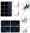
Fig. 2. The variant template compromises repeat addition processivity.
a Schematic showing the template-telomere mismatch after the addition of the variant telomere sequence. Graphic was created with BioRender.com and released under a Creative Commons Attribution-NonCommercial-NoDerivs 4.0 international license. b Variant templates used and the resulting telomere sequence. Changes to the sequence are shown in red. c Telomerase direct assay with WT, C50A, and C50/56A telomerase RNA templates. RAP was measured extending from both a wild-type (-GGTTAGx3) and variant (-GTTTAGx3) primer. * Indicates the location of the radiolabeled primer and the numbers indicate the number of hexanucleotide repeats that were added. d Telomerase direct assay with primer A5, (TTAGGGTTAGCGTTAGGG) with the addition of POT1-TPP1 processivity factors. Primer A5 blocks 3` POT1 binding while allowing for processive addition. * Indicates the location of the radiolabeled A5 primer and the numbers on the side of the gel indicate the number of telomeric repeats that were added. The A5 primer has two additional Gs at the three prime end so that only 4 nucleotides are added in the first cycle. e Quantification of the percentage of product that extended beyond one repeat from (c). Mean±s.d is shown, n = 3 biological replicates, and groups were compared with one-way ANOVA with Tukey’s multiple comparisons test. f Schematic showing the expected conformation of WT and C50A TR with the nascent telomere over the leucine zipperhead described in Wan et al. g Proportion of TTAGGG (blue/light blue) and TTAGGT (yellow/orange) consecutive repeats found in WGS data from the proband and his son. Lines are also shown for theoretical numbers of consecutive repeats for non-processive addition (gray line). h Model of in vivo processivity. i Probability of processive addition from the WGS data, mean±s.d is shown, n = 2 (proband and son). ns, non-significant p ≥0.05, ****p < 0.0001. Source data are provided as a Source Data file.
Fig. 3
Fig. 3. Incorporation and persistence of a variant telomere sequence in cells.
a Representative photomicrograph of interphase FISH with PNA probes to the wild-type and variant sequence in hTERT-RPE cells 6 days post-transduction with empty vector (EV), WT, C50A or C50/56A TR. b Quantification of the percentage of cells with 10 or more visible variant telomere foci in (a). c Mean intensity per cell of the variant telomeres in (a) normalized to C50A. For (b) and (c), n = 3 biological replicates and mean±s.d is shown and at least 60 cells were evaluated in each independent experiment (see Source Data for exact numbers). d Telomere restriction fragment (TRF) southern blot of HCT116 cells co-transduced with hTERT and either and empty vector (EV), WT, C50A, or C50/56A TR, 15 days post-transduction. e Quantification of TRF southern blot in (d), n = 3 biological replicates, mean ± s.d. is shown. f Representative photomicrograph of metaphase FISH of LOX Melanoma cells with the wild-type and variant sequence at 9- and 85-days post-transduction with C50A or C50/56A TR. g Quantification of variant telomere intensity per metaphase of (f) normalized to C50A Day 9. Total metaphases analyzed as follows: C50A Day 9 (18), C50/56A Day 9 (22), C50A Day 85 (25), C50/56A Day 85 (20), median±s.d. is shown. Groups were compared with a two-tailed Student’s unpaired t test. Groups in (b), (c), and (e) were compared with one-way ANOVA and Tukey’s multiple comparison test. For all pair-wise comparisons, ns, non-significant p ≥ 0.05, ****p < 0.0001. Source data are provided as a Source Data file.
Fig. 4
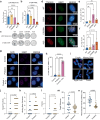
Fig. 4. Effects of variant sequence addition on DDR and chromosome stability.
a, b Proliferation assays for LOX Melanoma and hTERT-RPE cells following transduction with lentiviruses expressing the indicated TR. Data are from n = 3 biological replicates. Mean±s.d. is shown. c Representative images of GFP-positive cells in the proliferation assay 7- (LOX Melanoma) or 9-days (hTERT-RPE) after transduction. d Representative photomicrograph of 53BP1 and telomere FISH staining in hTERT-RPE cells transduced with variant TR, 6 days post-transduction. e Quantification of telomere-induced foci (TIFs), of 53BP1 colocalized with the telomere of the cells in (d). f Quantification of large nuclei (>200 μm2) of the cells in (d). For (e) and (f) data are n = 3 biological replicates and mean±s.d. is shown. g Representative photomicrograph of 53BP1 staining in the proband and control lymphoblast, with a 4 Gy irradiated control. h Quantification of 53BP1 foci from (g), n = 4 independent experiments, mean±s.d is shown. Groups were compared with by one-way ANOVA with Tukey’s multiple comparison test. i Representative images of chromosomal abnormalities quantified in lymphoblast metaphases. jm Quantification of chromosomal abnormalities per metaphase in lymphoblast cell lines, control n = 20, proband n = 22. Each point is a single metaphase, only the mean is shown. Groups were compared with a Mann–Whitney test. n Mean telomere intensity from the same samples as (j-m), only the mean is shown. Groups were compared with a Mann-Whitney test. For (a), (b), (e), and (f), groups were compared by one-way ANOVA with Dunnet’s multiple comparison test. For all comparisons, ns, non-significant, p ≥0.05 and ****p < 0.0001. Source data are provided as a Source Data file.
Fig. 5
Fig. 5. Telomere length and dynamics caused by a variant telomere sequence.
a, b FlowFISH telomere length measured in the proband seven years post-lung transplantation. Telomere length is shown relative to a nomogram of leukocyte telomere lengths from healthy controls. Lymphocyte (a) and granulocyte (b) telomere length are shown. c Telomere intensity distribution for individual telomeres for the control and proband cell lines for both interphase (top) and metaphase (bottom) chromosomes. Telomeres are normalized to the mean telomere intensity within each cell. d Average intensity of wild-type telomere probe for wild-type telomeres either associated or not associated with a variant telomere, averaged per cell (n = 61) or metaphase (n = 12). Intensity was normalized the same as (c). Groups were compared with Mann–Whitney test and the mean is shown. e Telomere intensity distribution for individual telomeres either associated or not associated with variant telomere foci for both interphase (top) and metaphase (bottom) chromosomes. Normalization was the same as (c). f Schematic showing the C-tailed PCR product for sequencing of the terminal telomere. g The percentage of telomeres that terminate in a wild-type vs variant sequence. h Occurrence of each permutation as a fraction of total telomere ends of the wild-type or variant sequence. The arrow indicates the permutation expected based on the biochemically predicated pause site for both wild-type and variant TR. Data for the wild-type sequences were determined from both the control and patient-derived lymphoblasts whereas the data for the variant sequence could be determined only for the patient-derived lymphoblasts. ****p < 0.0001. Source data are provided as a Source Data file.
Fig. 6
Fig. 6. Proposed mechanism for tolerance and telomere length maintenance.
a Graphic describing the opposing effects a variant telomere sequence has on telomere length maintenance. b A binding curve for POT1 generated from electromobility shift assays (EMSAs) for POT1 with single-stranded DNA oligonucleotides corresponding to either the G-strand wild-type telomere sequence (WT; n = 6), the variant TTAGGT sequence (C50A; n = 9), or a G to C substitution of the wild-type sequence (TTAGCG; n = 3). The latter is known to inhibit POT1 binding and serves as a negative control. Mean ± s.d. is shown. Statistical comparison between WT and TTAGGT binding is shown, groups are compared with two-way ANOVA with Tukey’s multiple comparison test. c Quantification of TRF Southern blot of HCT116 cells 13 days post-transduction. Cells were co-transduced with WT or C50/56A TR and POT1ΔOB. n = 3 for each group. Mean ± s.d. is shown, WT and C50/56A were compared by a two-tailed unpaired t test. d Model showing how shelterin might flexibly bind around the variant telomere sequence to effectively protect the telomere, while variant telomere sequence at the telomere end primes it for telomerase elongation due to decreased POT1 binding, attenuating the effect of reduced RAP from the variant sequence. The graphic was created with BioRender.com and released under a Creative Commons Attribution-NonCommercial-NoDerivs 4.0 international license. ns, non-significant, ***p < 0.001, ****p < 0.0001. Source data are provided as a Source Data file.

References

    1. Meyne J, Ratliff RL, Moyzis RK. Conservation of the human telomere sequence (TTAGGG)n among vertebrates. Proc. Natl. Acad. Sci. USA. 1989;86:7049–7053. doi: 10.1073/pnas.86.18.7049. - DOI - PMC - PubMed
    1. Bianchi A, et al. TRF1 binds a bipartite telomeric site with extreme spatial flexibility. EMBO J. 1999;18:5735–5744. doi: 10.1093/emboj/18.20.5735. - DOI - PMC - PubMed
    1. Loayza D, Parsons H, Donigian J, Hoke K, de Lange T. DNA binding features of human POT1 a nonamer 5′-TAGGGTTAG-3′ minimal binding site, sequence specificity, and internal binding to multimeric sites. J. Biol. Chem. 2004;279:13241–13248. doi: 10.1074/jbc.M312309200. - DOI - PubMed
    1. de Lange T. Shelterin-mediated telomere protection. Annu. Rev. Genet. 2018;52:223–247. doi: 10.1146/annurev-genet-032918-021921. - DOI - PubMed
    1. Wang F, et al. The POT1–TPP1 telomere complex is a telomerase processivity factor. Nature. 2007;445:506–510. doi: 10.1038/nature05454. - DOI - PubMed