. 2024 Jun 2;6(1):27.
doi: 10.1186/s42836-024-00248-0.

Stem anteversion is not affected by proximal femur geometry in robotic-assisted total hip arthroplasty

Affiliations

Stem anteversion is not affected by proximal femur geometry in robotic-assisted total hip arthroplasty

Andrea Marcovigi et al. Arthroplasty. .

Abstract

Background: In the present study, the surgeon aimed to align the stem at 5° to 25° in anteversion. The robotic technology was used to measure stem anteversion with respect to proximal femur anteversion at different levels down the femur.

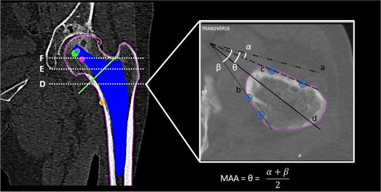
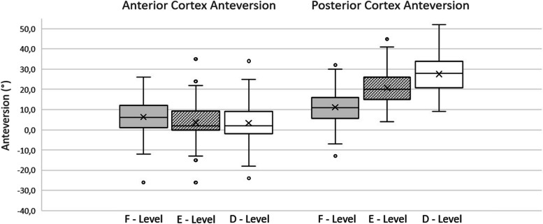
Methods: A total of 102 consecutive patients underwent robotic-arm-assisted total hip arthroplasty (RTHA). 3D CT-based preoperative planning was performed to determine femoral neck version (FNV), posterior cortex anteversion (PCA), anterior cortex anteversion (ACA), and femoral metaphyseal axis anteversion (MAA) at 3 different levels: D (10 mm above lesser trochanter), E (the midpoint of the planned neck resection line) and F (head-neck junction). The robotic system was used to define and measure stem anteversion during surgery.

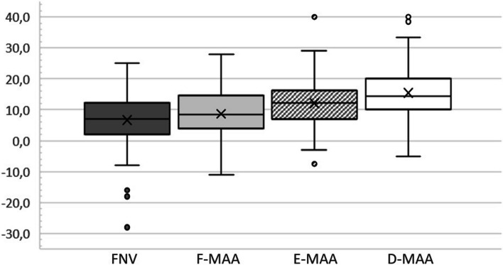
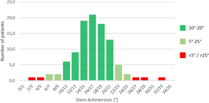
Results: Mean FNV was 6.6° (SD: 8.8°) and the mean MAA was consistently significantly higher than FNV, growing progressively from proximal to distal. Mean SV was 16.4° (SD: 4.7°). There was no statistically significant difference (P = 0.16) between SV and MAA at the most distal measured level. In 96.1% cases, the stem was positioned inside the 5°-25° anteversion range.

Conclusions: Femoral anteversion progressively increased from neck to proximal metaphysis. Aligning the stem close to femoral anteversion 10 mm above the lesser trochanter often led to the desired component anteversion.

Keywords: Combined anteversion; Robotic arm-assisted surgery; Stem anteversion; Total hip arthroplasty.


Conflict of interest statement

F.C. reports a relationship with Stryker Orthopaedics that includes: consulting or advisory and travel reimbursement. Royalties from Stryker Orthopaedics; M.P. reports a relationship with AB Medica s.p.a. that includes: employment; F.Z.: Guest Editor for Arthroplasty, and all authors were not involved in the journal’s review or decisions related to this manuscript. Other authors declare that they have no competing interests.

Figures

Fig. 1
Fig. 1
Metaphyseal anteversion measurement at D level: “a” is a line parallel to transepicondylar axis, passing through the point of intersection of “b” and “c” lines; “b” is the line passing through two different points on femoral posterior cortex and represents the posterior cortex axis; “c” is the line passing through two different points on femoral anterior cortex and represents the anterior cortex axis; “d” line is the bisector of the angle formed by anterior and posterior cortex axes and represents the metaphyseal axis. The α angle is equivalent to the angle formed by the anterior cortex axis and the transepicondylar axis, which is defined as anterior cortex anteversion (ACA). The β angle is equivalent to the angle formed by the posterior cortex axis and the transepicondylar axis, and is defined as posterior cortex anteversion (PCA). The θ angle is equivalent to the angle formed between the metaphyseal axis and the transepicondylar axis (metaphyseal axis anteversion: MAA) and, knowing the value of ACA and PCA, could be calculated with the simplified formula shown in the figure
Fig. 2
Fig. 2
Mean FNV compared with mean metaphyseal axis at F, E and D levels. Femoral anteversion characterized by a progressive increase from proximal to distal metaphysis
Fig. 3
Fig. 3
Anterior and posterior cortex mean values. Anterior cortex version was characterized by a slight decrease from level F to level E, while there was no statistically significant difference between E level and D level. Posterior cortex showed a progressive increase in anteversion from level F to level D
Fig. 4
Fig. 4
Stem anteversion distribution in the considered population

Similar articles

Cited by

References

    1. Harrison CL, Thomson AI, Cutts S, Rowe PJ, Riches PE. Research synthesis of recommended acetabular cup orientations for total hip arthroplasty. J Arthroplasty. 2014;29(2):377–382. doi: 10.1016/j.arth.2013.06.026. - DOI - PubMed
    1. Dorr LD, Malik A, Dastane M, Wan Z. Combined anteversion technique for total hip arthroplasty. Clin Orthop Relat Res. 2009;467:119–127. doi: 10.1007/s11999-008-0598-4. - DOI - PMC - PubMed
    1. Widmer KH, Zurfluh B. Compliant positioning of total hip components for optimal range of motion. J Orthop Res. 2004;22(4):815–821. doi: 10.1016/j.orthres.2003.11.001. - DOI - PubMed
    1. Yoshimine F. The safe-zones for combined cup and neck anteversions that fulfill the essential range of motion and their optimum combination in total hip replacements. J Biomech. 2006;39(7):1315–1323. doi: 10.1016/j.jbiomech.2005.03.008. - DOI - PubMed
    1. Hayashi S, Hashimoto S, Matsumoto T, Takayama K, Nishida K, Ishida K, Kuroda R. Stem anteversion mismatch to the anatomical anteversion causes loss of periprosthetic bone density after THA. J Orthop Surg. 2017;25(3):2309499017739478. doi: 10.1177/2309499017739478. - DOI - PubMed

LinkOut - more resources