. 2024 Jun 21;5(2):103100.
doi: 10.1016/j.xpro.2024.103100. Epub 2024 Jun 1.

Protocol for generating three-dimensional induced early ameloblasts using serum-free media and growth factors

Affiliations

Protocol for generating three-dimensional induced early ameloblasts using serum-free media and growth factors

Ammar Alghadeer et al. STAR Protoc. .

Abstract

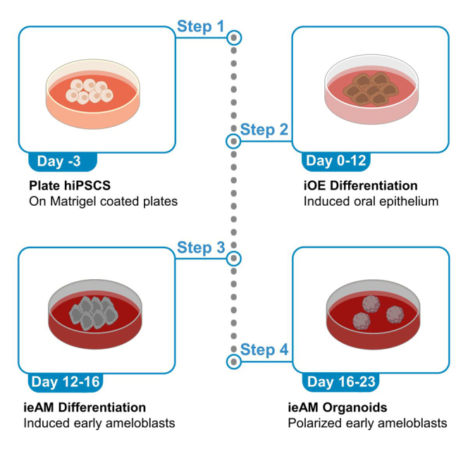
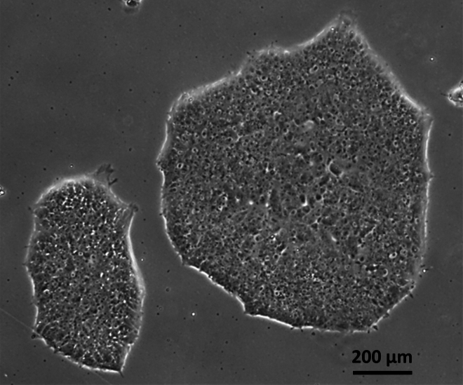
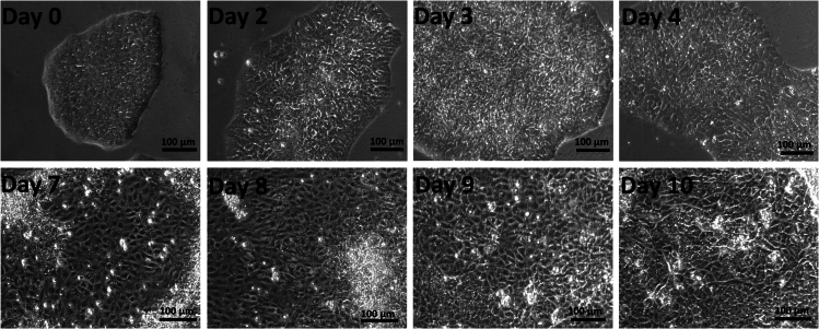
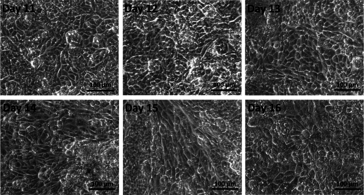
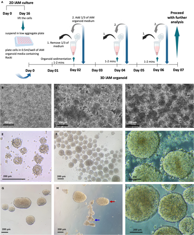
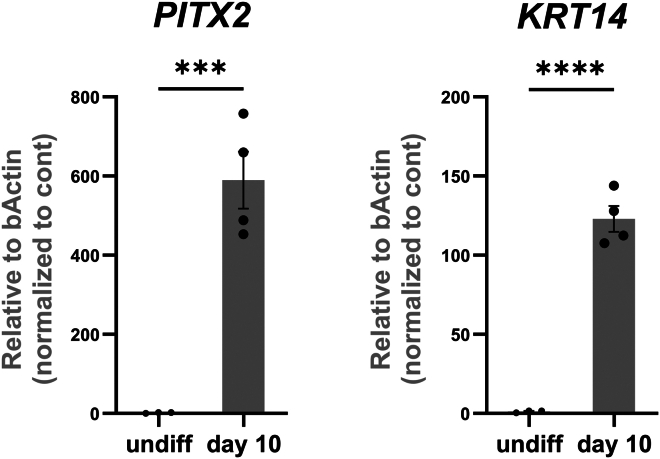
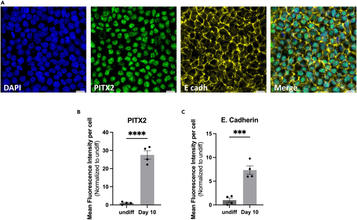
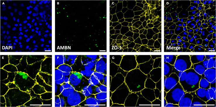
Adult humans cannot regenerate the enamel-forming cell type, ameloblasts. Hence, human induced pluripotent stem cell (hiPSC)-derived ameloblasts are valuable for investigating tooth development and regeneration. Here, we present a protocol for generating three-dimensional induced early ameloblasts (ieAMs) utilizing serum-free media and growth factors. We describe steps for directing hiPSCs toward oral epithelium and then toward ameloblast fate. These cells can form suspended early ameloblast organoids. This approach is critical for understanding, treating, and promoting regeneration in diseases like amelogenesis imperfecta. For complete details on the use and execution of this protocol, please refer to Alghadeer et al.1.

Keywords: Cell Differentiation; Developmental biology; Organoids; Stem Cells.


Conflict of interest statement

Declaration of interests A.A., A.P.P., J.M., and H.R.-B. are co-inventors on a patent application entitled “A Method to Direct the Differentiation of Human Induced Pluripotent Stem Cells into Early Ameloblasts” (PCT/US2022/053517 filed 12/20/2022).

Figures

None
Graphical abstract
Figure 1
Figure 1
Bright-field image of hiPSC colonies
Figure 2
Figure 2
Bright-field images of day 0 to day 10 iOE of in vitro differentiation A change in morphology is seen, starting from the periphery moving inwards. The cells’ shape changes from circular to polygonal and they increase in size. The nuclear-cytoplasmic ratio decreases, and the nuclei become darker. The cells become flatter, spread out, and assume a cobblestone morphology by day 10.
Figure 3
Figure 3
Bright-field images of day 11 to day 16 of iAM in vitro differentiation Here, the cells continue to change in morphology. The cells become elongated. The nuclei contract in size and become denser. Cytoplasmic granules become evident.
Figure 4
Figure 4
Formation of ieAM organoids (A) A schematic of the culture procedure for organoid formation and maintenance. (B‒D) Bright-field images of ieAM organoids on day 0 to day 2. (E‒G) Bright-field image of ieAM organoids on day 2 after washing (E), day 4 (F), and day 7 (G). (H) Bright-field image showing examples of healthy organoids (red arrows), and irregular organoids (blue arrows). (I and J) Bright-field images of day 7 ieAM organoids before wash (I) and after wash (J).
Figure 5
Figure 5
qRT-PCR of oral epithelium markers The cells at Day 10 of differentiation (iOE) show upregulated expression of oral epithelium markers PITX2 and KRT14 as assessed by qRT-PCR. ∗∗∗p < 0.001; ∗∗∗∗p < 0.0001; Graph error bars are the means ± SEM.
Figure 6
Figure 6
Immunofluorescence staining of day 10 iOE (A) The staining shows PITX2-positive nuclei (green) and E-cadherin-positive (yellow) membranes. Scale bars: 50 μm. (B and C) Quantification of the mean fluorescence intensity of day 10 iOE markers PITX2 (B) and E-cadherin (C) in comparison to undifferentiated hiPSCs. Quantification was done using Image J (Image J2 v2.14.0). For PITX2, nuclear signal intensity was quantified and normalized to DAPI signal intensity per cell. For E-cadherin, the overall signal intensity per field was normalized to DAPI overall signal intensity per field (n = 4). 329 cells out of 350 cells showed PITX2+ expression, hence 94% of cells successfully expressed PITX2. Graph error bars are the means ± SEM. Statistical significance was determined using student T-test; ∗∗∗p ≤ 0.001; ∗∗∗∗p ≤ 0.0001.
Figure 7
Figure 7
Immunofluorescence staining of AMBN at day 16 ieAM (A‒H) The staining shows AMBN-positive cells (green) and the tight junction marker ZO-1 (yellow) at low power magnification (A‒D), and high-power magnification (E‒F) and (G‒H). Scale bar: 20 μm.
Figure 8
Figure 8
Immunofluorescence staining of SP6 at day 16 ieAM The staining shows SP6-positive nuclei (red) and the tight junction marker ZO-1 (yellow). Scale bar: 20 μm.
Figure 9
Figure 9
QRT-PCR of early ameloblasts markers The cells at Day 16 of differentiation (iAM) show upregulated expression of early ameloblast markers AMBN and SP6 as assessed by QRT-PCR. ∗∗p < 0.01; ∗∗∗p < 0.001; ∗∗∗∗p < 0.0001; Graph error bars are the means ± SEM.
Figure 10
Figure 10
Immunofluorescence staining of medium-sized day 7 ieAM organoid showing polarized early ameloblasts Apical sides of these polarized epithelial cells (marked by ZO-1, green) produce a lumen. The lumen is indicated by a white arrow, and the dying cells are indicated by a yellow arrow.
Figure 11
Figure 11
Immunofluorescence staining of smaller-sized day 7 ieAM organoid showing polarized early ameloblasts toward a lumen marked by high expression of ZO-1 (yellow)

References

    1. Alghadeer A., Hanson-Drury S., Patni A.P., Ehnes D.D., Zhao Y.T., Li Z., Phal A., Vincent T., Lim Y.C., O’Day D., et al. Single-cell census of human tooth development enables generation of human enamel. Dev. Cell. 2023;58:2163–2180.e9. doi: 10.1016/j.devcel.2023.07.013. - DOI - PMC - PubMed
    1. Schindelin J., Arganda-Carreras I., Frise E., Kaynig V., Longair M., Pietzsch T., Preibisch S., Rueden C., Saalfeld S., Schmid B., et al. Fiji: an open-source platform for biological-image analysis. Nat. Methods. 2012;9:676–682. doi: 10.1038/nmeth.2019. - DOI - PMC - PubMed
    1. Schindelin J., Rueden C.T., Hiner M.C., Eliceiri K.W. The ImageJ ecosystem: An open platform for biomedical image analysis. Mol. Reprod. Dev. 2015;82:518–529. doi: 10.1002/mrd.22489. - DOI - PMC - PubMed

Publication types

MeSH terms

Substances

LinkOut - more resources