Review
. 2024;148(11-12):796-822.
doi: 10.1159/000539602. Epub 2024 May 31.

The SLC6A18 Transporter Is Most Likely a Na-Dependent Glycine/Urea Antiporter Responsible for Urea Secretion in the Proximal Straight Tubule: Influence of This Urea Secretion on Glomerular Filtration Rate

Affiliations
Review

The SLC6A18 Transporter Is Most Likely a Na-Dependent Glycine/Urea Antiporter Responsible for Urea Secretion in the Proximal Straight Tubule: Influence of This Urea Secretion on Glomerular Filtration Rate

Lise Bankir et al. Nephron. 2024.

Abstract

Background: Urea is the major end-product of protein metabolism in mammals. In carnivores and omnivores, a large load of urea is excreted daily in urine, with a concentration that is 30-100 times above that in plasma. This is important for the sake of water economy. Too little attention has been given to the existence of energy-dependent urea transport that plays an important role in this concentrating activity.

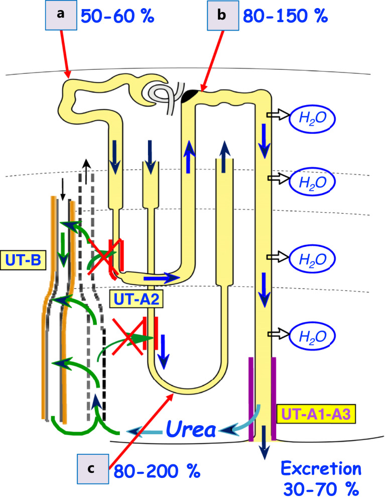
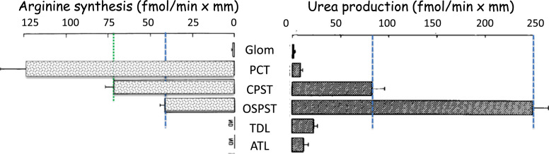
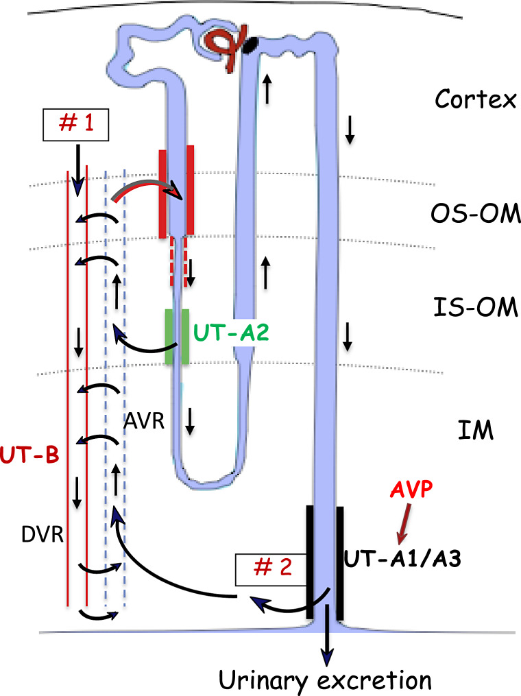
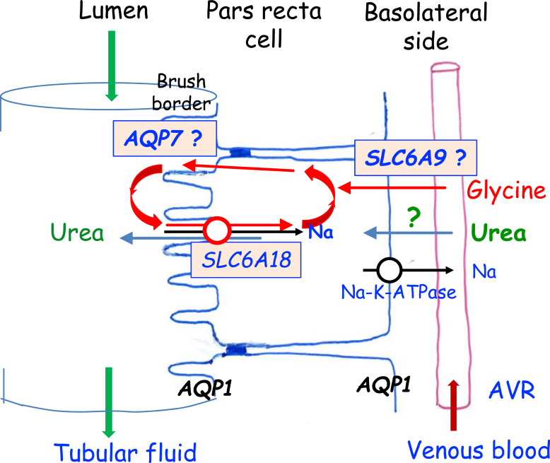
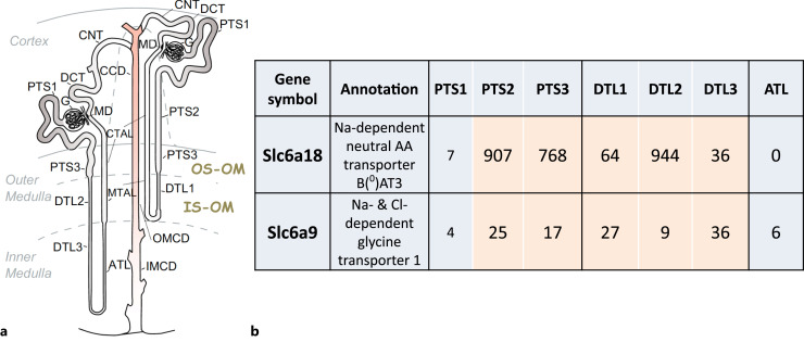
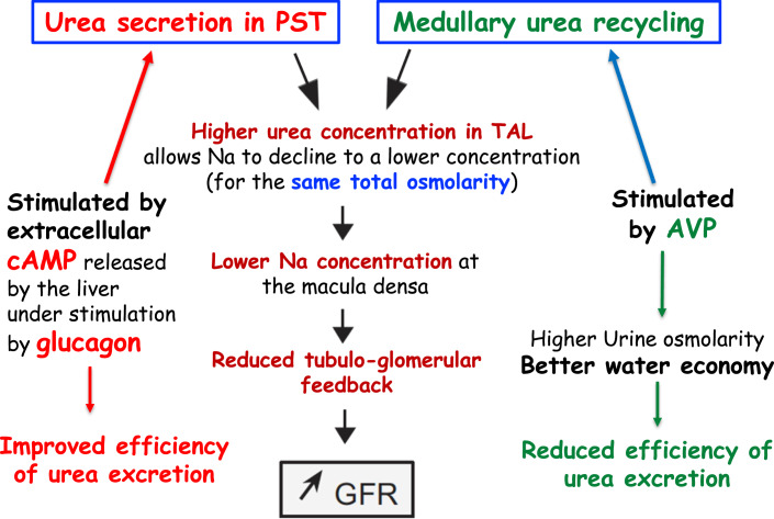
Summary: This review first presents functional evidence for an energy-dependent urea secretion that occurs exclusively in the straight part of the proximal tubule (PST). Second, it proposes a candidate transmembrane transporter responsible for this urea secretion in the PST. SLC6A18 is expressed exclusively in the PST and has been identified as a glycine transporter, based on findings in SLC6A18 knockout mice. We propose that it is actually a glycine/urea antiport, secreting urea into the lumen in exchange for glycine and Na. Glycine is most likely recycled back into the cell via a transporter located in the brush border. Urea secretion in the PST modifies the composition of the tubular fluid in the thick ascending limb and, thus, contributes, indirectly, to influence the "signal" at the macula densa that plays a crucial role in the regulation of the glomerular filtration rate (GFR) by the tubulo-glomerular feedback.

Key messages: Taking into account this secondary active secretion of urea in the mammalian kidney provides a new understanding of the influence of protein intake on GFR, of the regulation of urea excretion, and of the urine-concentrating mechanism.

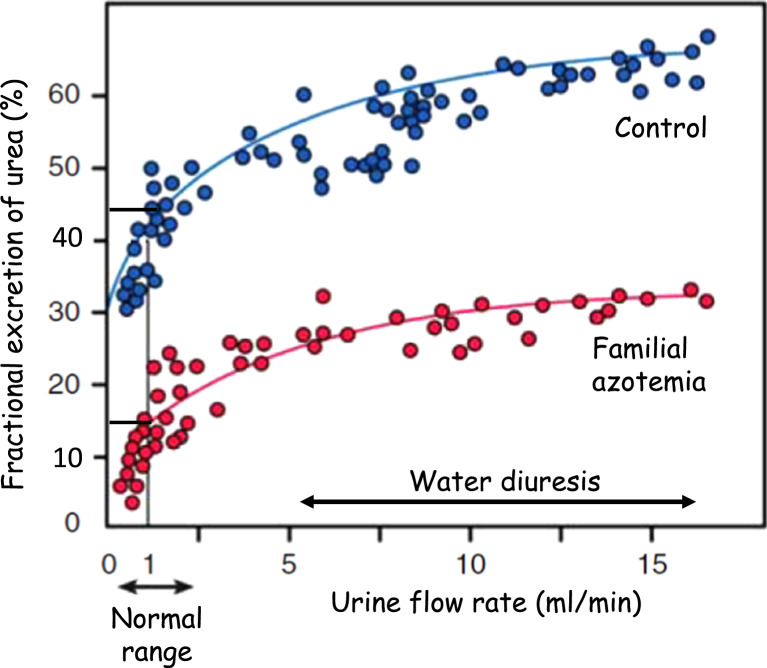
Keywords: Familial azotemia; Fractional excretion; Glomerular filtration rate; Glycine; Pars recta.


Conflict of interest statement

No conflict of interest for any of the authors.

Figures

Fig. 1.
Fig. 1.
Schematic representation of a short-looped nephron and the loop of Henle of a long-looped nephron showing urea transport in green and the percentages of filtered urea found in different sites along the nephron and collecting duct. The percentages shown are those observed in many micropuncture studies of dogs, rats, and several other rodents, and were usually based on measurements of 14C-radio-labelled urea [–, –193]. From 100% filtered in the glomeruli, 40–50% are reabsorbed in the proximal convoluted tubule accessible at the kidney surface, a percentage that is not influenced by vasopressin and/or the level of diuresis [42]. The fraction excreted in the urine varies from 30 to 70%. This fractional excretion is mainly dependent on the influence of vasopressin on the facilitated urea transporters UT-A1 and UT-A3 in the terminal IMCD that allows urea accumulation in the inner medullary interstitium, for the sake of water economy. But it reduces the efficiency of urea excretion (visible in Fig. 2). The most important observation in the context of this review is that the percentage of urea flowing in the early distal tubule is, in every study, largely higher than that found at the end of the corresponding late proximal tubule (or at the tip of Henle’s loop for long-looped nephrons. Lissamine green injected into a superficial proximal tubule allows the identification of more distal portions of the same nephron on the cortex surface. Many studies thus have well established that large amounts of urea are added into the loop of Henle. For a long time, this added urea was assumed to be “recycled” urea entering into the thin descending limb via UT-A2. However, two studies showed that mice with deletion of UT-A2 do not exhibit a significant urine-concentrating defect. This route thus cannot account for the observed urea addition in the loop of Henle (shown here by red crosses over two green arrows).
Fig. 2.
Fig. 2.
Familial azotemia is characterized by a several fold higher plasma urea concentration and largely reduced FEurea than normal, but without any other sign of kidney dysfunction. This figure shows that FEurea is markedly reduced over the whole range of urine flow rates compared to unaffected subjects. The sharp decline in FEurea in low urine flow rates is due to vasopressin-dependent urea reabsorption that occurs in the collecting duct. The shape of the relationship is the same in both groups, suggesting that collecting duct function is normal in familial azotemia. But the large shift down of the control curve suggests that a possible secretion of urea is missing in the affected subjects. Modified after [48].
Fig. 3.
Fig. 3.
a Results obtained from microperfusion experiments of isolated rabbit PST and analysis of the collected perfusate. A positive bath-to-lumen flux of urea was observed in PST from superficial (SF) and juxtamedullary (JM) nephrons. This flux was largely (and reversibly) inhibited by a reduction in bath temperature or the addition of a metabolic inhibitor (cyanide), thus showing that this urea flux is energy-dependent. Adapted from [22]. b Diagram of a nephron showing the sites of tubular fluid collection with micropipettes in late proximal and early distal tubules. The difference in urea flux between these two sites indicates urea movements in the loop of Henle. The red cross on the outer medullary PST shows the site of selective damage induced by cisplatin. c Net urea fluxes calculated from measurements of urea between the two sites of puncture. Each line represents data from one nephron (punctured in two sites), and thick lines the mean of all nephrons in each group. A net addition of urea was observed in control rats whereas a net urea reabsorption was observed in cisplatin-treated rats (in which the PST is damaged). This shows that urea addition in the loop of Henle takes place in the straight part of the proximal tubule. Adapted from [23].
Fig. 4.
Fig. 4.
Arterial (a) and venous (b) vascularization of the outer stripe of outer medulla (OS), showing scarcity of arterial capillaries and very abundant venous vasa recta. c Cross-section through the outer stripe of the outer medulla illustrating the high surface area of contact between the PSTs (p) and the numerous venous vasa recta (v). d = thick ascending limbs (= straight distal tubules), c = collecting ducts. Asterisks show arterial (descending) vasa recta. Reproduced from [30]. d 3-D representation of a PST cell showing the extremely large surface area of its basal membrane. Reproduced from [108].
Fig. 5.
Fig. 5.
Arginine synthesis (left) and urea production (right) in single pieces of micro-dissected nephron segments from rat kidney, studied in vitro. In these two independent studies, arginine was synthetized from radio-labeled citrulline, and urea was produced by hydrolysis of radio-labeled arginine, each added in vitro. Arginine synthesis is maximum in the PCT, and urea formation is maximum in the outer stripe PST (OSPST). Since these two reactions are present together in the cortical and OSPST, it is conceivable that newly synthetized arginine could be used to produce urea. Note, however, the different scales of the two graphs: in the OSPST, much more urea is formed than the locally synthetized arginine (about 250 vs. only 40 fmol/min per mm, respectively). Reproduced from [81] (left), and from [80] (right).
Fig. 6.
Fig. 6.
Diagram of a nephron (combination of short- and long-looped nephrons) showing the two sources of urea available for secretion in the PST. UT-B, UT-A2, and UT-A1/3 are facilitated urea transporters. DVR (in red) and AVR (dotted line) are descending (arterial) and ascending (venous) vasa recta. OS-OM and IS-OM, outer and inner stripes of the outer medulla, respectively; IM, inner medulla. Thick red lines show the site of urea secretion. Short curved arrows illustrate counter-current exchanges of urea between AVRs and DVRs. Thin straight arrows show the direction of tubular fluid flow. Note that DVRs expresses the facilitated urea transporter UT-B and that AVRs have a fenestrated endothelium. #1 shows urea entering the medullary circulation by DVRs issued from the efferent arteriole of juxtamedullary glomeruli after about 25% of the plasma has been filtered. It flows down in DVRs toward deeper regions of the medulla. #2 represents urea that was filtered, was carried in tubular fluid along the nephron, reached the terminal IMCD where it diffused in the inner medullary interstitium through vasopressin-stimulated UT-A1/3. Urea in the inner medullary interstitium is taken up by AVRs and may be secreted when the AVRs reach the level where SLC6A18 (shown by red lines) is expressed in the PST. Dotted red lines show the possible expression of SLC6A18 in thin descending limbs of long-looped nephrons (see text). The sub-segmentation of the thin limbs of short loops of Henle, with UT-A2 expressed only in their deep portion, has been well demonstrated by Nielsen et al. [154]. Urea from both sources (#1 and #2) can diffuse out of the thin limbs via UT-A2, ascend in AVRs and be secreted again in the PST, thus amplifying urea concentration in the nephron lumen and minimizing the loss of urea in the venous circulation.
Fig. 7.
Fig. 7.
a, b In situ hybridization on a cryosection of an adult rat kidney revealing the localization of mRNA of “ROSIT = SLC6A18. It reveals a strong and homogeneous labeling of the proximal tubules exclusively in the outer stripe of the outer medulla and the deep part of the medullary rays in the cortex. This localization corresponds to the S2 and S3 segments of the proximal tubule (magnification A ×14 and B ×50). Reproduced from [92].
Fig. 8.
Fig. 8.
Model of the proposed urea/glycine antiport represented in a PST (pars recta) cell. SLC6A18 is located in the luminal membrane (the brush border). Urea is secreted and glycine is reabsorbed along with one Na. Initially, the counter-transport of glycine and urea is initiated by the uptake of some glycine from the basal side of the cell. The glycine transporter SLC6A9 that is expressed in the pars recta could allow this uptake of glycine. Glycine is not secreted into the lumen but is only transferred to the brush border where it is continuously recycled between the brush border and the cell body, while urea is secreted against a Na atom. Because AQP7 is expressed only in the PST in addition to AQP1, it is possible to assume that it may play a special role there for recycling glycine, if this aquaglyceroporin can indeed transport glycine. Urea and glycine are supplied to the pars recta cells by blood flowing in the ascending vasa recta (AVR).
Fig. 9.
Fig. 9.
a Diagram of a short-looped and a long-looped nephron showing the localization of the different nephron segments and collecting system. b Localization of mRNA from SLC6A18 and SLC6A9 in proximal tubule and thin limb subsegments, provided in the database “ Solute Carrier (SLC) Transcript Expression along the Renal Tubule”. Numbers indicate mRNA levels in units of transcripts per million or TPM. https://esbl.nhlbi.nih.gov/Databases/SLC-kidney/. Note that SLC6A18 is expressed not only in PTS 2 and 3 but also to a relatively high level in the DTL of long-looped nephrons. PTS1, initial segment of the proximal tubule; PTS2, proximal straight tubule in cortical medullary rays; PTS3, last segment of the proximal tubule in the OS-OM. DTL1, short descending limb of the loop of Henle in the IS-OM; DTL2, long descending limb of the loop of Henle in IS-OM; DTL3, long descending limb of the loop of Henle in the IM; ATL, thin ascending limb of the loop of Henle.
Fig. 10.
Fig. 10.
a Concentrations of Na and Cl, and osmolarity in fluid collected in the early distal tubule of rats fed for 1 week a low (LP = 6%) or high (HP = 40%) protein diet. These results were obtained by analysis of fluid collected by micropuncture in anesthetized rats. The puncture site in the early distal tubule (ED) is very close to the macula densa and thus reflects the composition of tubular fluid that is known to regulate GFR by the “TGF.” In this experiment, both Na and Cl concentrations were significantly lower in HP than in LP rats, but remarkably, osmolarity was the same. This shows that the osmolarity of the early distal tubular fluid is not influenced by protein intake. Reproduced from [139]. b Interpretation of the data shown in a. Na and Cl concentrations and osmolarity are taken from data of Seney et al. [139]. Concentration of potassium and other solutes is assumed to be about 20 mmol/L in both groups. The “missing solute” between osmolarity and the concentration of electrolytes is assumed to be urea. It amounts to about 15 mmol/L in LP- and 45 mmol/L in HP-fed rats. High protein intake is known to increase the secretion of glucagon. The difference in urea concentration in early distal fluid between the two groups may result from a higher secretion of urea in the PST of HP rats under the stimulation by extracellular cAMP (released by the liver is response to glucagon). The lower concentrations of Na and Cl in HP-fed rats result from NaCl pumping in the TAL stimulated directly by glucagon.
Fig. 11.
Fig. 11.
Diagram depicting the mechanism by which intrarenal urea handling may influence the GFR. Both secondary active urea secretion in the PST and vasopressin-dependent urea recycling in the medulla influence the concentration of urea in the TAL. This leads to the sequence shown in the central part of this diagram. Urea recycling is influenced by vasopressin, and urea secretion is most likely influenced indirectly by glucagon (via extracellular cAMP). The vasopressin-dependent water economy leads to a reduction in the efficiency of urea excretion (see Fig. 2) The influence of glucagon on urea secretion partially compensates this effect.
Fig. 12.
Fig. 12.
Top: Steve Hebert and Bodil Schmidt-Nielsen in Mount Desert Island (Maine, USA), in Summer 1998. Bottom: Bodil Schmidt-Nielsen in her office at home in Mount Desert Island, in Summer 1998.

Similar articles

Cited by

References

    1. Bankir L, Roussel R, Bouby N. Protein- and diabetes-induced glomerular hyperfiltration: role of glucagon, vasopressin, and urea. Am J Physiol Renal Physiol. 2015;309(1):F2–23. - PubMed
    1. Bankir L, Ahloulay M, Bouby N, Trinh-Trang-Tan MM, Machet F, Lacour B, et al. . Is the process of urinary urea concentration responsible for a high glomerular filtration rate? J Am Soc Nephrol. 1993;4(5):1091–103. - PubMed
    1. Bankir L, Bouby N, Trinh-Trang-Tan MM, Ahloulay M, Promeneur D. Direct and indirect cost of urea excretion. Kidney Int. 1996;49(6):1598–607. - PubMed
    1. Gamble JL, McKhann CF, Butler AM, Tuthill E. An economy of water in renal function referable to urea. Am J Physiol. 1934;109(1):139–54.
    1. Fenton RA, Chou CL, Sowersby H, Smith CP, Knepper MA. Gamble’s “economy of water” revisited: studies in urea transporter knockout mice. Am J Physiol Renal Physiol. 2006;291(1):F148–54. - PubMed

LinkOut - more resources