

Native and tagged CENP-A histones are functionally inequivalent
- PMID: 38825690
- PMCID: PMC11145777
- DOI: 10.1186/s13072-024-00543-9
Native and tagged CENP-A histones are functionally inequivalent
Abstract
Background: Over the past several decades, the use of biochemical and fluorescent tags has elucidated mechanistic and cytological processes that would otherwise be impossible. The challenging nature of certain nuclear proteins includes low abundancy, poor antibody recognition, and transient dynamics. One approach to get around those issues is the addition of a peptide or larger protein tag to the target protein to improve enrichment, purification, and visualization. However, many of these studies were done under the assumption that tagged proteins can fully recapitulate native protein function.
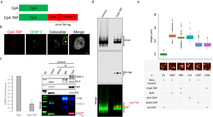
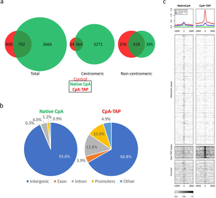
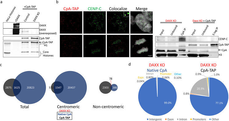
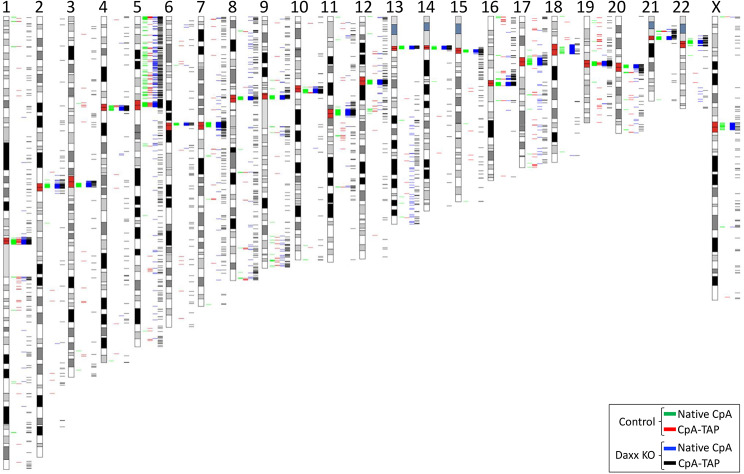
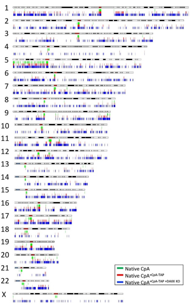
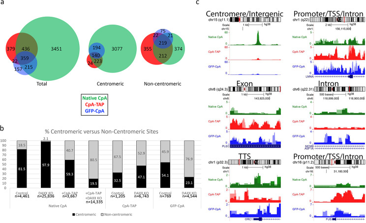
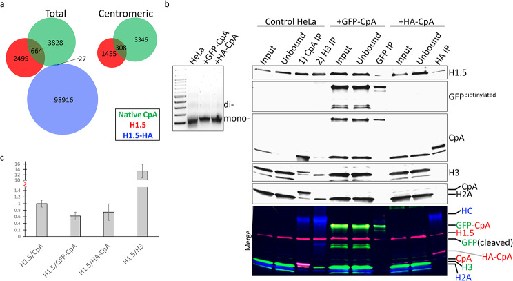
Results: We report that when C-terminally TAP-tagged CENP-A histone variant is introduced, it undergoes altered kinetochore protein binding, differs in post-translational modifications (PTMs), utilizes histone chaperones that differ from that of native CENP-A, and can partially displace native CENP-A in human cells. Additionally, these tagged CENP-A-containing nucleosomes have reduced centromeric incorporation at early G1 phase and poorly associates with linker histone H1.5 compared to native CENP-A nucleosomes.
Conclusions: These data suggest expressing tagged versions of histone variant CENP-A may result in unexpected utilization of non-native pathways, thereby altering the biological function of the histone variant.
Keywords: Atomic force microscopy; CENP-A; Centromere; Chromatin; Chromatin immuno-precipitation; Deep sequencing; Epitope tags; Immuno-fluorescence; Post-translational modifications.
© 2024. This is a U.S. Government work and not under copyright protection in the US; foreign copyright protection may apply.
Conflict of interest statement
The authors declare no competing interests.
Figures
Similar articles
-
Internal modifications in the CENP-A nucleosome modulate centromeric dynamics.Epigenetics Chromatin. 2017 Apr 4;10:17. doi: 10.1186/s13072-017-0124-6. eCollection 2017. Epigenetics Chromatin. 2017. PMID: 28396698 Free PMC article.
-
Identification of the Post-translational Modifications Present in Centromeric Chromatin.Mol Cell Proteomics. 2016 Mar;15(3):918-31. doi: 10.1074/mcp.M115.053710. Epub 2015 Dec 18. Mol Cell Proteomics. 2016. PMID: 26685127 Free PMC article.
-
Histone H4 Lys 20 monomethylation of the CENP-A nucleosome is essential for kinetochore assembly.Dev Cell. 2014 Jun 23;29(6):740-9. doi: 10.1016/j.devcel.2014.05.001. Dev Cell. 2014. PMID: 24960696 Free PMC article.
-
Critical histone post-translational modifications for centromere function and propagation.Cell Cycle. 2017 Jul 3;16(13):1259-1265. doi: 10.1080/15384101.2017.1325044. Epub 2017 Jun 9. Cell Cycle. 2017. PMID: 28598241 Free PMC article. Review.
-
Putting CENP-A in its place.Cell Mol Life Sci. 2013 Feb;70(3):387-406. doi: 10.1007/s00018-012-1048-8. Epub 2012 Jun 23. Cell Mol Life Sci. 2013. PMID: 22729156 Free PMC article. Review.
Cited by
-
Pioneer in Molecular Biology: Conformational Ensembles in Molecular Recognition, Allostery, and Cell Function.J Mol Biol. 2025 Jun 1;437(11):169044. doi: 10.1016/j.jmb.2025.169044. Epub 2025 Feb 25. J Mol Biol. 2025. PMID: 40015370 Review.
-
High-resolution analysis of human centromeric chromatin.Life Sci Alliance. 2025 Jan 23;8(4):e202402819. doi: 10.26508/lsa.202402819. Print 2025 Apr. Life Sci Alliance. 2025. PMID: 39848706 Free PMC article.
References
MeSH terms
Substances
LinkOut - more resources
Full Text Sources
Molecular Biology Databases
Research Materials
Miscellaneous
