

Orbital Metastasis as the First Manifestation of Hepatocellular Carcinoma, and Its Effective Treatment with Combined Dual Immunotherapy: A Case Report and Review of the Literature
- PMID: 38825807
- PMCID: PMC11163833
- DOI: 10.12659/AJCR.944002
Orbital Metastasis as the First Manifestation of Hepatocellular Carcinoma, and Its Effective Treatment with Combined Dual Immunotherapy: A Case Report and Review of the Literature
Abstract
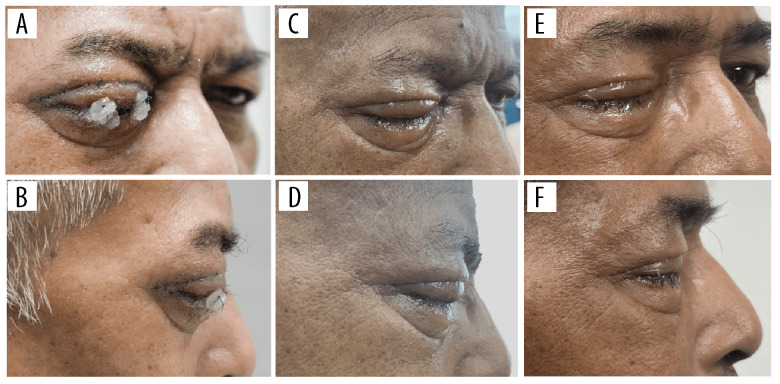
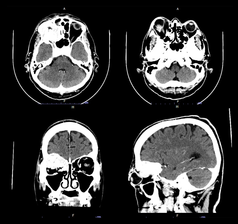
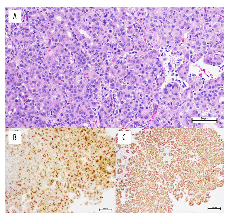
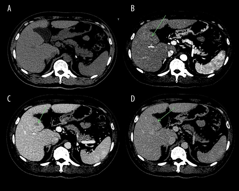
BACKGROUND Orbital metastasis originating from hepatocellular carcinoma (HCC), particularly as an initial manifestation in patients without a known history of HCC, is rare. Few reports exist on the treatment of patients having HCC with orbital metastasis using targeted therapy or immunotherapy. CASE REPORT We report a case of advanced-stage HCC in a 65-year-old man who first presented with progressive, painless blurred vision and proptosis of the right eye for 2 weeks. The patient had no history of chronic liver disease or cancer. Computed tomography revealed an enhancing hyperdense extraconal mass in the right orbit; a biopsy revealed metastatic HCC. Abdominal CT, which was performed to investigate the primary cancer, revealed a 1.2×1.6-cm arterial-enhancing nodule with venous washout in hepatic segment 5, associated with liver cirrhosis. The patient's serum alpha-fetoprotein level was 70.27 ng/dL. Chest computed tomography revealed lung metastasis. Thus, first-line systemic therapy combining durvalumab and tremelimumab was initiated alongside palliative radiotherapy targeting the right orbit, which began 1 week after the first dose of dual immunotherapy. The patient had significant clinical improvement, reduced proptosis, and serum alpha-fetoprotein levels. CONCLUSIONS Although orbital metastasis is a rare manifestation of HCC, physicians should recognize and consider aggressive investigations for early diagnosis, especially in patients with existing risk factors for HCC. Dual immunotherapy with durvalumab and tremelimumab in combination with radiotherapy can be considered a potential treatment option for managing advanced HCC with orbital metastasis.
Conflict of interest statement
Figures
Similar articles
-
Systemic treatments for metastatic cutaneous melanoma.Cochrane Database Syst Rev. 2018 Feb 6;2(2):CD011123. doi: 10.1002/14651858.CD011123.pub2. Cochrane Database Syst Rev. 2018. PMID: 29405038 Free PMC article.
-
Contrast-enhanced ultrasound for the diagnosis of hepatocellular carcinoma in adults with chronic liver disease.Cochrane Database Syst Rev. 2022 Sep 2;9(9):CD013483. doi: 10.1002/14651858.CD013483.pub2. Cochrane Database Syst Rev. 2022. PMID: 36053210 Free PMC article.
-
[Guidelines for the prevention and management of bronchial asthma (2024 edition)].Zhonghua Jie He He Hu Xi Za Zhi. 2025 Mar 12;48(3):208-248. doi: 10.3760/cma.j.cn112147-20241013-00601. Zhonghua Jie He He Hu Xi Za Zhi. 2025. PMID: 40050074 Chinese.
-
Systemic pharmacological treatments for chronic plaque psoriasis: a network meta-analysis.Cochrane Database Syst Rev. 2021 Apr 19;4(4):CD011535. doi: 10.1002/14651858.CD011535.pub4. Cochrane Database Syst Rev. 2021. Update in: Cochrane Database Syst Rev. 2022 May 23;5:CD011535. doi: 10.1002/14651858.CD011535.pub5. PMID: 33871055 Free PMC article. Updated.
-
Liver transplantation in a patient with metastatic hepatocellular carcinoma after downstaging with single-agent immunotherapy: the first case report.J Gastrointest Oncol. 2025 Jun 30;16(3):1321-1330. doi: 10.21037/jgo-24-764. Epub 2025 Jun 26. J Gastrointest Oncol. 2025. PMID: 40672077 Free PMC article.
Cited by
-
From Vision Changes to Hospice: The Rapid Progression of Orbital Metastasis in Hepatocellular Carcinoma.Cureus. 2025 Mar 9;17(3):e80288. doi: 10.7759/cureus.80288. eCollection 2025 Mar. Cureus. 2025. PMID: 40201885 Free PMC article.
References
-
- Sung H, Ferlay J, Siegel RL, et al. Global cancer statistics 2020: GLOBOCAN estimates of incidence and mortality worldwide for 36 cancers in 185 countries. Cancer J Clin. 2021;71:209–49. - PubMed
-
- Llovet JM, Kelley RK, Villanueva A, et al. Hepatocellular carcinoma. Nat Rev Dis Primers. 2021;7:6. - PubMed
-
- Srinivasan R, Krishnanand G. Cytologic diagnosis of metastatic hepatocellular carcinoma presenting as an orbital mass. A case report. Acta Cytol. 2007;51:83–85. - PubMed
-
- Katyal S, Oliver JH, Peterson MS, et al. Extrahepatic metastases of hepato-cellular carcinoma. Radiology. 2000;216:698–703. - PubMed
Publication types
MeSH terms
Substances
LinkOut - more resources
Full Text Sources
Medical
