. 2024 Jun 1;17(6):dmm050570.
doi: 10.1242/dmm.050570. Epub 2024 Jul 2.

Hyperactivation of MEK1 in cortical glutamatergic neurons results in projection axon deficits and aberrant motor learning

Affiliations

Hyperactivation of MEK1 in cortical glutamatergic neurons results in projection axon deficits and aberrant motor learning

George R Bjorklund et al. Dis Model Mech. .

Abstract

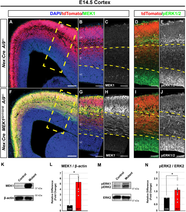
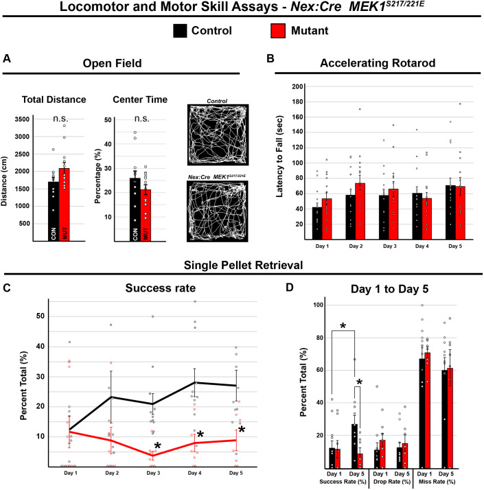
Abnormal extracellular signal-regulated kinase 1/2 (ERK1/2, encoded by Mapk3 and Mapk1, respectively) signaling is linked to multiple neurodevelopmental diseases, especially the RASopathies, which typically exhibit ERK1/2 hyperactivation in neurons and non-neuronal cells. To better understand how excitatory neuron-autonomous ERK1/2 activity regulates forebrain development, we conditionally expressed a hyperactive MEK1 (MAP2K1) mutant, MEK1S217/221E, in cortical excitatory neurons of mice. MEK1S217/221E expression led to persistent hyperactivation of ERK1/2 in cortical axons, but not in soma/nuclei. We noted reduced axonal arborization in multiple target domains in mutant mice and reduced the levels of the activity-dependent protein ARC. These changes did not lead to deficits in voluntary locomotion or accelerating rotarod performance. However, skilled motor learning in a single-pellet retrieval task was significantly diminished in these MEK1S217/221E mutants. Restriction of MEK1S217/221E expression to layer V cortical neurons recapitulated axonal outgrowth deficits but did not affect motor learning. These results suggest that cortical excitatory neuron-autonomous hyperactivation of MEK1 is sufficient to drive deficits in axon outgrowth, which coincide with reduced ARC expression, and deficits in skilled motor learning. Our data indicate that neuron-autonomous decreases in long-range axonal outgrowth may be a key aspect of neuropathogenesis in RASopathies.

Keywords: Axon; Connectivity; Cortex; Development; Kinase; RASopathy.


Conflict of interest statement

Competing interests The authors declare no competing or financial interests.

Figures

Fig. 1.
Fig. 1.
Selective MEK1 hyperactivation in immature glutamatergic neurons results in increased levels of pERK1/2 by E14.5. (A-J) Immunolabeling of embryonic day (E) 14.5 Nex:Cre; MEK1S217/221E; Ai9+/− mutant cortices showed a substantial increase of MEK1 expression in the intermediate zone (IZ) and cortical plate (CP) of mutants (F-H) compared to that in Nex:Cre; Ai9+/− controls (A-C) (n=4). tdTomato+ axons within the IZ of mutants (I,J, white arrows) exhibited a qualitative enrichment in levels of phosphorylated ERK1/2 (pERK1/2) relative to that in controls (D,E) (n=4). We did not detect a notable difference in the levels of pERK1/2 in the ventricular zone (VZ), where recombination is essentially absent in these mice. The integrated density (mean gray value×mm2) of the CP+IZ in panels C,H,E,J is 1.36, 4.70, 4.46 and 4.10, respectively. Scale bars: 100 µm (A,F); 60 µm (B-E,G-J). (K-N) Western blots of E14.5 dorsal cortical lysates (K,M) revealed a highly significant increase in MEK1 (L) (n=5, two-tailed unpaired Student's t-test, *P=0.002) and pERK2 (n=10, two-tailed unpaired Student's t-test, *P=0.005) levels in mutants compared to those in controls. Data are shown as mean±s.e.m.
Fig. 2.
Fig. 2.
Adult Nex:Cre; MEK1S217/221E cortical excitatory neurons exhibit compartment-specific changes in pERK1/2 levels. (A-H) An increase in MEK1 immunolabeling was apparent in the cortical grey matter and corpus collosum of adult Nex:Cre; MEK1S217/221E mice (C,D) relative to that in controls (A,B). Within the mutant grey matter, we noted a significant decrease in pERK1/2-labeled neurons in layers II/III (G,H, magenta arrowheads) compared to those in controls (E-F) (n=6, quantitation in Fig. S1A). The integrated density (mean gray value×mm2) of pERK1/2 labeling in cortical grey matter is 60.1 and 48.4 in panels F and H, respectively. In contrast, pERK1/2 immunolabeling was qualitatively increased in the corpus callosum (CC, dashed yellow outlines) of adult mutants (G,H, yellow arrows, the CC integrated density in H is 7.3) compared to that in controls (E,F, the CC integrated density in F is 2.5). ACC, anterior cingulate cortex; M1, primary motor cortex; M2, secondary motor cortex; st, striatum. (I-L) Representative high-resolution confocal images of NeuN/DAPI-labeled neurons in M1 layer V (I-L, dashed yellow outlines) revealed a decrease in cytoplasmic and nuclear levels of pERK1/2 in mutants (K,L) compared to those in controls (I,J) (n=4, individual neuron quantitation in Fig. S1B). (M-P) Confocal Airyscan imaging was used to better resolve individual TUJ1+ projections in the corpus callosum, likely derived from callosally projecting excitatory neurons. We detected a modest increase in pERK1/2 colocalization with TUJ1 in mutants (O,P, yellow arrows) compared to that in controls (M,N) (n=4, quantification in Fig. S2C). Scale bars: 500 µm (A-H); 10 µm (I-L); 2 µm (M,O); 1 µm (N,P).
Fig. 3.
Fig. 3.
Nex:Cre; MEK1S217/221E mice exhibit disrupted contralateral corticocortical and corticostriatal arborization. (A-D) The adeno-associated viral vector AAV:CAG-Flex-tdTomato was injected into the left primary motor cortex (M1) at postnatal day (P) 1 and brains were collected at P30, as shown in the rostral to caudal serial sections surrounding the injection site (A) and representative images of the injected region in the M1 of the control (B) and Nex:Cre; MEK1S217/221E (C) forebrains. Dashed white and yellow lines in B,C show the boundaries of M1 and the cortex, respectively. No significant difference (n.s.) in the average density of tdTomato-labeled cells was detected between Nex:Cre; MEK1S217/221E and control mice (mean±s.e.m., n=5, two-tailed unpaired Student's t-test, P=0.2839) (D). (E-L) Representative confocal images of contralateral corticocortical and corticostriatal (st, striatum) tdTomato-labeled axonal arborization in Nex:Cre; MEK1S217/221E (H,J) and control mice (E,G). Nex:Cre; MEK1S217/221E mice showed a 19.57±4.30% reduction of normalized intracortical axon arborization (I) compared to that in control mice (mean±s.e.m., n=5, two-tailed unpaired Student's t-test, *P=0.014) (F, quantified in K). Cortical axon arborization in the mutant dorsolateral striatum was reduced by 37.50±9.14% (J) compared to that in control mice (mean±s.e.m., n=5, two-tailed unpaired Student's t-test, *P=0.013) (G, quantified in L). Scale bars: 500 µm (B,C,E,H); 100 µm (F,G,I,J).
Fig. 4.
Fig. 4.
MEK1S217/221E reduces cortical ARC protein expression. (A,B) Western blots of adult cortical lysates (A) and quantification of band intensities (B) showed a significant reduction in ARC protein levels in Nex:Cre; MEK1S217/221E mutants compared to those in control mice (mean±s.e.m., n=11, two-tailed unpaired Student's t-test, *P<0.005). (C-F) Immunolabeling revealed a qualitative reduction of ARC in Nex:Cre; MEK1S217/221E cortices (E,F) compared to that in controls (C,D) (n=5). The integrated density (mean gray value×mm2) of ARC in D and E is 96.6 and 72.3, respectively. M1, primary motor cortex. Scale bars: 200 µm.
Fig. 5.
Fig. 5.
Motor skill acquisition is disrupted by MEK1 hyperactivation in cortical glutamatergic neurons. (A) Open-field testing of Nex:Cre; MEK1S217/221E (n=12) and control (n=12) mice revealed no significant difference (n.s.) in distance traveled (mean±s.e.m., two-tailed unpaired Student's t-test, P=0.075) or percentage of time spent in the center of the arena (mean±s.e.m., two-tailed unpaired Student's t-test, P=0.21). (B) Accelerating rotarod testing revealed a significant effect of day {two-way repeated measures ANOVA, [F(4, 27)=6.144, P<0.001]} over 5 days of testing, but no significant effect of genotype {mean±s.e.m., n=15 controls and 14 mutants, [F(1, 27)=0.246, P=0.624]}. (C,D) In the single-pellet retrieval task, the control success rate significantly improved over 5 days from 12.39±4.50 to 27.06±5.11% [mean±s.e.m., least significant difference (LSD) post hoc two-tailed unpaired t-test for day 1 versus day 5, *P=0.016, n=11]. The Nex:Cre; MEK1S217/221E mutant mice did not show significant improvement and exhibited a significantly lower success rate relative to that for controls during the final 3 days of testing {mean±s.e.m., n=11 controls and 8 mutants, two-way repeated measures ANOVA [F(1,16)=11.04, P=0.004], LSD post hoc test, *P<0.05}.
Fig. 6.
Fig. 6.
MEK1S214/221E expression in cortical layer V reduces corticospinal axon elongation in the spinal cord dorsal fasciculus. (A-G) Sections from Rbp4:Cre; Ai9+/− control (A-C) and Rbp4:Cre; MEK1S17/221E; Ai9+/− mutant (D-F) mice were labeled for descending cortical layer V-derived corticospinal axons (dashed yellow boxes). Analysis of cross-sectional lumbar segments revealed a statistically significant 28% reduction of tdTomato+ labeled pixels in the dorsal funiculus (df) of Rbp4:Cre; MEK1S217/221E; Ai9+/− mice (E,F, dashed yellow outlines) compared to those in Rbp4:Cre; Ai9+/− controls (B,C; quantification in G, mean±s.e.m., n=3, two-tailed unpaired Student's t-test, *P<0.05). The schematic shows a midsagittal outline of the mouse central nervous system, and lumbar segments L3-L5 are indicated. CC, central canal; dh, dorsal horn; vh, ventral horn. Scale bars: 200 µm (A,D); 20 µm (B,C,E,F).
Fig. 7.
Fig. 7.
Corticospinal, corticocortical and corticostriatal layer V projections are reduced in Rbp4:Cre; MEK1S217/221E mutants. (A-E) Unilateral AAV labeling of descending corticospinal axons derived from the primary motor cortex (M1) in Rbp4:Cre mice (A,C). Cross sections of cervical spinal cord segments reveal a reduction of descending tdTomato+ corticospinal axons in the dorsal funiculus of Rbp4:Cre; MEK1S217/221E mutants (D) compared to those in Rbp4:Cre control mice (B) when normalized to corticobulbar tract (CBT) labeling (E, mean±s.e.m., n=4, two-tailed unpaired Student's t-test, *P=0.036). The schematic shows a midsagittal outline of the mouse central nervous system, and the AAV injection site and cervical segments are indicated. (F-O) Virally labeled Rbp4:Cre; MEK1S217/221E axons (F,J) had significantly reduced arborization in the contralateral motor cortex (K) compared to that in Rbp4:Cre controls (G) when normalized to the number of transduced cells in M1 (quantification in N, mean±s.e.m., n=4, two-tailed unpaired Student's t-test, *P=0.019). Additionally, Rbp4:Cre; MEK1S217/221E mice had significantly reduced arborization of the dorsolateral striatum (L,M) compared to that in Rbp4:Cre controls (H,I) (quantification in O, mean±s.e.m., n=5, two-tailed unpaired Student's t-test, **P=0.007). Scale bars: 200 µm (A,C); 20 µm (B,D); 500 µm (F,H,J,L); 150 µm (G,I,K,M). CST, corticospinal tract; df, dorsal funiculus; dh, dorsal horn, iz, intermediate zone, st, striatum; vh, ventral horn.
Fig. 8.
Fig. 8.
MEK1S217/221E in cortical layer V glutamatergic neurons does not disrupt motor learning. (A) Open-field testing of Rbp4:Cre; MEK1S217/221E and control mice revealed that mutants exhibited a significant reduction in total distance traveled (mean±s.e.m., n= 22 controls and 11 mutants, two-tailed unpaired Student's t-test, *P=0.01) and percentage of time spent in the center of box (mean±s.e.m., n=22 controls and 11 mutants, two-tailed unpaired Student's t-test, *P=0.001). (B) Accelerating rotarod testing revealed no significant effect of genotype over 5 days of testing control and mutant mice {mean±s.e.m., n=26 controls and 12 mutants, [F(1,36)=0.007, P=0.933]}. (C,D) In the single-pellet retrieval task, there was no significant effect of genotype on daily success rate {C, mean±s.e.m., n=14 controls and 12 mutants, two-way repeated-measures ANOVA [F(1,24)=0.03, P=0.88]}. Both mutants and controls improved their success rate over 5 days {D, mean±s.e.m., n=14 controls and 12 mutants, two-way repeated-measures ANOVA [F(4,24)=5.81, P<0.01]}.

Similar articles

Cited by

References

    1. Alessi, D. R., Saito, Y., Campbell, D. G., Cohen, P., Sithanandam, G., Rapp, U., Ashworth, A., Marshall, C. J. and Cowley, S. (1994). Identification of the sites in MAP kinase kinase-1 phosphorylated by p74raf-1. EMBO. J. 13, 1610-1619. 10.1002/j.1460-2075.1994.tb06424.x - DOI - PMC - PubMed
    1. Altmüller, F., Pothula, S., Annamneedi, A., Nakhaei-Rad, S., Montenegro-Venegas, C., Pina-Fernández, E., Marini, C., Santos, M., Schanze, D., Montag, D.et al. (2017). Aberrant neuronal activity-induced signaling and gene expression in a mouse model of RASopathy. PLoS Genet. 13, e1006684. 10.1371/journal.pgen.1006684 - DOI - PMC - PubMed
    1. Anastasaki, C. and Gutmann, D. H. (2014). Neuronal NF1/RAS regulation of cyclic AMP requires atypical PKC activation. Hum. Mol. Genet. 23, 6712-6721. 10.1093/hmg/ddu389 - DOI - PMC - PubMed
    1. Andelfinger, G., Marquis, C., Raboisson, M.-J., Théoret, Y., Waldmüller, S., Wiegand, G., Gelb, B. D., Zenker, M., Delrue, M.-A. and Hofbeck, M. (2019). Hypertrophic cardiomyopathy in noonan syndrome treated by MEK-inhibition. J. Am. Coll. Cardiol. 73, 2237-2239. 10.1016/j.jacc.2019.01.066 - DOI - PMC - PubMed
    1. Angara, K., Pai, E. L.-L., Bilinovich, S. M., Stafford, A. M., Nguyen, J. T., Li, K. X., Paul, A., Rubenstein, J. L. and Vogt, D. (2020). Nf1 deletion results in depletion of the Lhx6 transcription factor and a specific loss of parvalbumin + cortical interneurons. Proc. Natl. Acad. Sci. USA. 117, 6189-6195. 10.1073/pnas.1915458117 - DOI - PMC - PubMed

MeSH terms

Substances