[Preprint]. 2024 May 22:2024.05.04.592527.
doi: 10.1101/2024.05.04.592527.

Liquid-like condensates that bind actin drive filament polymerization and bundling

Affiliations

Liquid-like condensates that bind actin drive filament polymerization and bundling

Caleb Walker et al. bioRxiv. .

Update in

Abstract

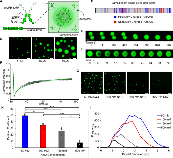
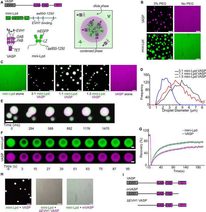
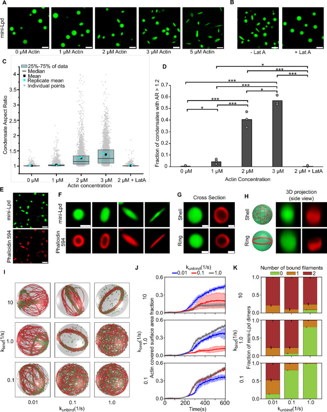
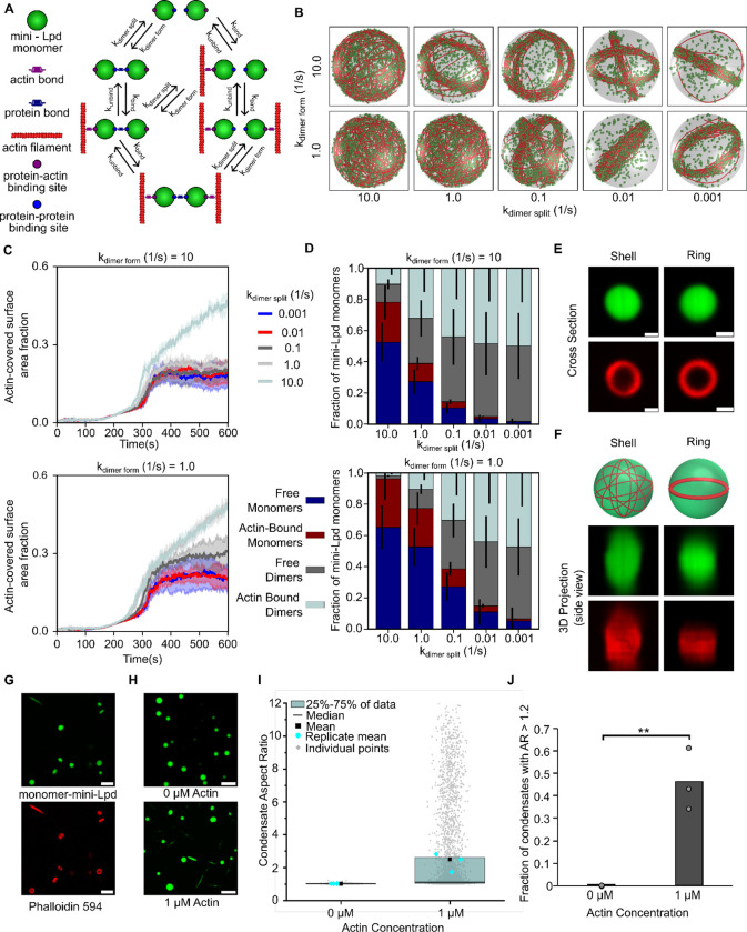
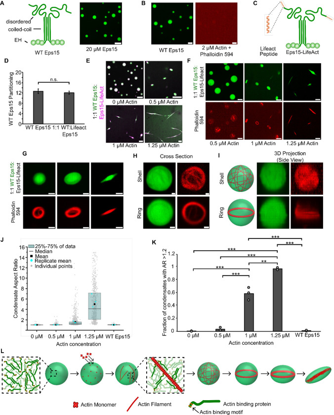
Liquid-like protein condensates perform diverse physiological functions. Previous work showed that VASP, a processive actin polymerase, forms condensates that polymerize and bundle actin. To minimize their curvature, filaments accumulated at the inner condensate surface, ultimately deforming the condensate into a rod-like shape, filled with a bundle of parallel filaments. Here we show that this behavior does not require proteins with specific polymerase activity. Specifically, we found that condensates composed of Lamellipodin, a protein that binds actin but is not an actin polymerase, were also capable of polymerizing and bundling actin filaments. To probe the minimum requirements for condensate-mediated actin bundling, we developed an agent-based computational model. Guided by its predictions, we hypothesized that any condensate-forming protein that binds actin could bundle filaments through multivalent crosslinking. To test this idea, we added an actin-binding motif to Eps15, a condensate-forming protein that does not normally bind actin. The resulting chimera formed condensates that drove efficient actin polymerization and bundling. Collectively, these findings broaden the family of proteins that could organize cytoskeletal filaments to include any actin-binding protein that participates in protein condensation.


Figures

Figure 1.
Figure 1.. A minimal version of Lamellipodin phase separates into liquid-like condensates
A) Left: Schematic depicting domains of mini-Lpd (LZ: Leucine Zipper). Right: schematic depicting condensate formation B) Amino acid sequence of amino acids 850–1250 of Lamellipodin with positively charged (blue) and negatively charged (red) amino acids highlighted. C) Condensates formed by mini-Lpd at increasing protein concentrations in a buffer containing 20 mM Tris (pH 7.4), 150 mM NaCl, and 5 mM TCEP. Scale bars are 5 μm. D) Time course of condensate fusion event for 10 μM mini-Lpd in buffer containing 20 mM Tris (pH 7.4), 50 mM NaCl, and 5 mM TCEP. Scale bar 2 μm. E) Representative images of fluorescence recovery after photobleaching of a mini-Lpd condensate. Scale bar 2 μm. F) Plot of average fluorescence recovery after photobleaching for mini-Lpd condensates. Lines are the average recovery +/− s.d. at each timepoint for each protein across n=6 independent samples. G) mini-Lpd condensates formed in buffers with increasing ionic strength. mini-Lpd concentration is 10 μM in all conditions. Scale bars are 5 μm. H) Quantification of the partitioning of mini-Lpd into condensates formed from 10 μM mini-Lpd under the conditions shown in G. Partition coefficient is defined at the ratio of protein intensity inside the condensates to that in the bulk solution. Bars represent the average across three independent experiments with at least three images quantified per experiment. One asterisk denotes p<.05, two asterisks denote p<.01, and three asterisks denote p<.001 using an unpaired, two-tailed t-test on the means of the replicates N=3. I) Distribution of condensate diameters for the conditions shown in G across three separate replicates for each condition.
Figure 2.
Figure 2.. Interactions between Lamellipodin and VASP mutually stabilize protein condensation.
A) Top: Schematic of domain organization in a VASP monomer (GAB: G-actin binding site; FAB: F-actin binding site; TET: Tetramerization domain) and a mini-Lpd monomer (LZ: Leucine Zipper dimerization motif) Bottom: Schematic of VASP tetramer, mini-Lpd dimer, and condensate formation with both proteins present. Right: Schematic depicting mini-Lpd and VASP co-partitioning into a condensate. B) Condensates form upon the inclusion of 3% w/v PEG in solution for both VASP (magenta) and mini-Lpd (green); however, neither protein forms condensates in buffer lacking PEG. Scale bars 5 μm. Protein concentrations were 20 μM for both the with and without PEG conditions. C) Panels showing representative images of mini-Lpd + VASP condensates formed at various mini-Lpd to VASP ratios. Scale bars 5 μm. D) Distribution of condensate diameters for each condition in C.E) Time course of a condensate fusion event between mini-Lpd (green) and VASP (magenta) condensates. Scale bar 2 μm. F) Representative images of fluorescence recovery after photobleaching of a mini-Lpd (green) and VASP (magenta) condensate. Scale bars 2 μm. G) Plot of average fluorescence recovery after photobleaching for mini-Lpd and VASP condensates formed in the absence of PEG. Lines are the average recovery +/− s.d. at each timepoint for each protein across n=9 independent samples. H) mini-Lpd added to the respective VASP mutant in a 1:1 ratio to test for condensate formation in the absence of PEG. Left – mini Lpd (green) and VASP (magenta), Middle - mini-Lpd (green) and VASPΔEVH1 (magenta), Right - mini-Lpd (green) and monomeric VASP (mVASP) (magenta). Scale bars 5 μm I) Diagrams depict domain structures of VASP mutants. All experiments were performed in a buffer containing 20 mM Tris (pH 7.4), 150 mM NaCl, and 5 mM TCEP in the absence of PEG, except where noted in panel B where 3% (w/v) PEG was included.
Figure 3.
Figure 3.. Condensates of VASP and mini-Lpd polymerize and bundle actin in the absence of crowding agents.
A) Condensates formed from 5 μM mini-Lpd (green) and 10 μM VASP (magenta) are increasingly deformed with the addition of increasing concentrations of G-actin (unlabeled). Scale bars 5 μm B) Phalloidin-iFluor-594 (red) staining of mini-Lpd (green) and VASP (unlabeled) condensates with 0.5 μM monomeric G-actin displaying rings and rods of polymerized actin within the protein condensates. Scale bars 5 μm. C) From left to right: representative confocal images depicting the progression of condensate deformation as actin polymerizes and bundles within the protein condensates. Scale bars 2 μm D) Cartoon depicting the mechanism of actin polymerization within protein condensates and the role it plays in condensate deformation. E) Distribution of condensate aspect ratios across the conditions in A, with at least 400 condensates analyzed for each condition, and more than 800 condensates were analyzed for the 0, 0.25, and 0.5 μM conditions. In the 0.75 μM actin condition values for aspect ratios above 10, corresponding to 4.8% of the data, are not displayed to better visualize distributions for all conditions. The mean indicates the mean of all data points in each condition, while the replicate mean is the mean of each of the individual replicates for each condition. F) Quantification of the fraction of elongated protein condensates, defined as condensates with aspect ratios > 1.2, across the conditions in A. Data are mean across three independent experiments with at least 400 condensates analyzed per condition, and more than 800 condensates were analyzed for the 0, 0.25, and 0.5 μM conditions. Overlayed gray circles denote the means of each replicate. One asterisk denotes p<.05, three asterisks denote p<.001 using an unpaired, two-tailed t-test on the means of the replicates N=3. All experiments were performed in a buffer containing 20 mM Tris (pH 7.4), 150 mM NaCl, and 5 mM TCEP in the absence of PEG.
Figure 4:
Figure 4:. Condensates of mini-Lpd polymerize and bundle actin.
A) The addition of actin at increasing concentrations to condensates formed from 5 μM mini-Lpd (green) results in increasingly deformed protein condensates. Scale bars 5 μm. B) Pretreatment of mini-Lpd condensates with 5 μM latrunculin A (LatA) prior to G-actin addition inhibits actin polymerization and results in spherical condensates. Scale bars 5 μm. C) Distribution of condensate aspect ratios across the conditions in A, with at least 1000 condensates analyzed for each condition. The mean is the mean of all data points in each condition, while the replicate mean is the mean of each of the individual replicates for each condition. D) Quantification of the fraction of high-aspect-ratio protein condensates, defined as condensates with aspect ratios > 1.2, across the conditions in A. Data are mean across three independent experiments with at least 1000 condensates analyzed per condition. Overlayed gray circles denote the means of each replicate. One asterisk denotes p<.05, two asterisks denote p<.01, and three asterisks denote p<.001 using an unpaired, two-tailed t-test on the means of the replicates, N=3. E) Phalloidin-iFluor-594 (red) staining of mini-Lpd condensates (green) with 5 μM monomeric G-actin (unlabeled) added displaying rings and rods of polymerized actin within the protein condensates. Scale bars 5 μm. F) Representative confocal cross-section images of the progression of condensate deformation as a result of actin polymerization. Scale bar 1 μm. G) Representative 2D confocal images of independent mini-Lpd condensates (green) containing peripheral actin, shown with phalloidin staining (red), in a shell of actin (top) and a ring of actin (bottom). Scale bars 1 μm. H) 3D reconstructions of the same mini-Lpd condensates shown in G demonstrating a shell of actin (top) and a ring of actin (bottom). I) Simulations show that bivalent crosslinker kinetics affect actin network organization in LLPS condensates. Representative final snapshots (t = 600 s) from simulations at various binding and unbinding rates within spherical condensates (R = 1 μm) containing 30 actin filaments (red) and 1000 bivalent crosslinkers (green spheres). Please refer to the Supplementary Methods section for a detailed description of the model. The binding rates of the bivalent crosslinkers are varied along each column, and unbinding rates are varied along each row. The polymerization rate at the plus (+) end is constant at 0.0103 μm/s, and neither end undergoes depolymerization. See also Supplementary Movie M2. For additional kinetic conditions, see Figure S4 and Supplementary Movie M3. J) Dynamics of the actin-covered surface area fraction for varied bivalent crosslinker binding and unbinding kinetics. (Data used: 10 replicates) K) Stacked bar graphs representing the fraction of bivalent crosslinkers bound to 0, 1, or 2 actin filaments for each condition. The error bars represent the standard deviation. For J and K, 10 replicates are considered per condition, and the data was obtained from the last 30 snapshots (5%) of each replicate. For analysis of additional kinetic conditions, see Figure S5.
Figure 5:
Figure 5:. Dynamic protein dimers bundle actin filaments.
A) Schematic showing the different possible binding interactions between the monomeric actin binding protein and actin, including both protein-protein interactions (dynamic dimerization) and protein-actin interactions. B) Simulations show that monovalent actin binding can lead to the formation of ring structures when monomers are allowed to interact, bind to form dimers, and function as transient bivalent crosslinkers. Representative final snapshots (t = 600 s) from simulations at various dimer formation and splitting rates within spherical condensates (R = 1 μm) containing 30 actin filaments (red) and 2000 monomers (green spheres) which can form a maximum of 1000 dimers. Please refer to the Supplementary Methods section for a detailed description of the model. The dimer formation rates of the monomers are varied along each column, and dimer splitting rates are varied along each row. Actin-binding kinetics were chosen from previous simulations (Fig. 4I) to correspond to ring-forming conditions for bivalent crosslinkers. The polymerization rate at the plus (+) end is constant at 0.0103 μm/s, and neither end undergoes depolymerization. Also see Supplementary Movie M4. C) Time series showing the mean (solid line) and standard deviation (shaded area) of simulated condensate surface that is covered with actin. The kdimer form value is shown on top of each subpanel while time series are colored by kdimer split values. Data used: 5 replicates. Please refer to supplementary methods for a detailed description. D) Stacked bar graph showing the distribution of condensate protein at different states namely, free monomers, free dimers, actin-bound monomers, and actin-bound dimers. Error bars show standard deviation. Data used: 5 replicates, data from last 30 snapshots. E) Representative 2D confocal cross sections of independent monomer mini-Lpd (green) condensates containing peripheral actin, shown with phalloidin staining (red), in both a shell of actin (top) and a ring of actin (bottom). Scale bars 1 μm. F) 3D reconstructions of the same condensates shown in E demonstrating a shell of actin (top) and a ring of actin (bottom). G) Phalloidin-iFluor-594 (red) staining of monomer-mini-Lpd condensates (green) with 5 μM monomeric G-actin (unlabeled) added displaying polymerized actin within the protein condensates. Scale bars 5 μm. H) The addition of actin to monomer-mini-Lpd also results in the deformation of protein condensates formed from 5 μM monomer-mini-Lpd. Scale bars 5 μm. I) Distribution of condensate aspect ratios across the conditions in H, with at least 1000 condensates analyzed for each condition. For the 1 μM actin condition values for aspect ratios above 10, corresponding to 2.97% of the data, are not displayed to better visualize distributions for all conditions. The mean is the mean of all data points in each condition, while the replicate mean is the mean of each of the individual replicates for each condition. J) Quantification of the fraction of high-aspect-ratio protein condensates, defined as condensates with aspect ratios > 1.2, across the conditions in H. Data are mean across three independent experiments with at least 1000 condensates analyzed per condition. Overlayed gray circles denote the means of each replicate. Two asterisks denote p<.01 using an unpaired, two-tailed t-test on the means of the replicates N=3. All experiments were performed in a buffer containing 20 mM Tris (pH 7.4), 50 mM NaCl, 5 mM TCEP, and 3% (w/v) PEG 8000.
Figure 6.
Figure 6.. Adding an actin-binding domain to an arbitrary condensate-forming protein is sufficient to confer the ability to polymerize and bundle actin filaments.
A) Left: Schematic depicting wild type Eps15 and its major domains. Right: 20 μM wild type Eps15 forms condensates in solution with 3% (w/v) PEG. Scale bar 5 μm. B) Wild-type Eps15 (green) condensates do not polymerize or bundle actin as indicated by the lack of condensate deformation and a lack of phalloidin-stained filaments. Scale bars 5 μm. C) Schematic depicting fusion of the Lifeact peptide to the C-terminus of Eps15. D) Partitioning of wild type Eps15 in condensates formed from only wild type Eps15 (self-partitioning) and condensates formed with 1:1 wild type Eps15 and Eps15-Lifeact. Six fields of view were analyzed for each condition with more than 6 condensates analyzed for partitioning per field of view. Error bars indicate standard deviation. E) Representative images of condensate deformation and actin polymerization upon the addition of increasing concentrations of monomeric actin to condensates formed from wild-type Eps15 and Eps15-Lifeact. Scale bars 5 μm. F) Representative images of phalloidin staining of condensates consisting of wild-type Eps15 and Eps15-Lifeact following actin addition. Scale bars 5 μm. G) Representative confocal cross-section images of the progressive, actin-driven deformation of condensates consisting of wild-type Eps15 and Eps15-Lifeact. Scale bars 2 μm. H) Representative 2D confocal images of independent 1:1 WT Eps15:Eps15-Lifeact condensates (green) containing peripheral actin, shown with phalloidin staining (red), in both a shell of actin (top) and a ring of actin (bottom). Scale bars 1μm. I) 3D reconstructions of the same condensates shown in H demonstrating a shell of actin (top) and a ring of actin (bottom). J) Distribution of condensate aspect ratios across the conditions in E, with at least 700 condensates analyzed for each condition. For the 1.25 μM actin condition, values for aspect ratios higher than 15, corresponding to 3.5% of the data, are not displayed to better visualize distributions for all conditions. The mean is the mean of all data points in each condition, while the replicate mean is the mean of each of the individual replicates for each condition. K) Quantification of the fraction of elongated protein condensates, defined as condensates with aspect ratios > 1.2, across the conditions in E. Data are mean +/− standard deviation across three independent experiments with at least 700 condensates analyzed in total per condition. Overlayed gray circles denote the means of each replicate. Two asterisks denote p<.01, and three asterisks denote p<.001 using an unpaired, two-tailed t-test on the means of the replicates N=3. L) Cartoon depicting the proposed mechanism of condensate-driven actin polymerization and bundling by condensates of actin-binding proteins. All experiments were performed in a buffer containing 20 mM Tris (pH 7.4), 150 mM NaCl, 5 mM TCEP, and 3% (w/v) PEG 8000.

Similar articles

References

    1. Fletcher D. A. & Mullins R. D. Cell mechanics and the cytoskeleton. Nature 463, 485–492 (2010). - PMC - PubMed
    1. Hinze C. & Boucrot E. Local actin polymerization during endocytic carrier formation. Biochem. Soc. Trans. 46, 565–576 (2018). - PubMed
    1. Pollard T. D. & Cooper J. A. Actin, a Central Player in Cell Shape and Movement. Science 326, 1208–1212 (2009). - PMC - PubMed
    1. Vasioukhin V., Bauer C., Yin M. & Fuchs E. Directed Actin Polymerization Is the Driving Force for Epithelial Cell–Cell Adhesion. Cell 100, 209–219 (2000). - PubMed
    1. Campellone K. G. & Welch M. D. A nucleator arms race: cellular control of actin assembly. Nat. Rev. Mol. Cell Biol. 11, 237–251 (2010). - PMC - PubMed

Publication types